A transcriptional corepressor of Stat1 with an essential LXXLL signature motif
- PMID: 11248056
- PMCID: PMC30631
- DOI: 10.1073/pnas.051489598
A transcriptional corepressor of Stat1 with an essential LXXLL signature motif
Abstract
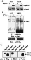
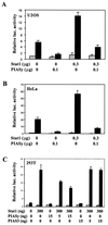
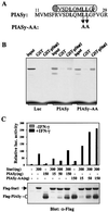
Interferon (IFN) treatment induces tyrosine phosphorylation and nuclear translocation of Stat1 (signal transducer and activator of transcription) to activate or repress transcription. We report here that a member of the protein inhibitor of activated STAT family, PIASy, is a transcriptional corepressor of Stat1. IFN treatment triggers the in vivo interaction of Stat1 with PIASy, which represses Stat1-mediated gene activation without blocking the DNA binding activity of Stat1. An LXXLL coregulator signature motif located near the NH(2) terminus of PIASy, although not involved in the PIASy-Stat1 interaction, is required for the transrepression activity of PIASy. Our studies identify PIASy as a transcriptional corepressor of Stat1 and suggest that different PIAS proteins may repress STAT-mediated gene activation through distinct mechanisms.
Figures


Similar articles
-
Distinct effects of PIAS proteins on androgen-mediated gene activation in prostate cancer cells.Oncogene. 2001 Jun 28;20(29):3880-7. doi: 10.1038/sj.onc.1204489. Oncogene. 2001. PMID: 11439351
-
Protein inhibitor of activated STAT Y (PIASy) and a splice variant lacking exon 6 enhance sumoylation but are not essential for embryogenesis and adult life.Mol Cell Biol. 2004 Jun;24(12):5577-86. doi: 10.1128/MCB.24.12.5577-5586.2004. Mol Cell Biol. 2004. PMID: 15169916 Free PMC article.
-
Repression of Smad transcriptional activity by PIASy, an inhibitor of activated STAT.Proc Natl Acad Sci U S A. 2003 Aug 19;100(17):9791-6. doi: 10.1073/pnas.1733973100. Epub 2003 Aug 6. Proc Natl Acad Sci U S A. 2003. PMID: 12904571 Free PMC article.
-
A putative protein inhibitor of activated STAT (PIASy) interacts with p53 and inhibits p53-mediated transactivation but not apoptosis.Apoptosis. 2001 Jun;6(3):221-34. doi: 10.1023/a:1011392811628. Apoptosis. 2001. PMID: 11388671
-
Convergence of interferon-gamma and progesterone signaling pathways in human endometrium: role of PIASy (protein inhibitor of activated signal transducer and activator of transcription-y).Mol Endocrinol. 2004 Aug;18(8):1988-99. doi: 10.1210/me.2003-0467. Epub 2004 May 20. Mol Endocrinol. 2004. PMID: 15155784
Cited by
-
PIAS proteins: pleiotropic interactors associated with SUMO.Cell Mol Life Sci. 2009 Sep;66(18):3029-41. doi: 10.1007/s00018-009-0061-z. Epub 2009 Jun 13. Cell Mol Life Sci. 2009. PMID: 19526197 Free PMC article. Review.
-
A conserved domain in the leader proteinase of foot-and-mouth disease virus is required for proper subcellular localization and function.J Virol. 2009 Feb;83(4):1800-10. doi: 10.1128/JVI.02112-08. Epub 2008 Dec 3. J Virol. 2009. PMID: 19052079 Free PMC article.
-
Physical and functional interactions of histone deacetylase 3 with TFII-I family proteins and PIASxbeta.Proc Natl Acad Sci U S A. 2002 Oct 1;99(20):12807-12. doi: 10.1073/pnas.192464499. Epub 2002 Sep 18. Proc Natl Acad Sci U S A. 2002. PMID: 12239342 Free PMC article.
-
Generation of a Quantitative Luciferase Reporter for Sox9 SUMOylation.Int J Mol Sci. 2020 Feb 13;21(4):1274. doi: 10.3390/ijms21041274. Int J Mol Sci. 2020. PMID: 32070068 Free PMC article.
-
Regulation of ULK1 Expression and Autophagy by STAT1.J Biol Chem. 2017 Feb 3;292(5):1899-1909. doi: 10.1074/jbc.M116.771584. Epub 2016 Dec 23. J Biol Chem. 2017. PMID: 28011640 Free PMC article.
References
Publication types
MeSH terms
Substances
Grants and funding
LinkOut - more resources
Full Text Sources
Molecular Biology Databases
Research Materials
Miscellaneous

