. 2001 Apr;75(7):3325-34.
doi: 10.1128/JVI.75.7.3325-3334.2001.

Detection of direct binding of human herpesvirus 8-encoded interleukin-6 (vIL-6) to both gp130 and IL-6 receptor (IL-6R) and identification of amino acid residues of vIL-6 important for IL-6R-dependent and -independent signaling

Affiliations

Detection of direct binding of human herpesvirus 8-encoded interleukin-6 (vIL-6) to both gp130 and IL-6 receptor (IL-6R) and identification of amino acid residues of vIL-6 important for IL-6R-dependent and -independent signaling

H Li et al. J Virol. 2001 Apr.

Abstract

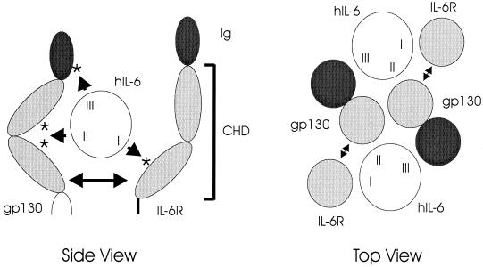
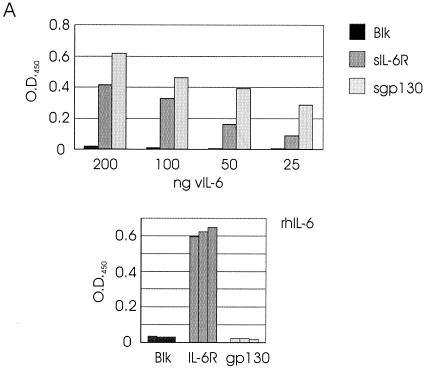
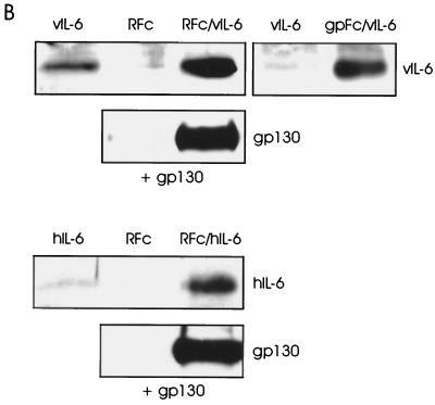
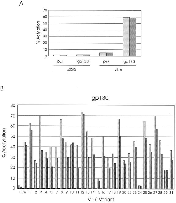
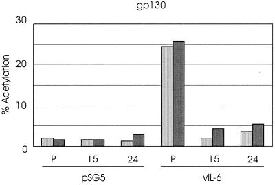
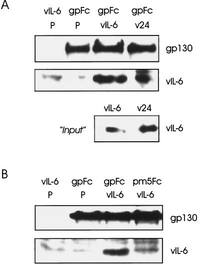
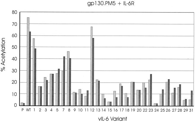
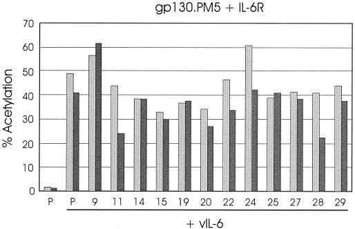
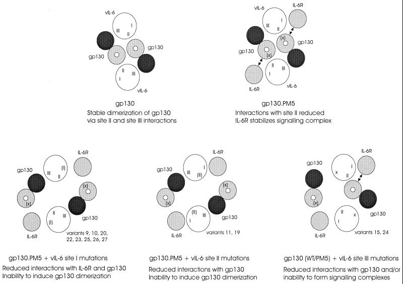
Human herpesvirus 8 (HHV-8) is associated with Kaposi's sarcoma, primary effusion lymphoma, and multicentric Castleman's disease; in all of these diseases, interleukin-6 (IL-6) has been implicated as a likely mitogenic and/or angiogenic factor. HHV-8 encodes a homologue of IL-6 (viral IL-6 [vIL-6]) that has been shown to be biologically active in several assays and whose activities mirror those of its mammalian counterparts. Like these proteins, vIL-6 mediates its effects through the gp130 signal transducer, but signaling is not dependent on the structurally related IL-6 receptor (IL-6R; gp80) subunit of the receptor-signal transducer complex. However, as we have shown previously, IL-6R can enhance vIL-6 signal transduction and can enable signaling through a gp130 variant (gp130.PM5) that is itself unable to support vIL-6 activity, indicating that IL-6R can form part of the signaling complex. Also, our analysis of a panel of vIL-6 mutants in transfection experiments in Hep3B cells (that express IL-6R and gp130) showed that most were able to function normally in this system. Here, we have used in vitro vIL-6-receptor binding assays to demonstrate direct binding of vIL-6 to both gp130 and IL-6R and vIL-6-induced gp130-IL-6R complex formation, and we have extended our functional analyses of the vIL-6 variants to identify residues important for IL-6R-independent and IL-6R-dependent signaling through native gp130 and gp130.PM5, respectively. These studies have identified residues in vIL-6 that are important for IL-6R-independent and IL-6R-mediated functional complex formation between vIL-6 and gp130 and that may be involved directly in binding to gp130 and IL-6R.


Figures

FIG. 1
FIG. 1
Diagrammatic representations of the cytokine receptor homology and immunoglobulin-like domains (CHD and Ig, respectively) of gp130 and IL-6R, their interactions with hIL-6, and complex formation resulting from these interactions. The figure on the left is a side view that indicates the general locations (asterisks) of receptor residues known to interact with hIL-6. Sites II and III of hIL-6 interact with distinct gp130 molecules to form, in association with IL-6R, the functional hexameric complex, as shown schematically in the right panel (top view); for IL-6R, only the hIL-6 site I-interacting CHD region is depicted (individual, lightly shaded circle), with CHD (site II-interacting) and Ig (site III-interacting) regions of gp130 indicated as adjoined lightly and darkly shaded circles, respectively. There is strong experimental evidence for an IL-6R–gp130 dimerization interface (27, 37), here indicated by double-headed arrows.
FIG. 2
FIG. 2
In vitro binding of vIL-6 to IL-6R and gp130. (A) ELISA techniques (see Materials and Methods) were used for the detection of binding by vIL-6 and hIL-6 of sgp130 and sIL-6R. For vIL-6, various amounts (25 to 200 ng) of bacterially produced, purified His6–vIL-6 fusion protein were applied to microassay plate wells coated with sIL-6R or sgp130 or to untreated wells (Blk). After incubation and washing, bound ligand was detected with vIL-6 rabbit antiserum (35) and HRP-conjugated α-rabbit-IgG secondary antibody. Visualization and quantitation was carried out using ABTS reagent and the determination of the OD450 (top panel). Analogous assays were undertaken with rhIL-6 using HRP-conjugated α-hIL-6 as the detection antibody. Experiments were performed in triplicate using 100 ng of ligand (bottom panel). (B) vIL-6 binding to IL-6R and gp130 was determined also by coprecipitation assays using transfected cell media containing sIL-6R–Fc (RFc) or sgp130-Fc (gpFc) together with media containing vIL-6 (see Materials and Methods). Protein A-Sepharose-precipitated material was analyzed by Western blot for the detection of vIL-6. Analogous experiments were undertaken with hIL-6. Inclusion of sgp130 in the sIL-6R–Fc/ligand-binding assays, followed by Western analysis to detect coprecipitated sgp130, demonstrated that vIL-6, in addition to hIL-6, could induce gp130–IL-6R complexing. Experiments using vIL-6 or hIL-6 in the absence of Fc fusion protein or sIL-6R-Fc plus sgp130 in the absence of ligand were included to control for nonspecific binding of proteins to protein A-Sepharose.
FIG. 2
FIG. 2
In vitro binding of vIL-6 to IL-6R and gp130. (A) ELISA techniques (see Materials and Methods) were used for the detection of binding by vIL-6 and hIL-6 of sgp130 and sIL-6R. For vIL-6, various amounts (25 to 200 ng) of bacterially produced, purified His6–vIL-6 fusion protein were applied to microassay plate wells coated with sIL-6R or sgp130 or to untreated wells (Blk). After incubation and washing, bound ligand was detected with vIL-6 rabbit antiserum (35) and HRP-conjugated α-rabbit-IgG secondary antibody. Visualization and quantitation was carried out using ABTS reagent and the determination of the OD450 (top panel). Analogous assays were undertaken with rhIL-6 using HRP-conjugated α-hIL-6 as the detection antibody. Experiments were performed in triplicate using 100 ng of ligand (bottom panel). (B) vIL-6 binding to IL-6R and gp130 was determined also by coprecipitation assays using transfected cell media containing sIL-6R–Fc (RFc) or sgp130-Fc (gpFc) together with media containing vIL-6 (see Materials and Methods). Protein A-Sepharose-precipitated material was analyzed by Western blot for the detection of vIL-6. Analogous experiments were undertaken with hIL-6. Inclusion of sgp130 in the sIL-6R–Fc/ligand-binding assays, followed by Western analysis to detect coprecipitated sgp130, demonstrated that vIL-6, in addition to hIL-6, could induce gp130–IL-6R complexing. Experiments using vIL-6 or hIL-6 in the absence of Fc fusion protein or sIL-6R-Fc plus sgp130 in the absence of ligand were included to control for nonspecific binding of proteins to protein A-Sepharose.
FIG. 3
FIG. 3
Signaling by vIL-6 variants through overexpressed gp130. (A) Dependence on overexpressed gp130 of high-level activation of the STAT binding site-containing pα2MCAT reporter plasmid was tested by comparing the levels of CAT expression in HEK293-T cells cotransfected with pSG5–vIL-6 plus pα2MCAT and either pEF-BOS–hgp130 or the empty pEF-BOS expression vector. Similar transfection experiments were performed in which pSG5 was substituted for pSG5–vIL-6 to ensure that gp130 overexpression alone did not influence pα2MCAT expression. (B) pSG5-cloned vIL-6 (WT) and vIL-6 variants (see Table 1), or pSG5 (P, negative control), were cotransfected into HEK293-T cells, together with pEF-BOS–hgp130 and pα2MCAT. Cells were harvested 2 days after transfection, and CAT activities present in cell extracts were assayed to determine the relative activities of each of the vIL-6 variants relative to wild-type vIL-6. The results of duplicate experiments are shown.
FIG. 4
FIG. 4
Inhibition of gp130-mediated vIL-6 signaling by functionally negative vIL-6 variants. Plasmids expressing vIL-6 variants 15 or 24, or pSG5 (P), were cotransfected with pEF-BOS–hgp130 and pα2MCAT and either pSG5–vIL-6 or pSG5. Results with pSG5 confirmed those obtained previously (Fig. 3B); these altered vIL-6 proteins are unable to signal through gp130. Coexpression of variants 15 and 24 with vIL-6 resulted in almost complete inhibition of vIL-6 signaling. The results of duplicate experiments are shown.
FIG. 5
FIG. 5
Interactions of vIL-6 and derivatives with gp130 and gp130.PM5. Media from cells transfected independently with vIL-6, vIL-6.24 (v24), sgp130-Fc (gpFc), and sgp130.PM5-Fc (pm5Fc) expression plasmids or pSG5 (negative control; P) were mixed, as indicated, precipitated with protein A-Sepharose, and size fractionated by SDS-PAGE, and component proteins were detected by Western blot analysis (see Materials and Methods). (A) The relative gp130 binding activities of vIL-6 and vIL-6.24 were determined from the amounts of coprecipitated vIL-6 protein. The amounts of sgp130-Fc protein precipitated were equivalent (top panel). The first lane shows no cross-reaction of the gp130 antibody with proteins binding (nonspecifically) to the protein A-Sepharose; there was a protein species, migrating slightly slower than vIL-6, that cross-reacted with the vIL-6 antiserum. The relative amounts of the vIL-6 proteins present in samples of the transfected cell media used in the binding assays are indicated (bottom panel). (B) Similar experiments using sgp130.PM5-Fc showed that vIL-6 was not coprecipitated with the altered receptor subunit but was coprecipitated with sgp130-Fc.
FIG. 6
FIG. 6
IL-6R-dependent signaling of vIL-6 variants through gp130.PM5. Transfection assays similar to those outlined in Fig. 3 were carried out to investigate the profile of activities of the vIL-6 variants, relative to pSG5 (P) and vIL-6 (WT) signaling through overexpressed gp130.PM5 and IL-6R. The results of duplicate experiments are shown.
FIG. 7
FIG. 7
Effects of functionally altered vIL-6 variants on vIL-6 signaling through gp130.PM5–IL-6R. Cotransfections of selected vIL-6 variants or pSG5 (P) with wild-type vIL-6 and gp130.PM5, IL-6R, and pα2MCAT were undertaken (see Materials and Methods). The CAT activities present in cell extracts from duplicate experiments are shown.
FIG. 8
FIG. 8
Models of vIL-6 interactions with gp130 and IL-6R, formation of functional signaling complexes, and effects of vIL-6 mutations on complex formation. Functional complexes, in which gp130 molecules (marked by white dots) are brought into close proximity, result from interactions of vIL-6 (e.g., via sites I and II) with different gp130 molecules. Amino acid changes affecting but not destroying interaction sites (e.g., the PM5 mutation in gp130, which occurs in one of two loci predicted to interact with vIL-6 site II) are indicated by parentheses [(X), (I), (II)]; site III changes in vIL-6 variants 15 and 24 (indicated by an “X” at the appropriate position in vIL-6) are presumed to abolish receptor interactions through this site.

Similar articles

Cited by

References

    1. Aoki Y, Jaffe E S, Chang Y, Jones K, Teruya-Feldstein J, Moore P S, Tosato G. Angiogenesis and hematopoiesis induced by Kaposi's sarcoma-associated herpesvirus-encoded interleukin-6. Blood. 1999;93:4034–4043. - PubMed
    1. Asou H, Said J W, Yang R, Munker R, Park D J, Kamada N, Koeffler H P. Mechanisms of growth control of Kaposi's sarcoma-associated herpes virus-associated primary effusion lymphoma cells. Blood. 1998;91:2475–2481. - PubMed
    1. Brakenhoff J P J, de Hon F D, Fontaine V, Ten Boekel E, Schooltink H, Rose-John S, Heinrich P C, Content J, Aarden L A. Development of a human interleukin-6 receptor agonist. J Biol Chem. 1994;269:86–93. - PubMed
    1. Bravo J, Staunton D, Heath J K, Jones E Y. Crystal structure of a cytokine-binding region of gp130. EMBO J. 1998;17:1665–1674. - PMC - PubMed
    1. Burger R, Neipel F, Fleckenstein B, Savino R, Ciliberto G, Kalden J R, Gramatzki G. Human herpesvirus type 8 interleukin-6 homologue is functionally active on human melanoma cell lines. Blood. 1998;91:1858–1863. - PubMed

Publication types

MeSH terms