. 2001 Mar 1;20(5):1033-41.
doi: 10.1093/emboj/20.5.1033.

Reversible inhibition of Hsp70 chaperone function by Scythe and Reaper

Affiliations

Reversible inhibition of Hsp70 chaperone function by Scythe and Reaper

K Thress et al. EMBO J. .

Abstract

Protein folding mediated by the Hsp70 family of molecular chaperones requires both ATP and the co-chaperone Hdj-1. BAG-1 was recently identified as a bcl-2-interacting, anti-apoptotic protein that binds to the ATPase domain of Hsp70 and prevents the release of the substrate. While this suggested that cells had the potential to modulate Hsp70-mediated protein folding, physiological regulators of BAG-1 have yet to be identified. We report here that the apoptotic regulator Scythe, originally isolated through binding to the potent apoptotic inducer Reaper, shares limited sequence identity with BAG-1 and inhibits Hsp70- mediated protein refolding. Scythe-mediated inhibition of Hsp70 is reversed by Reaper, providing evidence for the regulated reversible inhibition of chaperone activity. As Scythe functions downstream of Reaper in apoptotic induction, these findings suggest that Scythe/Reaper may signal apoptosis, in part through regulating the folding and activity of apoptotic signaling molecules.


Figures

None
Fig. 1. Scythe structurally resembles BAG family proteins. (A) The domains of several BAG family members along with Xenopus and human Scythe showing the relative positions of the ubiquitin-like motif (black) and C-terminal ‘BAG’ domain (striped). The complete open reading frame of BAG-3 has yet to be fully sequenced. C.e., Caenorhabditis elegans; S.p., Schizosaccharomyces pombe. (B) Alignment of the C-terminal BAG domains of the proteins in (A). Dark gray and light gray shading indicate identical and similar residues, respectively.
None
Fig. 2. Scythe binds Hsp70/Hsc70 in a BAG domain-dependent fashion. (AIn vitro translated Xenopus Hsc70 (IVT Hsc70) was added to extracts and incubated for 30 min at 4°C. GST or GST–Scythe C312 immobilized on glutathione–Sepharose beads was added to the extract and incubated for an additional 30 min. The beads were then washed three times with egg lysis buffer (ELB), resolved by SDS–PAGE and bands were visualized by autoradiography. (B) GST or the indicated GST fusion protein was immobilized on glutathione–Sepharose beads and incubated in the presence of Xenopus egg extract for 1 h at 4°C. The beads were then washed three times with ELB, resolved by SDS–PAGE and processed for western blotting with an anti-Scythe polyclonal antibody. (C) Antibodies against Xenopus Wee1 or Hsc70 were coupled to Protein A–Sepharose (PAS) beads and then incubated in Xenopus egg extract for 1 h at 4°C. Immunoprecipitates were washed three times with ELB, resolved by SDS–PAGE and processed for immunoblotting with an anti-Scythe polyclonal antibody. (D) 293T cells were transfected with 3 µg of either the indicated myc-tagged human Scythe construct (hScythe) or the parental myc plasmid alone (myc alone). Thirty-six hours after transfection, cells were lysed, centrifuged, and the supernatants incubated with a monoclonal myc antibody for 1 h at 4°C. PAS beads were then added and, after an additional 1 h incubation, the beads were pelleted, washed three times in lysis buffer, and bound proteins were resolved by SDS–PAGE. After western transfer, the blots were probed with an anti-Hsp70/Hsc70 monoclonal antibody. (E) The identical samples processed in (D) were run on a parallel SDS gel and proteins were stained with Coomassie Brilliant Blue. (F) His-tagged Scythe, His-tagged Scythe lacking the BAG domain (His-Scythe ΔC) or His-tagged BAG-1 was incubated in the presence of either full-length Hsp70 (FL Hsp70) or the ATPase domain of Hsp70 (ATPase) for 1 h at 4°C. His-tagged proteins were recovered using a nickel resin and bound proteins were separated by SDS–PAGE. After western transfer, the blots were probed with an anti-Hsp70 monoclonal antibody.
None
Fig. 3. Scythe functions as a negative regulator of Hsp70 chaperone activity. The inhibitory effect of full-length human Scythe (FL hScythe, 3.2 µM) and a truncated Scythe mutant lacking the ‘BAG’ domain (Scythe ΔC, 3.2 µM) on Hsp70-dependent refolding was examined by the percentage recovered activity of unfolded β-galactosidase (3.2 nM) diluted into a refolding buffer containing ATP (1 mM), Hsp70 (1.6 µM) and Hdj-1 (3.2 µM). As a control for spontaneous refolding, denatured β-galactosidase (3.2 nM) was diluted into refolding buffer containing 3.2 µM bovine serum albumin (BSA).
None
Fig. 4. Reaper specifically relieves Scythe-mediated inhibition of Hsp70. (A) The experiment shown in Figure 3 was repeated with 3.2 µM human Scythe in the presence of increasing concentrations of Reaper (3.2–12.8 µM) to examine the reversal of Scythe-mediated inhibition on Hsp70-mediated refolding. (B) The inhibitory effect of BAG-1 (1.6 µM) on refolding was observed in the presence or absence of 12.8 µM Reaper, demonstrating that Reaper-induced reversal is specific for Scythe. As a control for spontaneous refolding, denatured β-galactosidase (3.2 nM) was diluted into refolding buffer containing 3.2 µM BSA.
None
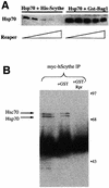
Fig. 5. Reaper specifically inhibits the physical association of Scythe and Hsp70. (A) His-Scythe (1 µM) or GST–BAG-1 (1 µM) was incubated with Hsp70 (1 µM) in refolding buffer. After complex formation, increasing concentrations (0, 2, 4, 8, 10 µM) of Reaper were added. Bound proteins were precipitated with either Ni+-agarose (His-Scythe) or glutathione–Sepharose (GST–BAG-1), washed, resolved by SDS–PAGE and processed for western blotting using Hsp70 monoclonal antibody 5a5. (B) 293T cells were transfected with 5 µg of myc-tagged human Scythe (myc-hScythe). Thirty-six hours after transfection, cells were lysed and centrifuged, and supernatants were incubated with recombinant GST or GST–Reaper (GST–Rpr) for 30 min at 4°C. Subsequently, the lysates were incubated with a monoclonal myc antibody for 1 h at 4°C. PAS beads were then added and, after an additional 1 h incubation, the beads were pelleted, washed three times in lysis buffer, and bound proteins were resolved by SDS–PAGE. After western transfer, the blots were probed with an anti-Hsc70 monoclonal antibody.
None
Fig. 6. BAG-deficient human Scythe is unable to protect against Reaper-induced apoptosis. (A) Recombinant Reaper (Rpr) protein (300 ng/µl) was added to Xenopus egg extracts in combination with equivalent amounts of recombinant full-length human Scythe (FL hScythe), BAG-deficient human Scythe (ΔC hScythe) or just the BAG domain of human Scythe (Scythe BAG). At the indicated times, 2 µl aliquots of extract were analyzed for caspase activity by cleavage of the artificial caspase substrate, DEVD-pNA. Following cleavage, released pNA was measured spectrophotometrically. (B) Samples were processed as in (A), but 15 µl aliquots were filtered through a 0.1 µM microfilter to remove particulate components, including mito chondria, and samples were processed for immunoblotting with an anti-cytochrome c monoclonal antibody. (C) Recombinant GST or GST–Reaper fusion proteins immobilized on glutathione–Sepharose beads were added to Xenopus egg extract and incubated at 4°C in the presence of equivalent amounts of either His-tagged full-length human Scythe (FL hScythe) or BAG-deficient Scythe (ΔC hScythe) proteins. After 1 h, the beads were pelleted, washed three times with ELB, resuspended in SDS sample buffer, and processed for immunoblotting using a monoclonal penta-His antibody.
None
Fig. 7. Model for Reaper/Scythe function. BAG-1 binds to the ATPase domain of Hsp70 and inhibits its ability to mediate protein folding. In the presence of a hypothetical ligand, BAG-1 is released from Hsp70, promoting release of native substrate. In the case of Scythe we hypothesize that a similar series of events occurs; however, Reaper serves as the ligand to trigger Scythe dissociation from the Hsp70 complex. According to this speculative model, ‘X’ is released in its native form and can then trigger mitochondrial cytochrome c release and caspase activation. The figure has been adapted from Bimston et al. (1998).

Similar articles

Cited by

References

    1. Adams J.M. and Cory,S. (1998) The Bcl-2 protein family: arbiters of cell survival. Science, 281, 1322–1326. - PubMed
    1. Adams M.D. et al. (2000) The genome sequence of Drosophila melanogaster. Science, 287, 2185–2195. - PubMed
    1. Bardelli A., Longati,P., Albero,D., Goruppi,S., Schneider,C., Ponzetto,C. and Comoglio,P.M. (1996) HGF receptor associates with the anti-apoptotic protein BAG-1 and prevents cell death. EMBO J., 15, 6205–6212. - PMC - PubMed
    1. Bimston D., Song,J., Winchester,D., Takayama,S., Reed,J.C. and Morimoto,R.I. (1998) BAG-1, a negative regulator of Hsp70 chaperone activity, uncouples nucleotide hydrolysis from substrate release. EMBO J., 17, 6871–6878. - PMC - PubMed
    1. Demand J., Luders,J. and Hohfeld,J. (1998) The carboxy-terminal domain of Hsc70 provides binding sites for a distinct set of chaperone cofactors. Mol. Cell. Biol., 18, 2023–2028. - PMC - PubMed

Publication types

MeSH terms