Top-down morphogenesis of colorectal tumors
- PMID: 11226292
- PMCID: PMC30191
- DOI: 10.1073/pnas.051629398
Top-down morphogenesis of colorectal tumors
Abstract
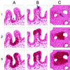
One of the fundamental tenets of oncology is that tumors arise from stem cells. In the colon, stem cells are thought to reside at the base of crypts. In the early stages of tumorigenesis, however, dysplastic cells are routinely found at the luminal surface of the crypts whereas the cells at the bases of these same crypts appear morphologically normal. To understand this discrepancy, we evaluated the molecular characteristics of cells isolated from the bases and orifices of the same crypts in small colorectal adenomas. We found that the dysplastic cells at the tops of the crypts often exhibited genetic alterations of adenomatous polyposis coli (APC) and neoplasia-associated patterns of gene expression. In contrast, cells located at the base of these same crypts did not contain such alterations and were not clonally related to the contiguous transformed cells above them. These results imply that development of adenomatous polyps proceeds through a top-down mechanism. Genetically altered cells in the superficial portions of the mucosae spread laterally and downward to form new crypts that first connect to preexisting normal crypts and eventually replace them.
Figures


Comment in
-
Initial transformed cells of colorectal adenoma: do they occur at the top of the crypt?J Gastroenterol. 2002;37(11):982-4. doi: 10.1007/s005350200165. J Gastroenterol. 2002. PMID: 12483257 No abstract available.
Similar articles
-
Clonality assessment and clonal ordering of individual neoplastic crypts shows polyclonality of colorectal adenomas.Gastroenterology. 2010 Apr;138(4):1441-54, 1454.e1-7. doi: 10.1053/j.gastro.2010.01.033. Epub 2010 Jan 25. Gastroenterology. 2010. PMID: 20102718
-
Clonality of dysplastic epithelium in colorectal adenomas from familial adenomatous polyposis patients.Cancer Res. 1997 Feb 1;57(3):355-61. Cancer Res. 1997. PMID: 9012454
-
APC mutation and the crypt cycle in murine and human intestine.Am J Pathol. 1997 Mar;150(3):833-9. Am J Pathol. 1997. PMID: 9060821 Free PMC article.
-
Multistep carcinogenesis in colorectal cancers.Southeast Asian J Trop Med Public Health. 1995;26 Suppl 1:190-6. Southeast Asian J Trop Med Public Health. 1995. PMID: 8629105 Review.
-
Colorectal cancer stem cells.ANZ J Surg. 2009 Oct;79(10):697-702. doi: 10.1111/j.1445-2197.2009.05054.x. ANZ J Surg. 2009. PMID: 19878163 Review.
Cited by
-
Biochemical and Metabolical Pathways Associated with Microbiota-Derived Butyrate in Colorectal Cancer and Omega-3 Fatty Acids Implications: A Narrative Review.Nutrients. 2022 Mar 9;14(6):1152. doi: 10.3390/nu14061152. Nutrients. 2022. PMID: 35334808 Free PMC article. Review.
-
Diverse tumorigenic pathways in ovarian serous carcinoma.Am J Pathol. 2002 Apr;160(4):1223-8. doi: 10.1016/s0002-9440(10)62549-7. Am J Pathol. 2002. PMID: 11943707 Free PMC article.
-
Design of the Building Research in CRC prevention (BRIDGE-CRC) trial: a 6-month, parallel group Mediterranean diet and weight loss randomized controlled lifestyle intervention targeting the bile acid-gut microbiome axis to reduce colorectal cancer risk among African American/Black adults with obesity.Trials. 2023 Feb 15;24(1):113. doi: 10.1186/s13063-023-07115-4. Trials. 2023. PMID: 36793105 Free PMC article. Clinical Trial.
-
Lef1 restricts ectopic crypt formation and tumor cell growth in intestinal adenomas.Sci Adv. 2021 Nov 19;7(47):eabj0512. doi: 10.1126/sciadv.abj0512. Epub 2021 Nov 17. Sci Adv. 2021. PMID: 34788095 Free PMC article.
-
Wnt activation disturbs cell competition and causes diffuse invasion of transformed cells through NF-κB-MMP21 pathway.Nat Commun. 2023 Nov 3;14(1):7048. doi: 10.1038/s41467-023-42774-6. Nat Commun. 2023. PMID: 37923722 Free PMC article.
References
-
- Garcia S B, Park H S, Novelli M, Wright N A. J Pathol. 1999;187:61–81. - PubMed
-
- Bach S P, Renehan A G, Potten C S. Carcinogenesis. 2000;21:469–476. - PubMed
-
- Cole J W, McKalen A. Cancer (Philadelphia) 1963;16:998–1002. - PubMed
-
- Lipkin M. Cancer (Philadelphia) 1974;34 Suppl.:878–888. - PubMed
-
- Maskens A P. Gastroenterology. 1979;77:1245–1251. - PubMed
Publication types
MeSH terms
Substances
Grants and funding
LinkOut - more resources
Full Text Sources
Medical

