. 2001 Mar;75(6):2710-28.
doi: 10.1128/JVI.75.6.2710-2728.2001.

Herpes simplex virus type 1 blocks the apoptotic host cell defense mechanisms that target Bcl-2 and manipulates activation of p38 mitogen-activated protein kinase to improve viral replication

Affiliations

Herpes simplex virus type 1 blocks the apoptotic host cell defense mechanisms that target Bcl-2 and manipulates activation of p38 mitogen-activated protein kinase to improve viral replication

G Zachos et al. J Virol. 2001 Mar.

Retraction in

Abstract

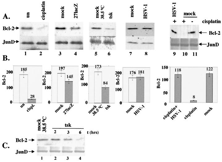
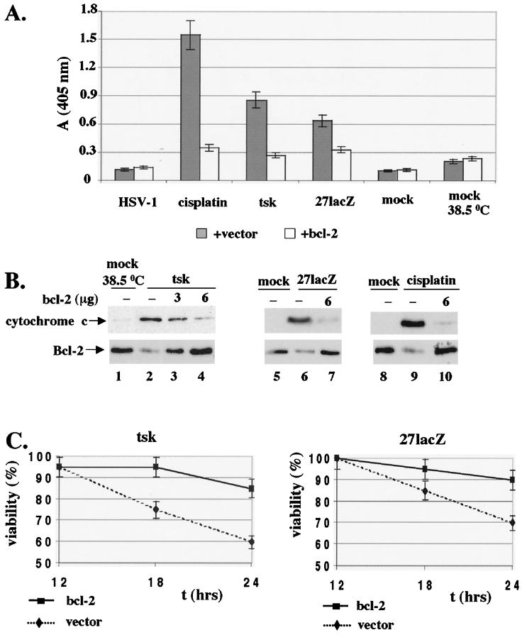
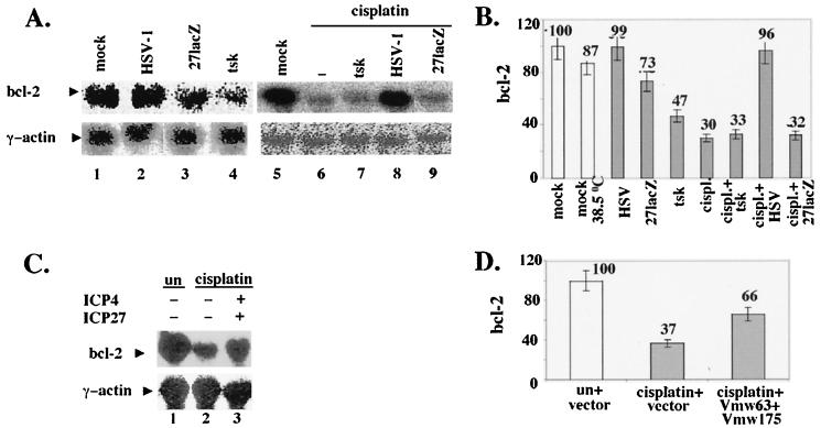
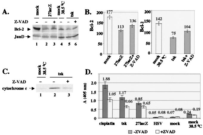
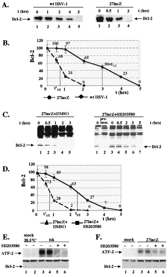
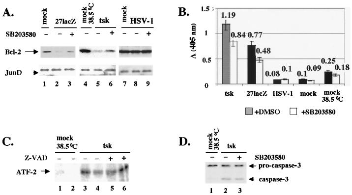
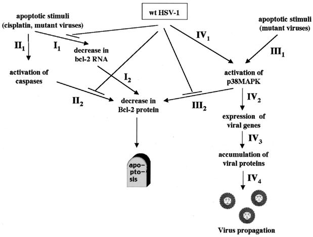
Wild-type (wt) herpes simplex virus type 1 (HSV-1) suppresses cell death. We investigated the apoptotic pathways triggered during infection with mutant viruses tsk and 27lacZ (which lack functional ICP4 and ICP27 viral proteins, respectively) and examined the mechanisms used by wt HSV-1 to protect against programmed cell death induced by the DNA-damaging compound cisplatin. In our studies, we used BHK and HeLa cells, with similar results. We suggest that a decrease in the levels of Bcl-2 protein is a key event during apoptosis induced by the mutant viruses and that Bcl-2 levels are targeted by (i) a decrease of bcl-2 RNA, (ii) caspase-related proteolysis, and (iii) p38 mitogen-activated protein kinase (p38MAPK)-dependent destabilization of Bcl-2 protein. We show that wt HSV-1, but not the mutant viruses, maintains bcl-2 RNA and protein levels during infection and protects from the cisplatin-induced decrease in bcl-2 RNA; our data suggest that both ICP27 and ICP4 are required for this function. Additionally, wt HSV-1 evades but does not actively block activation of caspases. Although wt HSV-1 induces p38MAPK activation during infection, it prevents p38MAPK-dependent destabilization of Bcl-2 and exploits p38MAPK stimulation to enhance transcription of specific viral gene promoters to increase viral yields.


Figures

FIG. 1
FIG. 1
Apoptosis triggered by cisplatin (30 μg/ml) and tsk (38.5°C) and 27lacZ viruses includes DNA degradation, activation of caspase-3 and -6, cytoplasmic release of cytochrome c, and decrease in cell viability. Cells (106) were mock infected or treated with these stimuli for 16 h, unless otherwise indicated. When both cisplatin and virus were added, cells were infected 5 h prior to addition of the drug. (A) Detection of cytoplasmic DNA by ELISA at 6 to 24 h posttreatment. Each value represents the mean ± standard deviation from three experiments performed with duplicate samples. (B) Left panel, cleavage of inactive pro-caspase-3 to active caspase-3 was detected by immunoblotting of total cell extracts. Right panel, Immunoblotting for pro-caspase-6. (C) Total cell extracts were immunoblotted for the intact (112-kDa) and cleaved (85-kDa) forms of PARP. (D) Cytosolic extracts were immunoblotted for cytochrome c. (E) Cell viability was measured by trypan blue exclusion in mock-infected cells and in cells treated with cisplatin and/ or mutant viruses.
FIG. 2
FIG. 2
Apoptosis triggered by cisplatin (30 μg/ml) and tsk (38.5°C) and 27lacZ viruses targets bcl-2 protein levels. Cells (106) were mock infected or treated with these stimuli for 16 h, unless otherwise indicated. When both cisplatin and virus were added, cells were infected 5 h prior to addition of the drug. (A) Total cell extracts were immunoblotted for Bcl-2 (upper panels) and JunD (lower panels). (B) Quantification of Bcl-2 levels presented in panel A. (C) Immunoblotting analysis of total cell extracts for Bcl-2 (upper panel) and JunD (lower panel) at several time points postinfection with tsk.
FIG. 3
FIG. 3
Overexpression of Bcl-2 protects cells from DNA damage, cytoplasmic cytochrome c release, and cell death induced by cisplatin (30 μg/ml) and tsk (38.5°C) and 27lacZ viruses. (A) Cells (5 × 10⁶) were transfected with 6 μg of bcl-2 plasmid or empty vector for 30 h, and apoptosis was induced with the stimuli for additional 16 h. Detection of cytoplasmic DNA by ELISA in cells mock infected or treated with wt HSV-1, tsk, 27lacZ, or cisplatin in the presence of bcl-2 plasmid or empty vector is shown. (B) Cells (5 × 10⁶) were transfected with 0 to 6 μg of bcl-2 plasmid, and the amount of transfected DNA was normalized to 6 μg with empty vector. At 30 h posttransfection, cells were mock infected, treated with cisplatin, or infected with tsk or 27lacZ for 16 h. Upper panel, cytosolic preparations were immunoblotted for cytochrome c. Lower panel, Total cell extracts were immunostained for Bcl-2. (C) Cell viability was measured by trypan blue exclusion in cells transfected with Bcl-2 or vector as for panel A and infected for 12 to 24 h with tsk (left panel) and 27lacZ (right panel).
FIG. 4
FIG. 4
Wt HSV-1, but not tsk and 27lacZ, maintains bcl-2 RNA levels during infection and treatment of cells with cisplatin, and transfection of ICP4 and ICP27 increases bcl-2 RNA levels during treatment of cells with cisplatin. (A) Cells (5 × 10⁶) were mock-infected; infected with wt HSV-1, tsk (38.5°C), or 27lacZ, or treated with cisplatin (30 μg/ml) for 16 h. When both cisplatin and virus were added, cells were infected 5 h prior to addition of the drug. Total RNA was analyzed by Northern blotting using bcl-2 (upper panels) and γ-actin (lower panels) DNA probes and visualized with a phosphorimager. (B) Quantitative analysis of Bcl-2 levels as shown in panel A. (C) Cells (5 × 10⁶) were cotransfected with 3 μg of ICP4- and 3 μg of ICP27-coding plasmids or transfected with 6 μg of empty vector for 30 h and then incubated in the absence or presence of cisplatin (30 μg/ml) for 16 h. Total RNA was analyzed by Northern blotting using bcl-2 (upper panel) and γ-actin (lower panel) DNA probes and visualized with a phosphorimager. (D) Quantitative analysis of Bcl-2 levels as shown in panel C.
FIG. 5
FIG. 5
Inhibition of caspases during infection with 27lacZ and tsk increases Bcl-2 levels and protects from cytoplasmic release of cytochrome c and DNA degradation. (A) Cells (106) were mock infected or infected with tsk (38.5°C) or 27lacZ in the presence or absence of Z-VAD-FMK. When Z-VAD-FMK was included, cells were treated with 25 μM Z-VAD-FMK 1 h prior to and 6 h after infection. Cells were harvested at 16 h postinfection, and extracts were immunoblotted for Bcl-2 (upper panel) and JunD (lower panel). (B) Quantification of Bcl-2 levels as shown in panel A. (C) Cells (106) were mock infected or infected with tsk for 16 h the presence or absence of Z-VAD-FMK, as described for panel A. Cytosolic fractions were immunoblotted for cytochrome c. (D) Cells (106) were mock infected or treated with cisplatin (30 μg/ml), tsk, 27lacZ, or wt HSV-1 in the presence or absence of Z-VAD-FMK, as described for panel A. Cytoplasmic DNA was detected by ELISA. Each value represents mean ± standard deviation from four independent experiments performed in duplicate.
FIG. 6
FIG. 6
Infection of cells with 27lacZ but not with the wt virus induces a p38MAPK-dependent decrease in Bcl-2 half-life, and p38MAPK kinase activity coimmunoprecipitates with Bcl-2 in cells infected with tsk and 27lacZ. (A) Left panel [35S]methionine pulse-chase analysis of Bcl-2 degradation rates during infection with wt HSV-1. Cells (5 × 10⁶) were infected with wt HSV-1 and at 7 h post-infection cells were labeled with [35S]methionine and either lysed (0 h) or cultured in medium containing unlabeled methionine prior to immunoprecipitation for Bcl-2. The gel was analyzed by autoradiography. Right panel, [35S]methionine pulse-chase analysis of Bcl-2 degradation rates during infection with 27lacZ. Cells (5 × 10⁶) were infected with 27lacZ, and at 7 h postinfection cells were analyzed as described for panel A. (B) Quantification of Bcl-2 levels from [35S]methionine pulse-chase analysis during infection with wt HSV-1 and 27lacZ as described for panel A. (C) Left panel, [35S]methionine pulse-chase analysis of Bcl-2 degradation rates during infection with 27lacZ in the presence of DMSO. Cells (5 × 10⁶) were treated with 30 μM DMSO for 1 h and infected with 27lacZ in the continuous presence of DMSO. At 7 h postinfection cells were labeled with [35S]methionine, and Bcl-2 levels were analysed as described for panel A. Right panel, [35S]methionine pulse-chase analysis of Bcl-2 degradation rates during infection with 27lacZ in the presence of SB203580. Cells (5 × 10⁶) were treated with 30 μM SB203580 for 1 h and infected with 27lacZ in the continuous presence of SB203580. At 7 h postinfection cells were labeled with [35S]methionine, and Bcl-2 levels were analyzed as described for panel A. Preimmune serum (pre-imm.) was used at 0 h postlabeling. (D) Quantification of Bcl-2 levels from [35S]methionine pulse-chase analysis during infection with 27lacZ in the presence of DMSO or SB203580, as described for panel C. (E) Cells (106) were pretreated with 30 μM SB203580 or DMSO for 1 h and mock infected or infected with tsk in the continuous presence of SB203580 or DMSO. In the upper panel, Bcl-2 was immunoprecipitated from total cell extracts and associated p38MAPK kinase activity was detected in immunocomplex kinase assays, using 1 μg of ATF-2 protein as the substrate. In the lower panel, total cell extracts from duplicate preparations similar to the ones analyzed in panel E, upper panel, were immunoblotted for Bcl-2. (F) Cells (106) were pretreated with 30 μM SB203580 or DMSO for 1 h, and mock infected or infected with 27lacZ, and analyzed as described for panel E. Upper panel, immunocomplex kinase assay for p38MAPK activity. Lower panel, immunoblotting of duplicate preparations for Bcl-2.
FIG. 7
FIG. 7
Inhibition of p38MAPK during infection with 27lacZ and tsk increases Bcl-2 levels and protects cells from DNA degradation, and activation of p38MAPK is independent of caspases. (A) Cells (106) were pretreated with 30 μM SB203580 or DMSO for 1 h and mock infected or infected with 27lacZ, tsk (38.5°C), or wt HSV-1. At 16 h postinfection, immunoblotting analysis of total cell extracts for Bcl-2 (upper panel) and JunD (lower panel) was performed. (B) Cells (106) were pretreated with 30 μM SB203580 or DMSO for 1 h and mock infected or infected with 27lacZ, tsk (38.5°C), or wt HSV-1 in the continuous presence of SB203580 or DMSO. At 16 h postinfection, cytoplasmic DNA was detected by ELISA. (C) Cells (106) were mock infected or infected with tsk in the presence or absence of Z-VAD-FMK. When Z-VAD-FMK was included, cells were treated with 25 μM Z-VAD-FMK 1 h prior to and 6 h after infection. At 8 h postinfection, p38MAPK was immunoprecipitated and immunocomplex kinase assays were performed using 1 μg of ATF-2 as the substrate. (D) Cells (106) were pretreated with 30 μM SB203580 or DMSO for 1 h and mock infected or infected with tsk (38.5°C) in the continuous presence of SB203580 or DMSO. At 16 h postinfection, total cell extracts were immunoblotted for caspase-3.
FIG. 8
FIG. 8
Inhibition of p38MAPK decreases levels of viral proteins and transcriptional activity of specific promoters. (A) Cells (106) were incubated with 30 μM SB203580 or DMSO for 1 h and infected with wt HSV-1 for 6 to 12 h in the continuous presence of SB203580 or DMSO. Immunoblotting of total cell extracts was performed for viral proteins ICP0 (upper panel) and ICP4 (lower panel) at the time points indicated. (B) Cells (106) were treated with 30 μM SB203580 or DMSO as described for panel A and infected with wt HSV-1. At 9 h postinfection, total cell extracts were analyzed for viral proteins ICP27 (upper panel) and UL42 (lower panel) by immunoblotting. (C) Cells (5 × 10⁶) were transfected with 3 μg of pIE1CAT or pIE3CAT. At 30 h posttransfection, cells were pretreated for 1 h with DMSO or SB203580 and infected with wt HSV-1 in the continuous presence of DMSO or SB203580. CAT activity was measured at 16 h postinfection.
FIG. 9
FIG. 9
Schematic model of the mechanisms used by wt HSV-1 to block Bcl-2-mediated apoptosis. Apoptotic stimuli (for example, infection of cells with tsk or 27lacZ mutant viruses) target the levels of the pro-survival protein Bcl-2 by three mechanisms (route I, decrease of Bcl-2 RNA; route II, caspase-dependent decrease of Bcl-2 protein levels; route III, p38MAPK-dependent destabilization of Bcl-2 protein). wt HSV-1 blocks all of these mechanisms to prevent apoptosis. In addition, wt HSV-1 benefits from activation of p38MAPK by enhanced expression of specific viral genes and improved viral growth (route IV).

Similar articles

Cited by

References

    1. Adams J M, Cory S. The Bcl-2 protein family: arbiters of cell survival. Science. 1998;281:1322–1326. - PubMed
    1. Allen K E, Everett R D. Mutations which alter the DNA binding properties of the herpes simplex virus type 1 transactivating protein Vmw175 also affect its ability to support virus replication. J Gen Virol. 1997;78:2913–2922. - PubMed
    1. Aubert M, Blaho J. The herpes simplex virus type 1 regulatory protein ICP27 is required for the prevention of apoptosis in infected human cells. J Virol. 1999;73:2803–2813. - PMC - PubMed
    1. Aubert M, O'Toole J, Blaho J. Induction and prevention of apoptosis in human HEp-2 cells by herpes simplex virus type 1. J Virol. 1999;73:10359–10370. - PMC - PubMed
    1. Conner J, Murray J, Cross A, Clements J B, Marsden H S. Intracellular localisation of herpes simplex virus type 1 ribonucleotide reductase subunits during infection of cultured cells. Virology. 1995;213:615–623. - PubMed

Publication types

MeSH terms