

Ross River virus glycoprotein-pseudotyped retroviruses and stable cell lines for their production
- PMID: 11222688
- PMCID: PMC115889
- DOI: 10.1128/JVI.75.6.2653-2659.2001
Ross River virus glycoprotein-pseudotyped retroviruses and stable cell lines for their production
Abstract
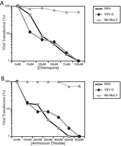
Pseudotyped retroviruses have important applications as vectors for gene transfer and gene therapy and as tools for the study of viral glycoprotein function. Recombinant Moloney murine leukemia virus (Mo-MuLV)-based retrovirus particles efficiently incorporate the glycoproteins of the alphavirus Ross River virus (RRV) and utilize them for entry into cells. Stable cell lines that produce the RRV glycoprotein-pseudotyped retroviruses for prolonged periods of time have been constructed. The pseudotyped viruses have a broadened host range, can be concentrated to high titer, and mediate stable transduction of genes into cells. The RRV glycoprotein-pseudotyped retroviruses and the cells that produce them have been employed to demonstrate that RRV glycoprotein-mediated viral entry occurs through endocytosis and that membrane fusion requires acidic pH. Alphavirus glycoprotein-pseudotyped retroviruses have significant advantages as reagents for the study of the biochemistry and prevention of alphavirus entry and as preferred vectors for stable gene transfer and gene therapy protocols.
Figures


Similar articles
-
Human immunodeficiency virus type 1-derived lentivirus vectors pseudotyped with envelope glycoproteins derived from Ross River virus and Semliki Forest virus.J Virol. 2004 Feb;78(3):1421-30. doi: 10.1128/jvi.78.3.1421-1430.2004. J Virol. 2004. PMID: 14722297 Free PMC article.
-
In vivo gene transfer using a nonprimate lentiviral vector pseudotyped with Ross River Virus glycoproteins.J Virol. 2002 Sep;76(18):9378-88. doi: 10.1128/jvi.76.18.9378-9388.2002. J Virol. 2002. PMID: 12186920 Free PMC article.
-
Human immunodeficiency virus type 1 vectors with alphavirus envelope glycoproteins produced from stable packaging cells.J Virol. 2005 Feb;79(3):1765-71. doi: 10.1128/JVI.79.3.1765-1771.2005. J Virol. 2005. PMID: 15650201 Free PMC article.
-
Generation of high-titer pseudotyped retroviral vectors with very broad host range.Methods Cell Biol. 1994;43 Pt A:99-112. doi: 10.1016/s0091-679x(08)60600-7. Methods Cell Biol. 1994. PMID: 7823872 Review.
-
Cell targeting by murine recombinant retroviruses.Bone Marrow Transplant. 1992;9 Suppl 1:139-42. Bone Marrow Transplant. 1992. PMID: 1504656 Review.
Cited by
-
HIV-1 interacts with human endogenous retrovirus K (HML-2) envelopes derived from human primary lymphocytes.J Virol. 2014 Jun;88(11):6213-23. doi: 10.1128/JVI.00669-14. Epub 2014 Mar 19. J Virol. 2014. PMID: 24648457 Free PMC article.
-
Targeted transgene expression in muller glia of normal and diseased retinas using lentiviral vectors.Invest Ophthalmol Vis Sci. 2007 Apr;48(4):1844-52. doi: 10.1167/iovs.05-1570. Invest Ophthalmol Vis Sci. 2007. PMID: 17389520 Free PMC article.
-
Human immunodeficiency virus type 1-derived lentivirus vectors pseudotyped with envelope glycoproteins derived from Ross River virus and Semliki Forest virus.J Virol. 2004 Feb;78(3):1421-30. doi: 10.1128/jvi.78.3.1421-1430.2004. J Virol. 2004. PMID: 14722297 Free PMC article.
-
Efficient functional pseudotyping of oncoretroviral and lentiviral vectors by Venezuelan equine encephalitis virus envelope proteins.J Virol. 2005 Jan;79(2):756-63. doi: 10.1128/JVI.79.2.756-763.2005. J Virol. 2005. PMID: 15613303 Free PMC article.
-
MARCH8 Targets Cytoplasmic Lysine Residues of Various Viral Envelope Glycoproteins.Microbiol Spectr. 2022 Feb 23;10(1):e0061821. doi: 10.1128/spectrum.00618-21. Epub 2022 Jan 12. Microbiol Spectr. 2022. PMID: 35019698 Free PMC article.
References
Publication types
MeSH terms
Substances
Grants and funding
LinkOut - more resources
Full Text Sources
Other Literature Sources
