. 2001 Mar;75(6):2544-56.
doi: 10.1128/JVI.75.6.2544-2556.2001.

A GP64-null baculovirus pseudotyped with vesicular stomatitis virus G protein

Affiliations

A GP64-null baculovirus pseudotyped with vesicular stomatitis virus G protein

J T Mangor et al. J Virol. 2001 Mar.

Abstract

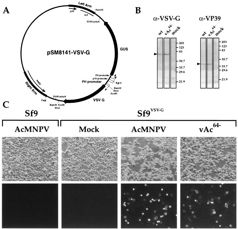
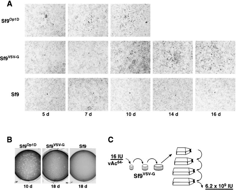
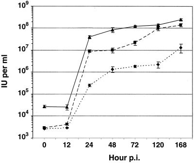
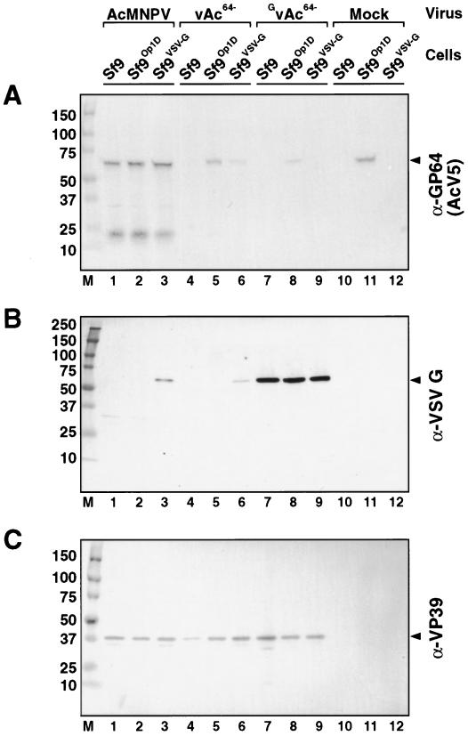
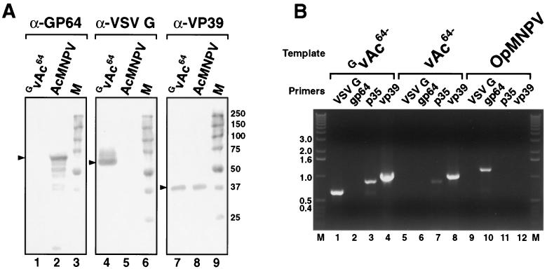
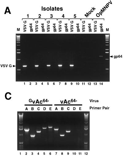
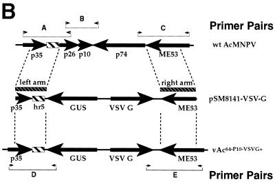
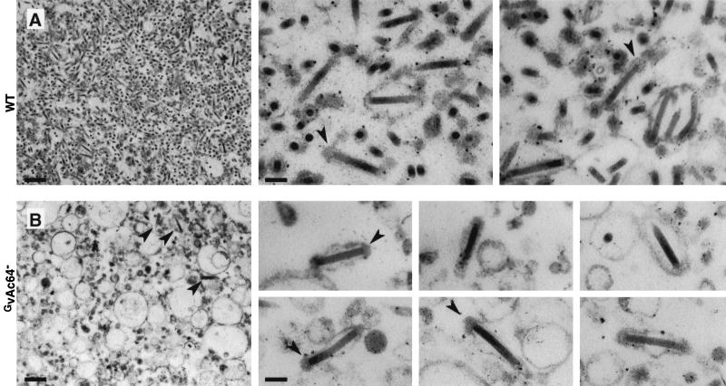
The Autographa californica multiple nucleopolyhedrovirus (AcMNPV) GP64 protein is an essential virion protein that is involved in both receptor binding and membrane fusion during viral entry. Genetic studies have shown that GP64-null viruses are unable to move from cell to cell and this results from a defect in the assembly and production of budded virions (BV). To further examine requirements for virion budding, we asked whether a GP64-null baculovirus, vAc(64-), could be pseudotyped by introducing a heterologous viral envelope protein (vesicular stomatitis virus G protein [VSV-G]) into its membrane and whether the resulting virus was infectious. To address this question, we generated a stably transfected insect Sf9 cell line (Sf9(VSV-G)) that inducibly expresses the VSV-G protein upon infection with AcMNPV Sf9(VSV-G) and Sf9 cells were infected with vAc(64-), and cells were monitored for infection and for movement of infection from cell to cell. vAc(64-) formed plaques on Sf9(VSV-G) cells but not on Sf9 cells, and plaques formed on Sf9(VSV-G) cells were observed only after prolonged intervals. Passage and amplification of vAc(64-) on Sf9(VSV-G) cells resulted in pseudotyped virus particles that contained the VSV-G protein. Cell-to-cell propagation of vAc(64-) in the G-expressing cells was delayed in comparison to wild-type (wt) AcMNPV, and growth curves showed that pseudotyped vAc(64-) was generated at titers of approximately 10(6) to 10(7) infectious units (IU)/ml, compared with titers of approximately 10(8) IU/ml for wt AcMNPV. Propagation and amplification of pseudotyped vAc(64-) virions in Sf9(VSV-G) cells suggests that the VSV-G protein may either possess the signals necessary for baculovirus BV assembly and budding at the cell surface or may otherwise facilitate production of infectious baculovirus virions. The functional complementation of GP64-null viruses by VSV-G protein was further demonstrated by identification of a vAc(64-)-derived virus that had acquired the G gene through recombination with Sf9(VSV-G) cellular DNA. GP64-null viruses expressing the VSV-G gene were capable of productive infection, replication, and propagation in Sf9 cells.


Figures

FIG. 1
FIG. 1
Construction and analysis of cell line Sf9VSV-G. (A) Plasmid pSM8141-VSV-G contains a VSV-G gene under the control of an AcMNPV polyhedrin (PH) promoter, and a GUS gene under the control of PH and p10 promoters from AcMNPV. Each gene cassette is terminated by a simian virus 40 (SV40) poly(A) cleavage and addition site. The two genes are flanked by left- and right-arm sequences from the AcMNPV p35-hr5 region and the me53 region, respectively. Plasmid pSM8141-VSV-G was transfected into Sf9 cells to generate a stably transfected cell line (Sf9VSV-G). (B) Western blot analysis of VSV-G protein induction in Sf9VSV-G cells infected with wt AcMNPV (wt) or a GP64-null virus (vAc64−). Sf9VSV-G cells were infected at an MOI of 1, harvested at 46 hp i, and then examined for VSV-G protein expression and VP39 protein expression, using MAbs (see Materials and Methods). The specificity of each MAb is indicated above each group of blots (α-VSV-G or α-VP39), and positions and molecular weights (in thousands) of protein size markers are indicated on the right. The positions of VSV-G and VP39 are indicated by an arrowhead on the left of each group of blots. (C) Immunofluorescent detection of VSV-G protein. SF9 or Sf9VSV-G cells (upper labels) were infected with wt AcMNPV or vAc64− (lower labels) at an MOI of 10 and then fixed at 40 hpi and immunostained with an anti-VSV-G MAb (P5D4) and goat anti-mouse IgG fluorescein isothiocyanate conjugate. Cells were examined and photographed by epifluorescence microscopy.
FIG. 2
FIG. 2
GP64-null virus propagation in Sf9VSV-G cells. (A) A monolayer of cells (Sf9Op1D, Sf9VSV-G, or Sf9) was infected with vAc64− at an MOI of approximately 6 × 10⁻⁵, and infected cells were identified at the indicated intervals (5, 7, 10, 14, or 16 days). Infected cells were identified by the presence of occlusion bodies, and these infected cells appear as dark cells against the background of lighter cells. (B) Plaque formation in vAc64− infected Sf9Op1D, Sf9VSV-G, and Sf9 cell monolayers was examined after 10, 18, and 18 days, respectively. (C) Schematic of vAc64− propagation in Sf9VSV-G cells. Sf9VSV-G cells (7.2 × 10⁵ cells) were infected with 16 IU of vAc64−, and cells and supernatants were passaged until all cells appeared to be infected (6 to 7 passages). Titration of the supernatant resulted in a final virus titer of 6.2 × 10⁹ IU. Control cells (Sf9 and Sf9Op1D) were also infected in parallel (see Results).
FIG. 3
FIG. 3
One-step growth curves. Growth curves are plotted for the GP64-null virus (vAc64−) in cells expressing VSV-G (Sf9VSV-G) (⧫) or OpMNPV GP64 (Sf9Op1D) (■). For comparison, a growth curve of wt AcMNPV infected Sf9 cells was generated in parallel and is also plotted (▴). Cells were infected and supernatants were collected at the indicated times postinfection, and virus yields were determined as TCID50 values on Sf9Op1D cells. Each data point represents three individual infections, and error bars represent standard error.
FIG. 4
FIG. 4
Western blot analysis of GP64, VSV-G, and VP39 proteins in infected cell lysates. Cells were infected with viruses at an MOI of 1, and lysates were harvested at 75 hpi. The viruses and cells used for each infection are indicated above panel A. Western blots were treated with either anti-GP64 MAb AcV5 (A), anti-VSV-G MAb P5D4 (B), or anti-VP39 MAb P10 (C).
FIG. 5
FIG. 5
Western blot and PCR analysis of virion preparations of wt AcMNPV, vAc64−, and GvAc64−. (A) Western blot analysis of GP64, VSV-G, and VP39 proteins in wt AcMNPV and GvAc64− virion preparations. Virus preparation GvAc64− represents the GP64-null virus (vAc64−) passaged in VSV-G expressing cells (Sf9VSV-G). Specificities of MAbs are indicated at the top (α-GP64, α-VSV-G, and α-VP39), and virion preparations are indicated above each lane (GvAc64− and AcMNPV). Marker lanes (M) are also indicated, and sizes of protein molecular weight standards (in thousands) are indicated on the right. (B) PCR analysis of viral DNAs isolated from purified virions of GvAc64− and vAc64−. Template viral DNA is indicated at the top, and gene specificities of primer pairs are indicated above individual lanes. Primer pairs were specific for VSV-G, OpMNPV gp64, AcMNPV p35, and AcMNPV vp39 genes. Lane M contains DNA size markers, and sizes (in kilobase pairs) are indicated on the left.
FIG. 6
FIG. 6
PCR analysis of virus preparations of GvAc64−. (A) PCR analysis of five GvAc64− virus preparations. Gene-specific primer pairs were used to examine DNAs from cell lysates of Sf9 cells infected with five GvAc64− virus preparations. DNA preparations were examined for the presence of the OpMNPV gp64 gene or the VSV-G gene. Isolation of individual preparations is described in the text and preparations are indicated as 1 to 5. The gene-specific primer pairs are indicated above individual lanes (VSV-G or gp64). (B) Strategy for PCR analysis of the p10 locus of AcMNPV in wt, vAc64−, and GvAc64− virus preparations. Large arrows show locations of AcMNPV ORFs in the p10 region (top line) or the predicted positions of the GUS and VSV-G ORFs integrated into the p10 region (bottom line). Small arrowheads show the locations of PCR primers on the wt AcMNPV genome (primer pairs A through C) and a predicted recombinant GvAc64− genome (vAc64−P10−VSVG+) (primer pairs D and E). Dashed lines indicate locations of the “arm” regions (potential recombination regions) present in the pSM8141-VSV-G plasmid. (C) PCR analysis of the p10 locus and the potential integration site of the VSV-G gene in vAc64− and GvAc64− virus preparations. DNAs from infected Sf9 cell lysates were used as templates for PCR analysis. Primer pairs specific for the wt AcMNPV p10 region in vAc64− (primer pairs A, B, and C) were compared with primer pairs specific for the predicted integration of VSV-G and GUS into the p10 region in viruses GvAc64− (primer pairs D and E).
FIG. 6
FIG. 6
PCR analysis of virus preparations of GvAc64−. (A) PCR analysis of five GvAc64− virus preparations. Gene-specific primer pairs were used to examine DNAs from cell lysates of Sf9 cells infected with five GvAc64− virus preparations. DNA preparations were examined for the presence of the OpMNPV gp64 gene or the VSV-G gene. Isolation of individual preparations is described in the text and preparations are indicated as 1 to 5. The gene-specific primer pairs are indicated above individual lanes (VSV-G or gp64). (B) Strategy for PCR analysis of the p10 locus of AcMNPV in wt, vAc64−, and GvAc64− virus preparations. Large arrows show locations of AcMNPV ORFs in the p10 region (top line) or the predicted positions of the GUS and VSV-G ORFs integrated into the p10 region (bottom line). Small arrowheads show the locations of PCR primers on the wt AcMNPV genome (primer pairs A through C) and a predicted recombinant GvAc64− genome (vAc64−P10−VSVG+) (primer pairs D and E). Dashed lines indicate locations of the “arm” regions (potential recombination regions) present in the pSM8141-VSV-G plasmid. (C) PCR analysis of the p10 locus and the potential integration site of the VSV-G gene in vAc64− and GvAc64− virus preparations. DNAs from infected Sf9 cell lysates were used as templates for PCR analysis. Primer pairs specific for the wt AcMNPV p10 region in vAc64− (primer pairs A, B, and C) were compared with primer pairs specific for the predicted integration of VSV-G and GUS into the p10 region in viruses GvAc64− (primer pairs D and E).
FIG. 7
FIG. 7
Electron micrographs of wt AcMNPV (A) and GvAc64− (B) virion preparations. Arrows indicate virions containing thickened regions near the termini. Bars on the panels at far left represent 500 nm, and bars on the rightmost panels represent 100 nm. Thin sections of virion preparations were stained with uranyl acetate and lead citrate and examined on a Philips 201 transmission electron microscope.

Similar articles

Cited by

References

    1. Barsoum J, Brown R, McKee M, Boyce F M. Efficient transduction of mammalian cells by a recombinant baculovirus having the vesicular stomatitis virus G glycoprotein. Hum Gene Ther. 1997;8:2011–2018. - PubMed
    1. Blissard G W, Wenz J R. Baculovirus GP64 envelope glycoprotein is sufficient to mediate pH dependent membrane fusion. J Virol. 1992;66:6829–6835. - PMC - PubMed
    1. Blum H, Beier H, Gross H J. Improved silver staining of plant proteins, RNA and DNA in polyacrylamide gels. Electrophoresis. 1987;8:93–99.
    1. Boyce F M, Bucher N L R. Baculovirus-mediated gene transfer into mammalian cells. Proc Natl Acad Sci USA. 1996;93:2348–2352. - PMC - PubMed
    1. Condreay J P, Witherspoon S M, Clay W C, Kost T A. Transient and stable gene expression in mammalian cells transduced with a recombinant baculovirus vector. Proc Natl Acad Sci USA. 1999;96:127–132. - PMC - PubMed

Publication types

Substances

LinkOut - more resources