Prostaglandin D2 selectively induces chemotaxis in T helper type 2 cells, eosinophils, and basophils via seven-transmembrane receptor CRTH2
- PMID: 11208866
- PMCID: PMC2193345
- DOI: 10.1084/jem.193.2.255
Prostaglandin D2 selectively induces chemotaxis in T helper type 2 cells, eosinophils, and basophils via seven-transmembrane receptor CRTH2
Abstract
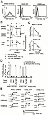
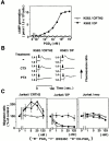
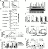
Prostaglandin (PG)D2, which has long been implicated in allergic diseases, is currently considered to elicit its biological actions through the DP receptor (DP). Involvement of DP in the formation of allergic asthma was recently demonstrated with DP-deficient mice. However, proinflammatory functions of PGD2 cannot be explained by DP alone. We show here that a seven-transmembrane receptor, CRTH2, which is preferentially expressed in T helper type 2 (Th2) cells, eosinophils, and basophils in humans, serves as the novel receptor for PGD2. In response to PGD2, CRTH2 induces intracellular Ca2+mobilization and chemotaxis in Th2 cells in a Galphai-dependent manner. In addition, CRTH2, but not DP, mediates PGD2-dependent cell migration of blood eosinophils and basophils. Thus, PGD2 is likely involved in multiple aspects of allergic inflammation through its dual receptor systems, DP and CRTH2.
Figures


Similar articles
-
Delta12-prostaglandin D2 is a potent and selective CRTH2 receptor agonist and causes activation of human eosinophils and Th2 lymphocytes.Prostaglandins Other Lipid Mediat. 2005 Jan;75(1-4):153-67. doi: 10.1016/j.prostaglandins.2004.11.003. Prostaglandins Other Lipid Mediat. 2005. PMID: 15789622
-
Differential modulation of human basophil functions through prostaglandin D2 receptors DP and chemoattractant receptor-homologous molecule expressed on Th2 cells/DP2.Clin Exp Allergy. 2004 Aug;34(8):1283-90. doi: 10.1111/j.1365-2222.2004.02027.x. Clin Exp Allergy. 2004. PMID: 15298571
-
Prostaglandin D2 causes preferential induction of proinflammatory Th2 cytokine production through an action on chemoattractant receptor-like molecule expressed on Th2 cells.J Immunol. 2005 Nov 15;175(10):6531-6. doi: 10.4049/jimmunol.175.10.6531. J Immunol. 2005. PMID: 16272307
-
Antagonists of the prostaglandin D2 receptor CRTH2.Drug News Perspect. 2008 Jul-Aug;21(6):317-22. doi: 10.1358/dnp.2008.21.6.1246831. Drug News Perspect. 2008. PMID: 18836589 Review.
-
[Prostaglandin D2 in allergy: PGD2 has dual receptor systems].Nihon Yakurigaku Zasshi. 2004 Jan;123(1):15-22. doi: 10.1254/fpj.123.15. Nihon Yakurigaku Zasshi. 2004. PMID: 14695454 Review. Japanese.
Cited by
-
The prostaglandin D₂ receptor CRTH2 regulates accumulation of group 2 innate lymphoid cells in the inflamed lung.Mucosal Immunol. 2015 Nov;8(6):1313-23. doi: 10.1038/mi.2015.21. Epub 2015 Apr 8. Mucosal Immunol. 2015. PMID: 25850654 Free PMC article.
-
Mast cell chemotaxis - chemoattractants and signaling pathways.Front Immunol. 2012 May 25;3:119. doi: 10.3389/fimmu.2012.00119. eCollection 2012. Front Immunol. 2012. PMID: 22654878 Free PMC article.
-
Indomethacin inhibits eosinophil migration to prostaglandin D2 : therapeutic potential of CRTH2 desensitization for eosinophilic pustular folliculitis.Immunology. 2013 Sep;140(1):78-86. doi: 10.1111/imm.12112. Immunology. 2013. PMID: 23582181 Free PMC article.
-
Anti-inflammatory role of PGD2 in acute lung inflammation and therapeutic application of its signal enhancement.Proc Natl Acad Sci U S A. 2013 Mar 26;110(13):5205-10. doi: 10.1073/pnas.1218091110. Epub 2013 Mar 11. Proc Natl Acad Sci U S A. 2013. PMID: 23479612 Free PMC article.
-
Evidence for asthma susceptibility genes on chromosome 11 in an African-American population.Hum Genet. 2003 Jul;113(1):71-5. doi: 10.1007/s00439-003-0934-4. Epub 2003 Mar 27. Hum Genet. 2003. PMID: 12664305
References
-
- Murphy P.M. The molecular biology of leukocyte chemoattractant receptors. Annu. Rev. Immunol. 1994;12:593–633. - PubMed
-
- Springer T.A. Traffic signals for lymphocyte recirculation and leukocyte emigrationthe multistep paradigm. Cell. 1994;76:301–314. - PubMed
-
- Robinson D.S., Hamid Q., Ying S., Tsicopoulos A., Barkans J., Bentley A.M., Corrigan C., Durham S.R., Kay A.B. Predominant TH2-like bronchoalveolar T-lymphocyte population in atopic asthma. N. Engl. J. Med. 1992;326:298–304. - PubMed
-
- Nagata K., Tanaka K., Ogawa K., Kemmotsu K., Imai T., Yoshie O., Abe H., Tada K., Nakamura M., Sugamura K. Selective expression of a novel surface molecule by human Th2 cells in vivo. J. Immunol. 1999;162:1278–1286. - PubMed
MeSH terms
Substances
LinkOut - more resources
Full Text Sources
Other Literature Sources
Molecular Biology Databases
Miscellaneous

