. 2001 Feb 15;20(4):661-71.
doi: 10.1093/emboj/20.4.661.

A reversible component of mitochondrial respiratory dysfunction in apoptosis can be rescued by exogenous cytochrome c

Affiliations

A reversible component of mitochondrial respiratory dysfunction in apoptosis can be rescued by exogenous cytochrome c

V K Mootha et al. EMBO J. .

Abstract

Multiple apoptotic pathways release cytochrome c from the mitochondrial intermembrane space, resulting in the activation of downstream caspases. In vivo activation of Fas (CD95) resulted in increased permeability of the mitochondrial outer membrane and depletion of cytochrome c stores. Serial measurements of oxygen consumption, NADH redox state and membrane potential revealed a loss of respiratory state transitions. This tBID-induced respiratory failure did not require any caspase activity. At early time points, re-addition of exogenous cytochrome c markedly restored respiratory functions. Over time, however, mitochondria showed increasing irreversible respiratory dysfunction as well as diminished calcium buffering. Electron microscopy and tomographic reconstruction revealed asymmetric mitochondria with blebs of herniated matrix, distended inner membrane and partial loss of cristae structure. Thus, apoptogenic redistribution of cytochrome c is responsible for a distinct program of mitochondrial respiratory dysfunction, in addition to the activation of downstream caspases.


Figures
None
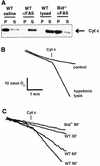
Fig. 1. Outer membrane permeability and loss of cytochrome c in mitochondria from Fas-activated hepatocytes. (A) Western blot analysis of the mitochondrial pellet (P) and supernatant (S) fractions from liver mitochondria of wild-type and Bid–/– mice 90 min after injection of saline or anti-Fas antibody. Hypotonically lysed control mitochondria are included for comparison of complete release. (B) Mitochondrial outer membrane permeability assay as developed by Colombini and colleagues (Lee et al., 1994). Exogenous cytochrome c will stimulate respiration if the cytochrome c can traverse the outer membrane and access cytochrome c oxidase. Hypotonically lysed mitochondria (mitoplasts) have a ruptured outer membrane and display a burst in oxygen consumption in response to added cytochrome c, demonstrating the utility of the assay. (C) Oxygen consumption following the addition of exogenous cytochrome c to mitochondria isolated from liver of wild-type mice at 30, 60 or 90 min after injection of anti-Fas antibody, as well as mitochondria from Bid–/– mice 90 min after Fas treatment.
None
Fig. 2. Overview of mitochondrial oxidative phosphorylation and respiratory states. Abbreviations: UQ, ubiquinone; c, cytochrome c; IM, mitochondrial inner membrane; OM, mitochondrial outer membrane; Δψm, mitochondrial membrane potential; VDAC, voltage-dependent anion channel; KCN, potassium cyanide; VO2, oxygen consumption.
None
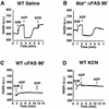
Fig. 3. Loss of NADH redox transitions in mitochondria damaged by the Fas pathway resembling cyanide-treated mitochondria. (A) Mitochondria from wild-type mice injected with saline were monitored for NADH fluorescence after the addition of carbon substrate (5 mM glutamate + 5 mM malate), a pulse of ADP (100 nmol) and finally cyanide (KCN, 1 mM). (B) Mitochondria from Bid–/– liver isolated 90 min after injection of anti-Fas antibody respond to carbon substrate (glutamate + malate) and ADP, similar to wild-type controls in (A). (C) Fas-activated mitochondria from wild-type mice were provided carbon substrate (glutamate + malate) and two subsequent additions of ADP (100 nmol), and NADH fluorescence was plotted. (D) Control wild-type mitochondria were incubated with cyanide (1 mM) at time 0. Carbon substrate (glutamate + malate) and two subsequent ADP (100 nmol) additions were made, and NADH fluorescence was monitored.
None
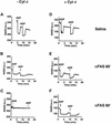
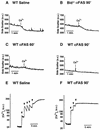
Fig. 4. NADH respiratory transitions rescued by exogenous cytochrome c. Freshly isolated mitochondria were incubated initially with complex I carbon substrate (5 mM glutamate + 5 mM malate). Liver mitochondria were obtained from mice following saline injection (A and D), or 60 min (B and E) and 90 min (C and F) after injection of anti-Fas antibody. Mitochondria are shown either in the absence (A–C) or presence (D–F) of 10 µM cytochrome c. (A) Mitochondria from saline-treated mice undergo classic NADH respiratory transitions in response to added ADP (100 nmol) pulses. (B) Sixty minutes after injection, the NADH redox transitions are blunted and take longer. (C) Ninety minutes after treatment, there is no response to added ADP and no recovery in NADH levels is observed. (D) An experiment identical to that in (A), except that it was performed in the presence of 10 µM cytochrome c, demonstrates that saline-treated control mitochondria continue to undergo NADH transitions even in the presence of cytochrome c. Cytochrome c quenches NADH fluorescence, so the y-axis has been rescaled in experiments performed in the presence of cytochrome c (D–F). (E) Addition of cytochrome c deepens the state 3 NADH redox transient and improves the recovery when compared with mitochondria seen in (B). (F) Mitochondria isolated 90 min after treatment previously (E) did not undergo NADH transitions but, with exogenous cytochrome c, now undergo robust redox transitions, indicating that the respiratory chain can still be rescued.
None
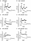
Fig. 5. Fas-induced defects in mitochondrial respiration and membrane potential partially rescued by exogenous cytochrome c. Freshly isolated mitochondria (after injection of saline or anti-Fas antibody) were placed in a respiratory chamber in which oxygen consumption (gray) and TPP+ concentration (black) were measured simultaneously. (A) Control mitochondria develop a membrane potential in response to an added carbon substrate (5 mM glutamate + 5 mM malate). Upon addition of ADP (100 nmol), mitochondria transiently depolarize and undergo a burst of oxygen consumption (state 3 respiration) until added ADP is converted to ATP. These mitochondria continue to respond to added ADP, demonstrating intact respiratory control. (BBid–/– liver mitochondria isolated 90 min after injection of anti-Fas antibody also exhibit high respiratory control and membrane potential responses to carbon substrate and ADP, indistinguishable from normal wild-type mitochondria in (A). (C) Mitochondria obtained 60 min after injection of anti-Fas antibody exhibit minimal responsiveness to added carbon substrate (glutamate + malate), but a slight membrane depolarization is seen with ADP addition. (D) Fas-pathway-damaged mitochondria as in (C) were incubated with 10 µM exogenous cytochrome c at time 0. Membrane potential now responds to carbon substrate as well as two pulses of ADP. Oxygen consumption also responds to ADP pulses, indicating partial recovery of respiratory control. (E) Mitochondria obtained 90 min after Fas activation display a membrane potential which does not increase with addition of glutamate + malate. Oxygen consumption and membrane potential fail to respond to ADP additions, indicating a loss of respiratory control. (F) Fas-pathway-damaged mitochondria as in (E) were incubated with 10 µM exogenous cytochrome c at time 0. Membrane potential now increases in response to glutamate + malate, while membrane potential drops with a concomitant increase in oxygen consumption after addition of ADP, indicating some recovery of respiratory control by adding cytochrome c. Later in the experiment, after oxygen consumption has slowed and membrane potential has recovered modestly, the mitochondria are still able to depolarize in response to a second pulse of ADP.
None
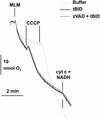
Fig. 6. Caspase inhibition does not prevent tBID-induced respiratory defect. Mitochondria isolated from untreated wild-type mouse livers (MLM) were incubated with 320 pmol tBID/mg mitochondrial protein (black line) or tBID plus 50 µM zVAD-fmk (dark gray line), or were left untreated (light gray line) in respiratory buffer. Oxygen consumption was measured with a Clarke-type electrode. Uncoupled respiration was started by adding 200 pmol carbonyl cyanide m-chlorophenylhydrazone (CCCP)/mg mitochondrial protein (lines). Where indicated, 20 µmol NADH/mg mitochondrial protein and 10 µmol cytochrome c/mg mitochondrial protein were added.
None
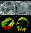
Fig. 7. Electron microscopy and tomographic reconstruction of Fas-injured mitochondria. (A) Representative field in a liver section obtained 90 min after anti-Fas antibody treatment, demonstrating mitochondria with large blebs (arrows). (B) Cross-sectional slice (6 nm thickness) from the electron tomographic reconstruction of a semi-thick (0.22 µM) section of a mitochondrion with a bleb. (C and D) Two surface-rendered views of the reconstructed mitochondrion, showing ruptured outer membrane (red), inner surface membrane (IM) with large bleb (yellow) and selected cristae (green), with narrow tubular junctions to the IM space (arrows).
None
Fig. 8. Mitochondrial swelling and loss of calcium buffering capacity. (A) Ninety degree side scatter provides a measure of mitochondrial size for control mitochondria purified from livers of saline-injected mice. Control mitochondria swell in response to a pulse of calcium (20 nmol CaCl2). (B) The ability to swell in response to Ca2+ is preserved in mitochondria from Bid–/– mice isolated 90 min after injection of anti-Fas antibody. (C) Sixty minutes after injection of Fas antibody, wild-type liver mitochondria exhibit a blunted swelling response to calcium addition. (D) By 90 min, mitochondria are initially more swollen and exhibit no response to added calcium. (E) Extramitochondrial calcium was measured fluorimetrically with the calcium indicator Calcium Green 5N. Control mitochondria can buffer multiple pulses (10 nmol CaCl2) of added calcium (arrowheads). (F) Mitochondria isolated 90 min after Fas activation cannot buffer even a single pulse of calcium.

Similar articles

Cited by

References

    1. Amarante-Mendes G.P., Finucane,D.M., Martin,S.J., Cotter,T.G., Salvesen,G.S. and Green,D.R. (1998) Anti-apoptotic oncogenes prevent caspase-dependent and independent commitment for cell death. Cell Death Differ., 5, 298–306. - PubMed
    1. Baffy G., Miyashita,T., Williamson,J.R. and Reed,J.C. (1993) Apoptosis induced by withdrawal of interleukin-3 (IL-3) from an IL-3-dependent hematopoietic cell line is associated with repartitioning of intracellular calcium and is blocked by enforced Bcl-2 oncoprotein production. J. Biol. Chem., 268, 6511–6519. - PubMed
    1. Basanez M.G., Nechushtan,A., Drozhinin,O., Chanturiya,A., Choe,E., Tutt,S., Wood,K.A., Hsu,Y., Zimmerberg,J. and Youle,R.J. (1999) Bax, but not Bcl-xL, decreases the lifetime of planar phospholipid bilayer membranes at subnanomolar concentrations. Proc. Natl Acad. Sci. USA, 96, 5492–5497. - PMC - PubMed
    1. Bernardi P. (1999) Mitochondrial transport of cations: channels, exchangers and permeability transition. Physiol. Rev., 79, 1127–1155. - PubMed
    1. Cai J. and Jones,D.P. (1998) Superoxide in apoptosis. Mitochondrial generation triggered by cytochrome c loss. J. Biol. Chem., 273, 11401–11404. - PubMed

Publication types