. 2001 Feb;75(4):1581-93.
doi: 10.1128/JVI.75.4.1581-1593.2001.

Reactivation of the human cytomegalovirus major immediate-early regulatory region and viral replication in embryonal NTera2 cells: role of trichostatin A, retinoic acid, and deletion of the 21-base-pair repeats and modulator

Affiliations

Reactivation of the human cytomegalovirus major immediate-early regulatory region and viral replication in embryonal NTera2 cells: role of trichostatin A, retinoic acid, and deletion of the 21-base-pair repeats and modulator

J L Meier. J Virol. 2001 Feb.

Abstract

Inactivity of the human cytomegalovirus (HCMV) major immediate-early regulatory region (MIERR), which is composed of promoter, enhancer, unique region, and modulator, is linked to lack of HCMV replication in latently infected cells and in other nonpermissive cell types, including human embryonal NTera2 carcinoma (NT2) cells. I refined the embryonal NT2 cell model to enable characterization of the unknown mechanistic basis for silencing of HCMV MIERR-dependent transcription and viral replication in nonpermissive cells. These infected NT2 cells contain nonreplicating viral genomes with electrophoretic mobility equivalent to a supercoiled, bacterial artificial chromosome of comparable molecular weight. MIERR-dependent transcription is minimal to negligible. Increasing the availability of positive-acting transcription factors by retinoic acid (RA) treatment after infection is largely insufficient in reactivating the MIERR. In contrast, trichostatin A (TSA), a histone deacetylase inhibitor, reactivates MIERR-dependent transcription. Contrary to prior findings produced from transfected MIERR segments, deletion of the 21-bp repeats and modulator from the MIERR in the viral genome does not relieve MIERR silencing. To demonstrate that MIERR silencing likely results from enhancer inactivity, I examined an HCMV with a heterologous MIERR promoter that is enhancer dependent but exempt from IE2 p86-mediated negative autoregulation. This heterologous promoter, like its neighboring native MIERR promoter, exhibits immediate-early transcriptional kinetics in fibroblasts. In embryonal NT2 cells, the heterologous MIERR promoter is transcriptionally inactive. This silence is relieved by TSA but not by RA. Remarkably, TSA-induced reactivation of MIERR-dependent transcription from quiescent viral genomes is followed by release of infectious virus. I conclude that a mechanism of active repression imposes a block to MIERR-dependent transcription and viral replication in embryonal NT2 cells. Because TSA overcomes the block, viral gene silencing may involve histone deacetylase-based modification of viral chromatin, which might account for the covalently closed circular conformation of quiescent HCMV genomes.


Figures
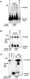
FIG. 1
FIG. 1
HCMV genomes in uninduced and RA-induced NT2 cells differ in structure. (A) Analysis of HCMV rΔ-640/-1108SVgfp genomes in uninduced NT2 cells at 48 h p.i. rΔ-640/-1108SVgfp was isolated by centrifugation through a sorbital cushion, and uninduced NT2 cells were grown in stem cell conditions (see Materials and Methods). Mock (MOC)- or rΔ-640/-1108SVgfp-infected cells (MOI of 50) were washed thrice after viral absorption (1.5 h). RA (10 μM), PFA (400 μg/ml), or nothing (−) was added (Add) to the growth medium. Less than 0.01% of infected NT2 cells without additive emitted green fluorescence at 48 h p.i. (HPI). Cells (106/sample) were harvested, washed, and subjected to Gardella gel electrophoresis and Southern blot analysis using a 32P-labeled HCMV-specific probe. The autoradiogram was exposed for 240 h. Isolated WT virions (VIR) were mixed with uninfected cells (106) to determine mobility of the 240-kbp linear genome among cellular chromosomes. Positions of immobile and 240-kbp linear viral genomes are shown. (B) Comparison of mobility of a 250-kbp BAC with WT genomes in uninduced NT2 cells. Infection was performed as described for panel A except that the source of WT was filtered (0.4-μm-pore-size-filter) crude viral stock (MOI of 10). After viral adsorption, PFA (400 μg/ml) was added to or omitted from the growth medium. At 48 h p.i. (HPI), WT- and mock (MOC)-infected cells (106/sample) were subjected to Gardella gel electrophoresis and Southern blot analysis using a 32P-labeled BAC- or HCMV-specific probe. Isolated virions (VIR) and bacteria containing a 250-kbp BAC were each mixed with uninfected cells (106) to mark positions of 240-kbp linear and 250-kbp CCC molecules, respectively. Autoradiography exposure was 8 and 144 h for BAC- and HCMV-specific probes, respectively. (C) Analysis of WT genomes in RA-induced NT2 cells at 24 and 48 h p.i. (HPI). NT2 cells were pretreated with RA (10 μM) for 7 days prior to infection; RA was omitted during infection. WT- and mock (MOC)-infected RA-induced NT2 cells (106/sample) were subjected to Gardella gel electrophoresis and Southern blot analysis as described for panel B. After viral absorption, PFA (400 μg/ml) was added to or omitted from the growth medium. Isolated virions (VIR) or bacteria containing a 250-kbp BAC were prepared for analysis as described for panel B. Autoradiography exposure was 18 h for BAC- and HCMV-specific probes.
FIG. 2
FIG. 2
Effect of TSA or RA on viral MIERR- and US3 promoter-dependent transcription in preinfected embryonal NT2 cells. (A) Analysis of IE1 RNA in WT- and mock-infected NT2 cells at 24 h p.i. RA (10 μM) or TSA (100 ng/ml) was added to or omitted from growth medium at 1.5 h p.i. as described in Materials and Methods. Isolated RNA (25 μg/sample) was subjected to RPA using both viral IE1- and cellular actin-specific riboprobes. Arrows indicate positions of protected unspliced and spliced IE1 and actin RNAs. (B) Analysis of US3 RNA at 24 h p.i. Isolated RNAs used for panel A were also subjected to RPA, with US3- and actin-specific riboprobes. Std, standard; nt, nucleotides.
FIG. 3
FIG. 3
Timing and durability of TSA-induced reactivation of MIERR-dependent transcription in embryonal NT2 cells. (A) Analysis of IE1 RNA in WT-infected NT2 cells at 4, 24, and 48 h p.i. (HPI). TSA (100 ng/ml) was added to or omitted from growth medium at 1.5 h p.i. and removed from the medium at 24 h p.i. RNA (25 μg/sample) was isolated at the indicated times p.i. and subjected to RPA using both viral IE1- and cellular actin-specific riboprobes. Arrows indicate positions of protected spliced IE1 and actin RNAs. nt, nucleotides; Std, standard
FIG. 4
FIG. 4
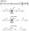
Schematic diagram of HCMVs lacking the MIERR distal enhancer and modulator or modulator alone. The MIERR of WT virus is composed of promoter (+1 to -64), enhancer (ENH; -65 to -550), unique region (-551 to -749), and modulator (MOD; -750/-1140). HCMVs rΔ-300/-1108Egfp and rΔ-582/-1108Egfp have deletions in the MIERR from base positions -300 to -1108 and -582 to -1108, respectively (56). Each of them also has adenovirus E1b TATA box, gfp ORF, and SV40 early intron and polyadenylation signal inserted at the site of deletion. Three copies of the 21-bp repeats (black bars) are located in the MIERR distal enhancer (-300 to -550), which was deleted from rΔ-300/-1108Egfp. Positions of MIE and putative UL128 genes (open boxes) are shown. Depicted above is the HCMV genome and its unique long (UL) and short (US), internal repeat long (IRL) and short (IRS), terminal repeat long (TRL) and short (TRS), and a-sequence (as) components.
FIG. 5
FIG. 5
MIERR-dependent transcription is not increased by deletion of the 21-bp repeats and modulator. (A) Analysis of viral IE1 RNAs in WT-, rΔ-582/-1108Egfp-, rΔ-300/-1108Egfp-, or mock-infected HFF cells at 8 h p.i. Infections (MOI of 3) were performed in parallel with equivalent input viral titers as described in Materials and Methods. Isolated RNA was subjected to RPA, using IE1- and actin-specific riboprobes. Arrows point to positions of protected unspliced and spliced IE1 and actin RNAs. Std, standard; nt, nucleotides. (B) Analysis of viral IE1 RNAs in WT-, rΔ-582/-1108Egfp-, rΔ-300/-1108Egfp-, or mock-infected RA-pretreated or untreated NT2 cells at 24 h p.i. Viral stocks used for panel A were used to infect NT2 cells (MOI of 3). Isolated RNA (25 μg/sample) was subjected to RPA, using both IE1- and actin-specific riboprobes. (C) TSA-induced reactivation of IE1 RNA production in NT2 cells preinfected with WT or rΔ-300/-1108Egfp. Infections (MOI of 3) were performed in parallel with viral inocula used for experiments shown in panels A and B. TSA (100 ng/ml) was added 1.5 h p.i. RNA (25 μg/sample) was isolated at 24 h p.i. and subjected to RPA, using IE1- and actin-specific riboprobes.
FIG. 6
FIG. 6
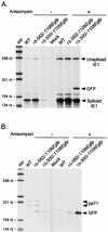
The heterologous MIERR promoter of rΔ-300/-1108Egfp exhibits IE transcriptional kinectics. (A) Analysis of viral IE1 and GFP RNAs in WT-, rΔ-582/-1108Egfp-, rΔ-300/-1108Egfp- or mock-infected HFF cells at 8 h p.i. in the presence or absence of anisomycin (100 μM). Infections were performed in parallel at an MOI of 0.05. Isolated RNA (20 μg/sample) was subjected to RPA, using both IE1- and GFP-specific riboprobes. Arrows indicate positions of protected unspliced and spliced IE1 and GFP RNAs. (B) Analysis of pp71 (UL82) and GFP RNAs of these viruses at 8 h p.i. RNAs used for Fig. 5A were also subjected to RPA, with pp71- and GFP-specific riboprobes. Arrows indicate positions of protected pp71 and GFP RNAs. std, standard; nt, nucleotides.
FIG. 7
FIG. 7
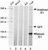
TSA reactivates both heterologous and native MIERR promoters of rΔ-300/-1108Egfp in embryonal NT2 cells. (A) Analysis of IE1 and GFP RNAs in WT- and mock-infected embryonal NT2 cells at 24 h p.i. RA (10 μM) or TSA (100 ng/ml) was added to or omitted from growth medium at 1.5 h p.i. Isolated RNA was subjected to RPA, using both IE1- and GFP-specific riboprobes. Samples were normalized for the amount of cellular actin RNA as determined by RPA (data not shown). Arrows indicate positions of protected unspliced and spliced IE1 and GFP RNAs. std, standard; nt, nucleotides.
FIG. 8
FIG. 8
Percentages of infected embryonal NT2 cells with reactivated MIERR as determined by flow cytometry. rΔ-300/-1108Egfp - and WT-infected embryonal NT2 cells (MOI of 3) were analyzed by flow cytometry at 72 h p.i. RA (10 μM) or TSA (100 ng/ml) was added to or omitted (No Add) from growth medium at 24 h p.i. Ten thousand live cell events in each experimental group were acquired for analysis. For rΔ-300/-1108Egfp, the proportion of TSA-treated and untreated infected cells excluding propidium iodide varied by 21%. FSC, forward scatter.
FIG. 9
FIG. 9
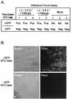
Infectious focus assay of TSA-induced preinfected NT2 cells. (A) Scoring of infectious focus assays for rΔ-300/-1108Egfp- and rΔ-582/-1108Egfp-infected NT2 cells (MOI of 3). Infected embryonal NT2 cells were induced with TSA (100 ng/ml) at 24 h p.i. and flow sorted into GFP-positive (+GFP) and -negative (−GFP) groups at 72 h p.i. Mock, +GFP, or −GFP NT2 cells (102) were cocultivated with subconfluent HFF cells (105). The cultures were monitored for evidence of infectious foci for 30 days by light and fluorescent microscopes. Infectious focus assays were performed in triplicate except for rΔ-582/-1108Egfp, which was tested in duplicate. Infectious focus assays were scored as positive (Pos) or negative (Neg). NA, not applicable. (B) Confocal microscopy of infectious focus. Embryonal NT2 cells were infected with rΔ-300/-1108Egfp, treated with TSA (100 ng/ml) at 24 h p.i., and flow sorted into GFP-positive (+GFP) and -negative (−GFP) groups at 72 h p.i. Segregated infected NT2 cells were cocultivated with HFF cells as described above. Inverted confocal microscopy was performed a 21 days of culture. The asterisk marks the location of a colony of NT2 cells within the HFF monolayer.

Similar articles

Cited by

References

    1. Adler S T, McVoy M A. Human cytomegalovirus DNA replicates after early circularization by concatemer formation, and inversion occurs within the concatemer. J Virol. 1994;68:1040–1051. - PMC - PubMed
    1. Ahn J-H, Jang W J, Hayward G S. The human cytomegalovirus IE2 and UL112–113 proteins accumulate in viral DNA replication compartments that initiate from periphery of promyelocytic leukemia protein-associated nuclear bodies (PODs or ND10) J Virol. 1999;73:10458–10471. - PMC - PubMed
    1. Akugan E, Ziegler M, Grez M. Determinants of retrovirus gene expression in embryonal carcinoma cells. J Virol. 1991;65:382–388. - PMC - PubMed
    1. Andrews P W. Retinoic acid induces neuronal differentiation of a cloned human embryonal carcinoma cell line in vitro. Dev Biol. 1984;103:285–293. - PubMed
    1. Andrews P W, Damjanov I, Simon D, Banting G S, Carlin C, Dracopoli N C, Fogh J. Pluripotent embryonal carcinoma clones derived from the human teratocarcinoma cell line Tera-2. Differentiation in vivo and in vitro. Lab Investig. 1984;50:147–162. - PubMed

Publication types