. 2001 Feb 5;152(3):483-90.
doi: 10.1083/jcb.152.3.483.

DIABLO promotes apoptosis by removing MIHA/XIAP from processed caspase 9

Affiliations

DIABLO promotes apoptosis by removing MIHA/XIAP from processed caspase 9

P G Ekert et al. J Cell Biol. .

Abstract

MIHA is an inhibitor of apoptosis protein (IAP) that can inhibit cell death by direct interaction with caspases, the effector proteases of apoptosis. DIABLO is a mammalian protein that can bind to IAPs and antagonize their antiapoptotic effect, a function analogous to that of the proapoptotic Drosophila molecules, Grim, Reaper, and HID. Here, we show that after UV radiation, MIHA prevented apoptosis by inhibiting caspase 9 and caspase 3 activation. Unlike Bcl-2, MIHA functioned after release of cytochrome c and DIABLO from the mitochondria and was able to bind to both processed caspase 9 and processed caspase 3 to prevent feedback activation of their zymogen forms. Once released into the cytosol, DIABLO bound to MIHA and disrupted its association with processed caspase 9, thereby allowing caspase 9 to activate caspase 3, resulting in apoptosis.


Figures
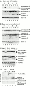
Figure 1
Figure 1
(A) MIHA and Bcl-2 inhibit caspase 9 and caspase 3 processing in response to UV radiation but not serum withdrawal, and MIHA binds to endogenous, processed caspase 3. NT 2 cells stably transfected with MIHA (M) or LacZ (C) were exposed to 100 J/m2 UV radiation and lysates were harvested at 6 h. Kinetics of caspase processing was followed using Western blot immunoblotted with anti-caspase 3 and anti-caspase 9 antibodies. Full-length caspase 9 and the cleaved 37-kD fragment of caspase 9, full-length caspase 3, and the 20- and 17-kD large subunits are indicated. Almost complete processing of caspase 9 was observed in control cells, and this was inhibited by the stable expression of MIHA. Caspase 3 processing was also inhibited by MIHA, since most caspase 3 in the MIHA line remained in the unprocessed form (lane 7) compared with control cells (lane 8). (B) NT2 cells stably transfected with MIHA (M) or LacZ (C) were grown under serum-free conditions and lysates were harvested and immunoblotted with anti-caspase 3 and anti-caspase 9 antibodies. Caspase 3 cleavage occurred in both MIHA- and LacZ-expressing cells. Over the time course, most caspase 9 remained in the full-length rather than cleaved form. (C) NT2 cells stably expressing MIHA, LacZ, or Bcl-2 were exposed to 100 J/m2 UV radiation and the lysates were harvested 24 h later and immunoblotted with anti-caspase 9 and anti-caspase 3 antibodies. MIHA lines inhibited caspase 9 and caspase 3 cleavage but not to the same degree as Bcl-2. In all the Western blots, anti-HSP70 immunoblotting was done as loading controls. (D) Flag-tagged MIHA was immunoprecipitated from the lysates of NT2 cells stably expressing MIHA or LacZ that were cultured in serum-containing media (C), or exposed to 100 J/m2 (UV), or were cultured in serum-free media for 24 h (−S), and the immunoprecipitate was probed with anti-caspase 3 and anti-caspase 9 antibodies. Processed endogenous caspase 3 coimmunoprecipitated with MIHA after serum withdrawal and UV radiation. No association with caspase 9 in either its zymogen or processed form was detected.
Figure 5
Figure 5
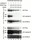
DIABLO inhibits the binding of processed caspase 9 to MIHA. 293T cells were transfected as indicated. 48 h after transfection, the lysates were harvested and MIHA was immunoprecipitated. Both the lysates and the immunoprecipitates (IP, indicated on the left) were immunoblotted with anti-caspase 9, anti-DIABLO, and anti-Flag antibody. MIHA coimmunoprecipitated processed, but not full-length, caspase 9 (lane 1). This association persisted when the MIHA/DIABLO ratio was 4:1 (lane 2) but was significantly inhibited by DIABLO at lower ratios (lanes 3–6). Endogenous and transfected DIABLO were able to associate with MIHA. The full-length (50-kD) and processed forms (37-kD) of caspase 9 were present in the lysates. Transfected DIABLO was expressed in both an unprocessed (25-kD) and processed (23-kD) form. The processed form coimmunoprecipitated with MIHA.
Figure 2
Figure 2
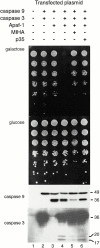
Expressing a truncated form of Apaf-1, procaspase 9 and procaspase 3 in S. cerevisiae can kill yeast, and this can be inhibited by MIHA. S. cerevisiae clones expressing the constructs indicated at the top were grown under inducing (galactose) or noninducing (glucose) conditions. Transformants expressing truncated Apaf-1, procaspase 9, and procaspase 3 were killed (galactose, lane 4) unless protected by either MIHA or p35 (lanes 5 and 6). The same transformants were induced in liquid medium for 8 h, and lysates were immunoblotted with anti-caspase 9 and anti-caspase 3 antibodies (bottom two panels). Apaf-1&1–530 caused processing of caspase 9 but did not kill yeast (lane 3). Procaspase 3 was cleaved by processed caspase 9 (lane 4), which resulted in death of the yeast. Both caspase 3 and caspase 9 processing was inhibited by MIHA and to a lesser degree, p35.
Figure 3
Figure 3
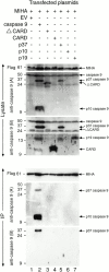
MIHA binds to processed, active caspase 9 and not to the zymogen form of caspase 9. 293T cells were transfected with Flag-tagged MIHA and the indicated constructs (EV, empty vector; ΔCARD, caspase 9 lacking the prodomain; CARD, prodomain only; p37, prodomain and large subunit; p10, small subunit only; p19, large subunit only). MIHA was immunoprecipitated and the lysates and immunoprecipitate (IP) were probed with the antibodies indicated on the left. Two caspase 9 antibodies were used: A, which recognizes the p10 and ΔCARD as well as full-length caspase 9 and the p37; and B, which recognizes the CARD and the p19 as well as the p37 fragment of caspase 9 and full-length caspase 9. FL, anti-Flag antibody. MIHA coimmunoprecipitated the processed (37-kD) and p10 subunit of caspase 9 (lane 2) but only when caspase 9 was transfected as a full-length construct and underwent processing within the cell (lane 2). None of the individual components of caspase 9 immunoprecipitated with MIHA, suggesting that MIHA binds only to the processed enzyme in its active conformation.
Figure 4
Figure 4
MIHA acts downstream of mitochondrial cytochrome c and DIABLO release. NT2 cells stably transfected with LacZ, Bcl-2, or MIHA were exposed to UV radiation for the indicated times. The cells were harvested and membrane and cytosolic fractions were separated. The two fractions were then immunoblotted with anti-caspase 3, anti-caspase 9, anti-cytochrome c and anti-DIABLO antibodies. Control (LacZ) cells show translocation of cytochrome c and DIABLO to the cytosolic fraction and cleavage of cytosolic caspase 3 and caspase 9 (lanes 1–6). All these processes were inhibited in cells expressing Bcl-2 (lanes 7–12). MIHA inhibited processing of caspase 9 and caspase 3, but cytochrome c and DIABLO translocated from membrane to cytosolic fractions to the same degree as in control cells (lanes 13–18).

Similar articles

Cited by

References

    1. Claveria C., Albar J.P., Serrano A., Buesa J.M., Barbero J.L., Martinez A.C., Torres M. Drosophila grim induces apoptosis in mammalian cells. EMBO (Eur. Mol. Biol. Organ.) J. 1998;17:7199–7208. - PMC - PubMed
    1. Clem R.J., Miller L.K. Control of programmed cell death by the baculovirus genes p35 and IAP. Mol. Cell. Biol. 1994;14:5212–5222. - PMC - PubMed
    1. Crook N.E., Clem R.J., Miller L.K. An apoptosis inhibiting baculovirus gene with a zinc finger like motif. J. Virol. 1993;67:2168–2174. - PMC - PubMed
    1. Day C.L., Dupont C., Lackmann M., Vaux D.L., Hinds M.G. Solution structure and mutagenesis of the caspase recruitment domain (CARD) from Apaf-1. Cell Death Differ. 1999;6:1125–1132. - PubMed
    1. Deveraux Q.L., Takahashi R., Salvesen G.S., Reed J.C. X-linked IAP is a direct inhibitor of cell-death proteases. Nature. 1997;388:300–304. - PubMed

Publication types

MeSH terms