. 2001 Feb;75(3):1487-506.
doi: 10.1128/JVI.75.3.1487-1506.2001.

Origin-independent assembly of Kaposi's sarcoma-associated herpesvirus DNA replication compartments in transient cotransfection assays and association with the ORF-K8 protein and cellular PML

Affiliations

Origin-independent assembly of Kaposi's sarcoma-associated herpesvirus DNA replication compartments in transient cotransfection assays and association with the ORF-K8 protein and cellular PML

F Y Wu et al. J Virol. 2001 Feb.

Abstract

Six predicted Kaposi's sarcoma virus herpesvirus (KSHV) proteins have homology with other well-characterized herpesvirus core DNA replication proteins and are expected to be essential for viral DNA synthesis. Intact Flag-tagged protein products from all six were produced from genomic expression vectors, although the ORF40/41 transcript encoding a primase-helicase component proved to be spliced with a 127-bp intron. The intracellular localization of these six KSHV replication proteins and the mechanism of their nuclear translocation were investigated. SSB (single-stranded DNA binding protein, ORF6) and PPF (polymerase processivity factor, ORF59) were found to be intrinsic nuclear proteins, whereas POL (polymerase, ORF9), which localized in the cytoplasm on its own, was translocated to the nucleus when cotransfected with PPF. PAF (primase-associated factor, ORF40/41), a component of the primase-helicase tripartite subcomplex together with PRI (primase, ORF56) and HEL (helicase, ORF44), required the presence of all five other replication proteins for efficient nuclear translocation. Surprisingly, even in the absence of a lytic cycle replication origin (ori-Lyt) and any known initiator or origin binding protein, the protein products of all six KSHV core replication genes cooperated in a transient cotransfection assay to form large globular shaped pseudo-replication compartments (pseudo-RC), which excluded cellular DNA. These pseudo-RC structures were confirmed to include POL, SSB, PRI, and PAF but did not contain any newly synthesized DNA. Similar to the human cytomegalovirus system, the peripheries of these KSHV pre-RC were also found to be surrounded by punctate PML oncogenic domains (PODs). Furthermore, by transient cotransfection, the six KSHV core replication machinery proteins successfully replicated a plasmid containing EBV ori-Lyt in the presence of the Epstein-Barr virus-encoded DNA binding initiator protein, ZTA. The KSHV-encoded K8 (ORF-K8) protein, which is a distant evolutionary homologue to ZTA, was incorporated into pseudo-RC structures formed by transient cotransfection with the six core KSHV replication genes. However, unlike ZTA, K8 displayed a punctate nuclear pattern both in transfected cells and at early stages of lytic infection and colocalized with the cellular PML proteins in PODs. Finally, K8 was also found to accumulate in functional viral RC, detected by incorporation of pulse-labeled bromodeoxyuridine into newly synthesized DNA in both tetradecanoyl phorbol acetate-induced JSC-1 primary effusion lymphoblasts and in KSHV lytically infected endothelial cells.


Figures
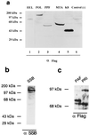
FIG. 1
FIG. 1
Western blot analysis of proteins expressed by eight KSHV DNA replication and replication-associated genes in transient DNA transfection assays. (a) Protein products from Flag-tagged expression plasmids encoding HEL, POL, PPF, MTA, and K8 were transfected into Vero cells. After 48 h, whole-cell protein extracts were electrophoretically fractionated on SDS– 10% polyacrylamide gels, and Western blot analysis was performed by incubating the membrane with a specific MAb or PAb followed by ECL color development. (b) Protein products of untagged pSG5 plasmid vector encoding KSHV SSB detected with rabbit anti-SSB PAb, (c) Protein products of Flag-tagged expression plasmids encoding PAF and PRI detected with mouse anti-Flag MAb.
FIG. 2
FIG. 2
A spliced ORF40 and ORF41 transcript is induced in TPA-treated BCBL-1 PEL cells. (a) Diagram of the genomic organization of ORF40 and ORF41 between coordinates 60308 to 62444 in KSHV. The locations and orientations of the PCR primers used (→) and the sizes of PCR products from spliced and unspliced cDNA templates are shown. (b) Photograph of an ethidium bromide-stained 1% agarose gel showing the separated RT-PCR products. Lanes 1 and 4, no RT-PCR product detected from DNase-treated total RNA isolated from BCBL-1 PEL cell line before TPA induction; lanes 2 and 5, RT-PCR products from RNA isolated 48 h after TPA induction; lanes 3 and 6, PCR products from the total DNA isolated from BCBL-1 cells. For lanes 1 to 3, PCR primers covering the intron region were used; for lanes 4 to 6, PCR primers covering the entire coding region of ORF40/41 were used. (c) Amino acid sequence alignment of KSHV ORF40/41, rhesus rhadinovirus (RRV) ORF40/41, HVS ORF40/41, and EBV BBLF2/3.
FIG. 2
FIG. 2
A spliced ORF40 and ORF41 transcript is induced in TPA-treated BCBL-1 PEL cells. (a) Diagram of the genomic organization of ORF40 and ORF41 between coordinates 60308 to 62444 in KSHV. The locations and orientations of the PCR primers used (→) and the sizes of PCR products from spliced and unspliced cDNA templates are shown. (b) Photograph of an ethidium bromide-stained 1% agarose gel showing the separated RT-PCR products. Lanes 1 and 4, no RT-PCR product detected from DNase-treated total RNA isolated from BCBL-1 PEL cell line before TPA induction; lanes 2 and 5, RT-PCR products from RNA isolated 48 h after TPA induction; lanes 3 and 6, PCR products from the total DNA isolated from BCBL-1 cells. For lanes 1 to 3, PCR primers covering the intron region were used; for lanes 4 to 6, PCR primers covering the entire coding region of ORF40/41 were used. (c) Amino acid sequence alignment of KSHV ORF40/41, rhesus rhadinovirus (RRV) ORF40/41, HVS ORF40/41, and EBV BBLF2/3.
FIG. 3
FIG. 3
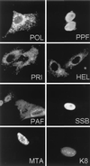
Intracellular localization of single transfected KSHV DNA replication and replication-associated proteins detected with IFA. Equal samples of plasmid DNA (0.2 μg) were used for each gene in transient transfection assays in Vero cells. All except SSB and K8, were detected with mouse anti-Flag MAb and donkey FITC or rhodamine-labeled anti-mouse IgG. SSB and K8 were detected with SSB or K8-specific rabbit PAb and donkey FITC or rhodamine-labeled anti-rabbit IgG.
FIG. 4
FIG. 4
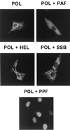
Contribution of PPF but not PAF, PRI, HEL, or SSB to nuclear translocation of POL. The Flag-tagged POL expression plasmid was paired with other untagged replication genes, cotransfected into Vero cells, and then detected with mouse anti-Flag and rhodamine-labeled anti-mouse IgG. POL alone shows cytoplasmic staining but was efficiently translocated into the nucleus in the presence of PPF.
FIG. 5
FIG. 5
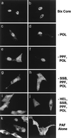
All six viral replication gene products are required for full nuclear translocation of PAF in cotransfected Vero cells. Flag-tagged PAF plasmids were used along with various combinations of the other untagged replication genes, and mouse anti-Flag and rhodamine-labeled anti-mouse IgG were used to visualize the PAF intracellular localization. (a and b) Two separate single-label frames showing cells cotransfected with the whole set of plasmids encoding POL, PPF, PRI, PAF, HEL, and SSB; (e to j) omission experiments showing cotransfection of all plasmids except those encoding POL (c and d), PPF and POL (e and f), SSB, POL, and PPF (g and h), and HEL, SSB, PPF, and POL (i and j). Sequential omission of the other replication genes reverts PAF to a cytoplasmic localization similar to that obtained by transfection with PAF alone (k and m).
FIG. 6
FIG. 6
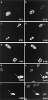
All six core KSHV DNA replication proteins are required for the assembly of complete RC-like structures in contransfected Vero cells, and active DNA synthesis occurs within KSHV RC assembled in the presence of EBV ori-Lyt and ZTA. (a to f) Double-label IFA demonstrating colocalization of POL, PRI, and PAF with SSB in large pseudo-RC in transient assembly assays. SSB was detected by IFA with FITC-labeled anti-SSB rabbit PAb, whereas POL, PRI, and PAF Flag-tagged fusion proteins were detected with rhodamine-labeled anti-Flag mouse MAb. (a and b) Flag-tagged POL cotransfected with the untagged plasmids encoding PPF, PRI, PAF, HEL, and SSB; (c and d) Flag-tagged PRI cotransfected with all five other untagged plasmids; (e and f) Flag-tagged PAF cotransfected with all five other untagged plasmids. The incompletely transfected cell at the upper right in panels c and d displays nuclear diffuse SSB and cytoplasmic PRI. Deliberate omission of any of the six replication genes also disrupted RC formation (not shown). (g to j) Evidence for newly synthesized DNA within KSHV RC formed in Vero cells cotransfected with the complete set of untagged KSHV core replication expression plasmids (POL, PPF, PRI, PAF, HEL, and SSB) plus EBV ori-Lyt and the ZTA DNA binding protein. Cells were pulse labeled for 30 min with BrdU prior to double-label screening for RC formation with an anti-SSB PAb and for active DNA synthesis with an anti-BrdU MAb. (g and i) Anti-KSHV SSB antibody to identify intranuclear RC containing core viral DNA replication proteins. (h and j) Anti-BrdU antibody to identify sites of ongoing DNA synthesis in the same cells (arrows indicate structures that resemble complete viral DNA RC). The anti-BrdU antibody also detected typical background speckled BrdU incorporation patterns found in the 25% of untransfected cells in S phase.
FIG. 7
FIG. 7
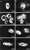
Nuclear DNA staining reveals that the RC-like structures assembled by cotransfection of the six core KSHV DNA replication genes exclude or displace cellular chromatin. (a, c, and g) RC-like structures visualized with anti-SSB PAb and FITC or rhodamine-labeled anti-rabbit IgG; (e and f) RC-like structures formed by Flag-POL fusion protein product detected with anti-Flag MAb and rhodamine-labeled anti-mouse IgG; (b, d, f, and h) nuclear DNA in the same cells detected by adding mounting solution contain the intercalating fluorescent dye DAPI.
FIG. 8
FIG. 8
Successful replication of EBV ori-Lyt in cotransfected Vero cells receiving all six essential KSHV viral replication proteins plus EBV ori-Lyt and ZTA. Southern blotting to detect unmethylated EBV ori-Lyt progeny DNA (Challberg assay) was performed with size-fractionated total DNA from cotransfected Vero cells receiving various combinations of replication gene plasmids. Lanes 1 and 5, EBV POL, PPF, PRI, PAF, HEL, and SSB core genes plus ZTA and EBV ori-Lyt; lanes 2 and 6, all EBV expression plasmids except EBV POL; lanes 3 and 7, KSHV POL, PPF, PRI, PAF, HEL, and SSB core genes plus KSHV MTA, EBV ZTA, and EBV ori-Lyt; lanes 4 and 8, all KSHV plasmids plus ZTA and ori-Lyt except for the plasmid encoding KSHV POL. Each DNA sample (10 μg) was digested with BamHI to excise and detect all EBV ori-Lyt DNA (lanes 1 to 4) or digested with BamHI and DpnI to degrade methylated input plasmid DNA and detect only amplified unmethylated ori-Lyt-containing progeny DNA in transfected cells (lanes 5 to 8). A 32P-labeled 5.4-kb BamHI DNA fragment containing the ori-Lyt target sequences isolated from pEF52 was used as the hybridization probe.
FIG. 9
FIG. 9
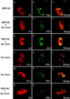
Formation of early KSHV pre-RC incorporating cotransfected ORF-K8 initiates from the periphery of cellular PODs. (a to c) Double-label IFA showing that transfected K8 targets to PODs by itself; detection with rhodamine-labeled anti-Myc MAb and FITC-labeled anti-PML PAb. (d to f) Double-label IFA showing that SSB forms nuclear punctate pre-RC foci associated with PODs when cotransfected with the six core replication genes and K8; detection with FITC-labeled anti-SSB PAb and rhodamine-labeled anti-Myc MAb (K8). (g to i) K8 accumulates in the pre-RC formed by the six core proteins; detection with rhodamine-labeled anti-Myc MAb and FITC-labeled anti-SSB PAb. (j to o) Small pre-RC initiating from the periphery of PODs (j to l), and mature pre-RC closely surrounded by PODs (m to o); detection with rhodamine-labeled anti-Flag MAb (POL) and FITC-labeled anti-PML PAb. (p to r) K8 accumulated in pre-RC surrounded by PODs; detection with rhodamine-labeled anti-Myc MAb and FITC-labeled anti-PML PAb.
FIG. 10
FIG. 10
Visualization and characterization of viral DNA RC in KSHV-infected DMVEC and TPA-induced JSC-1 PEL cells. (a to d) Double-label IFA detection of ORF-K8 accumulated in RC at 72 h after TPA treatment of JSC-1 PEL cells using FITC anti-K8 PAb and rhodamine anti-PPF MAb. (e to h) Double-label IFA detection of K8 colocalized with newly synthesized DNA in TPA-treated PEL cell RC after a 30-min BrdU pulse-label experiment using rhodamine anti-K8 PAb and FITC anti-BrdU MAb. (i to l) Double-label IFA detection of replication compartments surrounded by PODs in TPA-treated PEL cells, using rhodamine-labeled anti-PPF MAb and FITC-labeled anti-PML PAb. (m to o) Double-label IFA detection of K8 accumulated in RC formed in KSHV-infected DMVEC, using rhodamine anti-K8 PAb and FITC anti-BrdU MAb (30 min of BrdU pulse-labeling). (p to r) Double-label IFA detection of PPF in infected DMVEC RC surrounded by PODs, using rhodamine anti-PPF MAb and FITC anti-PML PAb.
FIG. 11
FIG. 11
Close association of the KSHV ORF-K8 and LANA proteins in punctate patterns in interchromatin spaces in a KSHV-infected DMVEC spindle cell nucleus undergoing early stages of the transition from latent to lytic cycle gene expression. The four photomicrograph panels show the same nucleus with different combinations of DAPI staining plus FITC or rhodamine IFA. (a) DAPI staining alone (blue) of an infected DMVEC monolayer spindle cell showing several interchromatin gaps. (b) Combined DAPI staining plus IFA with anti-K8 rabbit PAb (FITC, green) showing multiple small K8-positive punctate domains presumably representing PODs that are predominantly localized within some of the interchromatin spaces. (c) Combined DAPI staining plus IFA with anti-LANA mouse MAb (rhodamine, red) showing LANA punctate domains that are also in interchromatin spaces and presumably associated with latent state plasmid KSHV genomes (3). (d) Triple merge combining DAPI, FITC IFA (K8), and rhodamine IFA (LANA), showing close juxaposition and partial colocalization of the K8 punctate domains with each residual LANA punctate domain in the interchromatin gaps.

Similar articles

Cited by

References

    1. Ahn J H, Brignole III E J, Hayward G S. Disruption of PML subnuclear domains by the acidic IE1 protein of human cytomegalovirus is mediated through interaction with PML and may modulate a RING finger-dependent cryptic transactivator function of PML. Mol Cell Biol. 1998;18:4899–4913. - PMC - PubMed
    1. Ahn J H, Jang W J, Hayward G S. The human cytomegalovirus IE2 and UL112–113 proteins accumulate in viral DNA replication compartments that initiate from the periphery of promyelocytic leukemia protein-associated nuclear bodies (PODs or ND10) J Virol. 1999;73:10458–10471. - PMC - PubMed
    1. Ballestas M E, Chatis P A, Kaye K M. Efficient persistence of extrachromosomal KSHV DNA mediated by latency-associated nuclear antigen. Science. 1999;284:641–644. - PubMed
    1. Burkham J, Coen D M, Weller S K. ND10 protein PML is recruited to herpes simplex virus type 1 prereplicative sites and replication compartments in the presence of viral DNA polymerase. J Virol. 1998;72:10100–10107. - PMC - PubMed
    1. Bush M, Yager D R, Gao M, Weisshart K, Marcy A I, Coen D M, Knipe D M. Correct intranuclear localization of herpes simplex virus DNA polymerase requires the viral ICP8 DNA-binding protein. J Virol. 1991;65:1082–1089. - PMC - PubMed

Publication types

MeSH terms