. 2001 Feb;75(3):1312-24.
doi: 10.1128/JVI.75.3.1312-1324.2001.

The membrane M protein carboxy terminus binds to transmissible gastroenteritis coronavirus core and contributes to core stability

Affiliations

The membrane M protein carboxy terminus binds to transmissible gastroenteritis coronavirus core and contributes to core stability

D Escors et al. J Virol. 2001 Feb.

Abstract

The architecture of transmissible gastroenteritis coronavirus includes three different structural levels, the envelope, an internal core, and the nucleocapsid that is released when the core is disrupted. Starting from purified virions, core structures have been reproducibly isolated as independent entities. The cores were stabilized at basic pH and by the presence of divalent cations, with Mg(2+) ions more effectively contributing to core stability. Core structures showed high resistance to different concentrations of detergents, reducing agents, and urea and low concentrations of monovalent ions (<200 mM). Cores were composed of the nucleoprotein, RNA, and the C domain of the membrane (M) protein. At high salt concentrations (200 to 300 mM), the M protein was no longer associated with the nucleocapsid, which resulted in destruction of the core structure. A specific ionic interaction between the M protein carboxy terminus and the nucleocapsid was demonstrated using three complementary approaches: (i) a binding assay performed between a collection of M protein amino acid substitution or deletion mutants and purified nucleocapsids that led to the identification of a 16-amino-acid (aa) domain (aa 237 to 252) as being responsible for binding the M protein to the nucleocapsid; (ii) the specific inhibition of this binding by monoclonal antibodies (MAbs) binding to a carboxy-terminal M protein domain close to the indicated peptide but not by MAbs specific for the M protein amino terminus; and (iii) a 26-residue peptide, including the predicted sequence (aa 237 to 252), which specifically inhibited the binding. Direct binding of the M protein to the nucleoprotein was predicted, since degradation of the exposed RNA by RNase treatment did not affect the binding. It is proposed that the M protein is embedded within the virus membrane and that the C region, exposed to the interior face of the virion in a population of these molecules, interacts with the nucleocapsid to which it is anchored, forming the core. Only the C region of the M protein is part of the core.


Figures

FIG. 1
FIG. 1
Effect of chemical agents on virus core structure. Purified cores were treated with different chemical agents using the concentrations indicated in the figure. Representative electron microscopy images of purified cores stained with 2% uranyl acetate after the indicated treatments are shown (left). a, the effect of virus core incubation with Triton X-100 concentrations higher than 2% could not be evaluated due to interference with electron microscopy. ∗, structure not detected after treatment with any concentration of the indicated chemical agent below the maximum value shown within each column.
FIG. 2
FIG. 2
Treatment of TGEV cores with ionic agents. Purified cores were treated with increasing concentrations of NaCl and KCl (A) or guanidine isothiocyanate (B). For the upper left panels, the protein composition was analyzed by SDS-PAGE using 5 to 20% gradient gels and silver staining. The arrows to the right of the panel indicate the positions of TGEV structural proteins. The core structure observed by electron microscopy after negative staining (2% uranyl acetate) is shown for three representative treatments (bottom left panel). The right panel shows the percentage of the core-associated M protein after each treatment in relation to the M protein of untreated cores. The amount of each viral protein was estimated by densitometry using the N protein as an internal control.
FIG. 3
FIG. 3
Treatment of TGEV cores with nonionic agents. Purified cores were treated with increasing concentrations of a nonionic detergent, Triton X-100 (A) or 2-mercaptoethanol (B). The upper left panel shows that the protein composition was analyzed by SDS-PAGE using 5 to 20% gradient gels and silver staining. The positions of the TGEV structural proteins are indicated (arrows to the right of panel). The core structure is shown by electron microscopy of negatively stained specimens (2% uranyl acetate) after three representative treatments (bottom left). The percentage of the core-associated M protein after each treatment in relation to the M protein of untreated cores is also shown (right panel). The amount of virus protein was estimated by densitometry using the N protein as an internal control. The effect of detergent on core structure could not be evaluated when concentrations of Triton X-100 higher than 2% were used, due to interference with electron microscopy.
FIG. 4
FIG. 4
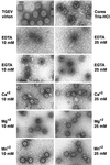
Effect of divalent cations on the core structure. Representative electron micrographs of purified TGEV virions stained with 2% uranyl acetate are shown (topmost left). The rest of the panels show electron microscopy images of cores incubated in 100 mM Tris-HCl (pH 8) for 15 min at room temperature in the presence of cation chelating agents (EDTA or EGTA) or divalent cations. Bar, 100 nm.
FIG. 5
FIG. 5
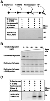
Interaction of 35S-labeled M protein with purified nucleocapsids. (A) Scheme of the assay developed to study the binding of the M protein to a nucleocapsid based on protein G-Sepharose beads coated with MAb 3D.C10 specific for the N protein (αN MAb) and purified nucleocapsids (top). Wild-type M protein and luciferase were transcribed in vitro and labeled with [35S]methionine/cysteine. M protein or luciferase was incubated with the nucleocapsid complex, and the bound proteins were analyzed by SDS-PAGE and fluorography (lower part of panel A). + and −, presence and absence of the indicated component in the assay. (B) Inhibition of binding of the M protein to a nucleocapsid by increasing concentrations of unlabeled proteins. The bound M protein was analyzed by SDS-PAGE and fluorography. (C) Effect of nucleocapsid immunocomplexes' incubation with RNase (60 μg/ml for 60 min at 37°C) on recognition of the M protein in the assay described for panel A.
FIG. 6
FIG. 6
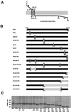
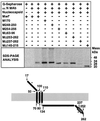
Generation of M gene mutants by site-directed mutagenesis. (A) Model of M protein topology with amino terminus-exo carboxy terminus-endo topology in the virion membrane. Predicted glycosylation sites are indicated by asterisks. Numbered arrows indicate the amino acid positions in the model. (B) Scheme of the M protein mutants generated. Numbers below the bars indicate the mutated amino acid (substitution mutants) or flanking amino acids in the deletion mutants. The mutated amino acid or the flanking amino acids of each deletion are indicated above the bars. Substitutions or deletions are indicated by open boxes within bars. Mutant names are indicated in the left column. (C) The mutant genes were expressed in a rabbit reticulocyte lysate in the presence of [35S]methionine/cysteine and were analyzed by SDS-PAGE and autoradiography.
FIG. 7
FIG. 7
Mapping of the domains recognized by the M protein-specific MAbs. Carboxy-terminal deletion mutants were expressed in a rabbit reticulocyte lysate in the presence of [35S]methionine/cysteine and were immunoprecipitated with M-specific MAbs. The immunoprecipitated proteins were analyzed by SDS-PAGE and fluorography. The M protein domains recognized by the MAbs used are indicated within the bar. Numbers on the top of the bar indicate amino acid positions.
FIG. 8
FIG. 8
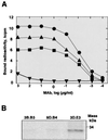
Relative avidity of M-specific MAbs. (A) Binding of MAbs 3D.E3 (●), 3B.B3 (■), and 9D.B4 (▴) and polyclonal serum rabbit anti-GUS (▾) to the TGEV M protein as determined by RIA to estimate the MAb relative avidity for the M protein. (B) Analysis by SDS-PAGE and autoradiography of the M protein immunoprecipitated by these MAbs.
FIG. 9
FIG. 9
Inhibition of binding of the M protein to nucleocapsids by M-specific MAbs. Inhibition of binding of the M protein to nucleocapsids by MAbs 9D.B4, 3D.E3, and 3B.B3 (specific for the M protein carboxy-terminal domain) and by MAbs 1A6 and 25.22 (specific for the amino-terminal domain of the M protein) was analyzed as described for Fig. 5. Labeled M protein was incubated with the protein G-Sepharose-MAb 3D.C10 complex coated with the viral nucleocapsid in the presence of increasing concentrations of MAb. Bound M protein was analyzed by SDS-PAGE and fluorography. Control immunoprecipitations were carried out in the presence of the S-specific MAb 5B.H1.
FIG. 10
FIG. 10
Binding of M protein mutants to purified nucleocapsids. The binding of 35S-labeled M protein mutants to viral nucleocapsids was performed as indicated for Fig. 5. Bound M protein was analyzed by SDS-PAGE and fluorography. + and −, presence and absence of the component in the reaction mixture. The scheme (bottom) illustrates the topology of the M protein with the conformation amino terminus-exo, carboxy terminus-endo within the virus envelope. Predicted glycosylation sites are indicated by asterisks. Numbered arrows indicate the approximate position of the amino acids in the model. wt, wild type.
FIG. 11
FIG. 11
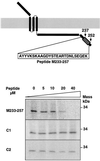
Inhibition of binding of the M protein to nucleocapsids by using synthetic peptides. The localization of the M233-257 peptide within the M protein is illustrated (top panel). Inhibition of binding of the M protein to nucleocapsids (Fig. 5) by increasing concentrations of the indicated synthetic peptides was analyzed by SDS-PAGE and fluorography.

Similar articles

Cited by

References

    1. Almazan F, González J M, Pénzes Z, Izeta A, Calvo E, Plana-Durán J, Enjuanes L. Engineering the largest RNA virus genome as an infectious bacterial artificial chromosome. Proc Natl Acad Sci USA. 2000;97:5516–5521. - PMC - PubMed
    1. Ansorge W. Fast and sensitive detection of protein and DNA bands by treatment with potassium permanganate. J Biochem Biophys Methods. 1985;11:13–20. - PubMed
    1. Ballesteros M L, Sánchez C M, Enjuanes L. Two amino acid changes at the N-terminus of transmissible gastroenteritis coronavirus spike protein result in the loss of enteric tropism. Virology. 1997;227:378–388. - PMC - PubMed
    1. Bass S H, Mulkerrin M G, Wells J A. A systematic mutational analysis of hormone-binding determinants in the human growth hormone receptor. Proc Natl Acad Sci USA. 1991;88:4498–4502. - PMC - PubMed
    1. Baudoux P, Carrat C, Besnardeau L, Charley B, Laude H. Coronavirus pseudoparticles formed with recombinant M and E proteins induce alpha interferon synthesis by leukocytes. J Virol. 1998;72:8636–8643. - PMC - PubMed

Publication types

Substances

LinkOut - more resources