. 2001 Jan;75(1):323-40.
doi: 10.1128/JVI.75.1.323-340.2001.

Essential role played by the C-terminal domain of glycoprotein I in envelopment of varicella-zoster virus in the trans-Golgi network: interactions of glycoproteins with tegument

Affiliations

Essential role played by the C-terminal domain of glycoprotein I in envelopment of varicella-zoster virus in the trans-Golgi network: interactions of glycoproteins with tegument

Z H Wang et al. J Virol. 2001 Jan.

Abstract

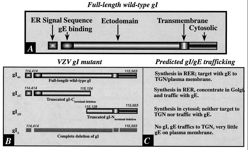
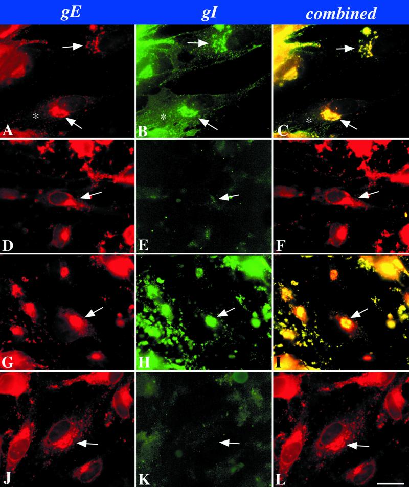
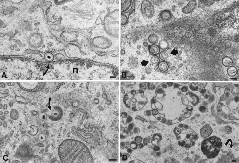
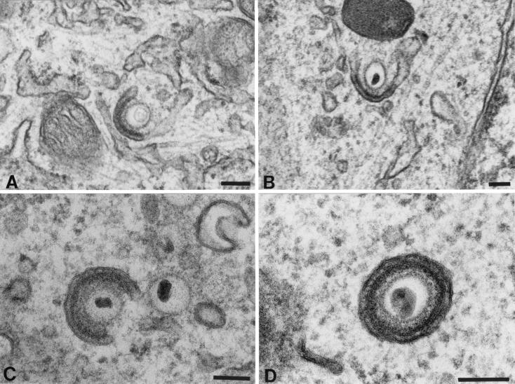
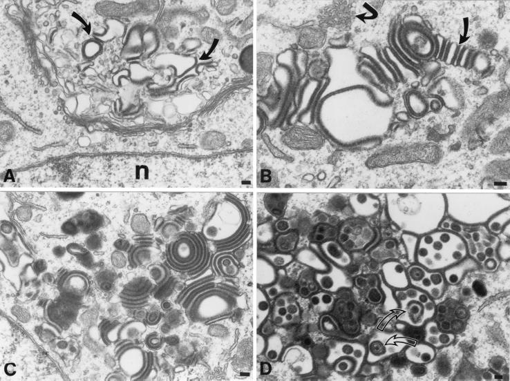
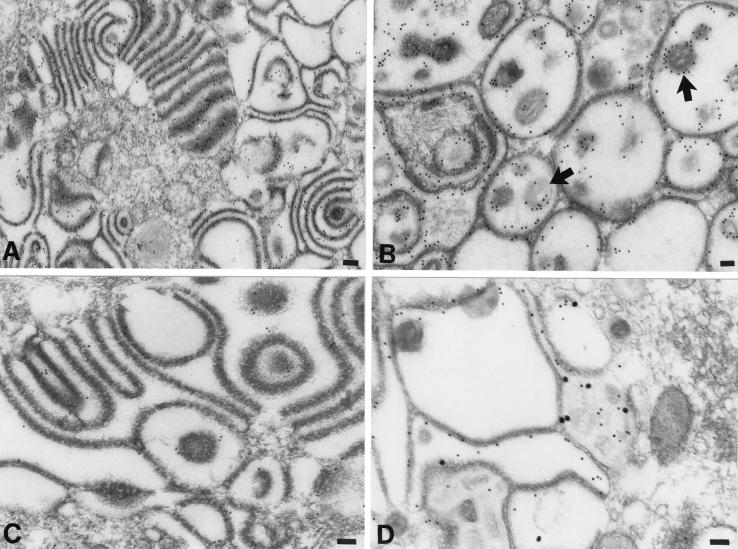
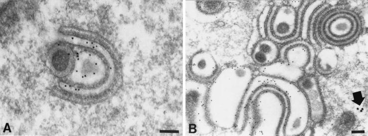
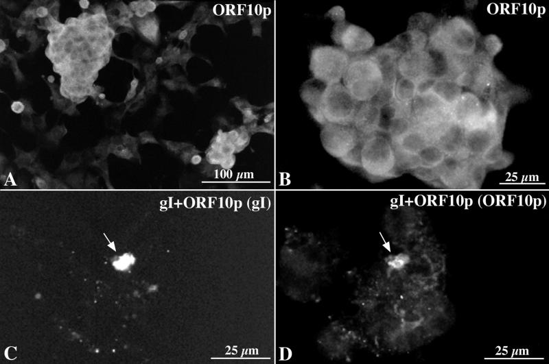
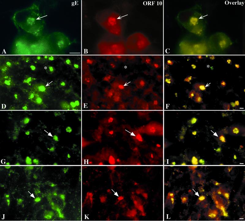
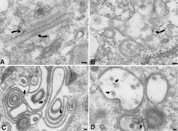
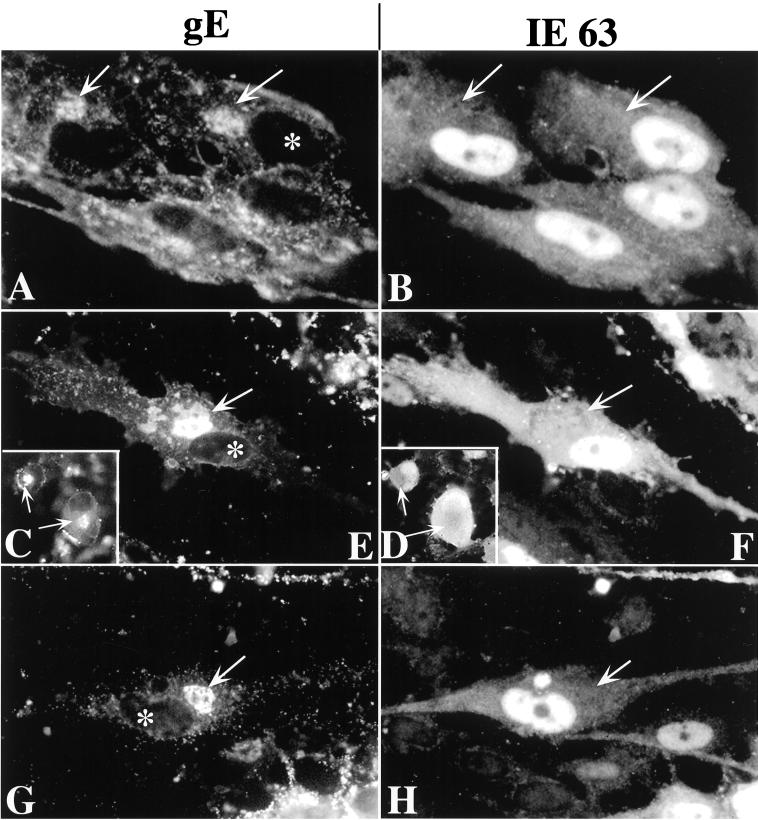
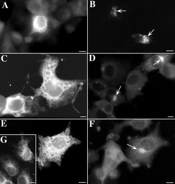
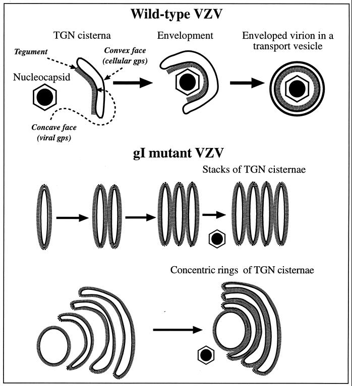
Varicella-zoster virus (VZV) is enveloped in the trans-Golgi network (TGN). Here we report that glycoprotein I (gI) is required within the TGN for VZV envelopment. Enveloping membranous TGN cisternae were microscopically identified in cells infected with intact VZV. These sacs curved around, and ultimately enclosed, nucleocapsids. Tegument coated the concave face of these sacs, which formed the viral envelope, but the convex surface was tegument-free. TGN cisternae of cells infected with VZV mutants lacking gI (gI(Delta)) or its C (gI(DeltaC))- or N-terminal (gI(DeltaN))-terminal domains were uniformly tegument coated and adhered to one another, forming bizarre membranous stacks. Viral envelopment was compromised, and no virions were delivered to post-Golgi structures. The TGN was not gI-immunoreactive in cells infected with the gI(Delta) or gI(DeltaN) mutants, but it was in cells infected with gI(DeltaC) (because the ectodomains of gI and gE interact). The presence in the TGN of gI lacking a C-terminal domain, therefore, was not sufficient to maintain enveloping cisternae. In cells infected with intact VZV or with gI(Delta), gI(DeltaN), or gI(DeltaC) mutants, ORF10p immunoreactivity was concentrated on the cytosolic face of TGN membranes, suggesting that it interacts with the cytosolic domains of glycoproteins. Because of the gE-gI interaction, cotransfected cells that expressed gE or gI were able to target truncated forms of the other to the TGN. Our data suggest that the C-terminal domain of gI is required to segregate viral and cellular proteins in enveloping TGN cisternae.


Figures

FIG. 1
FIG. 1
(A) The domains of gI are shown diagrammatically. Note that the signal sequence, which is presumably responsible for the biosynthesis of gI in the RER, is at the N-terminal end of the molecule. Because signal peptides are cleaved cotranslationally, this domain is not found in the completed integral membrane protein. (B) The domains of the tested mutant forms of gI are compared to those of the full-length intact gI. The numbers refer to base pairs in the VZV genome at which the sequence encoding the indicated domain begins or ends. (C) The predicted patterns of gI trafficking in cells infected with the corresponding virions diagrammed in panel B. These predictions were tested in immunocytochemical experiments. (i) gIwt. The trafficking of gI in cells infected with intact VZV has previously been described (30) and is thus the expected pattern in cells infected with intact virions. Since gI and gE form a complex in the RER, they would be expected to traffic together during post-RER stages of intracellular transport. (ii) gIΔC. Despite the deletion of its C-terminal domain, the gIΔC mutant protein would still be expected to be synthesized in the RER because it contains a signal sequence; however, since the transmembrane domain is lacking, the gIΔC mutant should be completely translocated to the lumen of the RER and lack a membranous anchor. Although gIΔC is thus analogous to a secreted protein, the complex formed with gE at its N-terminal would be expected to cause the gIΔC mutant to be transported along with its normal gE partner. (iii) gIΔN. The deletion of the N-terminal domain of gI (gIΔN) would be anticipated to prevent its biosynthesis in the RER because of the lack of a signal sequence. The deletion of the N-terminal domain of gI would also be expected to prevent interactions of gIΔN with gE. The elimination of these interactions would not be expected to interfere with the targeting of gE to the TGN because gE has its own TGN targeting sequence and patch (37). (iv) gI130. The total deletion of gI would, of course, be expected to eliminate gI immunoreactivity. Because of the endocytosis signal in the sequence of gE (22), relatively little gE would be expected to be retained on the plasma membrane unless it were induced to remain there because it is complexed with gI. gI is not retrieved to the TGN when it traffics to the plasma membrane in transfected cells that express only gI (, ; but see also reference 21). The retention of gE in the plasma membrane might thus occur in cells infected with virions carrying gIwt but not in those carrying gIΔN or gIΔ. Because the gIΔC protein is not membrane anchored, it also would not be expected to interfere with the endocytosis of the gE to which it is bound.
FIG. 2
FIG. 2
Immunocytochemical localization of gI and gE immunoreactivities in cells infected with intact (Ellen) or mutant VZV as shown in Fig. 1. (A to C) Wild-type VZV. (D to F) gIΔ. (G to I) gIΔC. (J to L) gIΔN. The arrows point to the location of the TGN in the infected cells. The asterisk indicates plasma membrane immunostaining. Bar, 10 μm.
FIG. 3
FIG. 3
Cellular locations of nucleocapsids and enveloped virions in HELF cells infected with intact (Ellen) VZV. (A) An enveloped virion lies in the PC-RER (arrow). The virion is located at a point where the RER is continuous with the outer membrane of the nuclear envelope. The thickness and electron density of the viral envelope are greater than those of adjacent cellular membranes. n, nucleus. (B) Enveloped nucleocapsids in the RER fuse with RER membranes, releasing unenveloped nucleocapsids into the cytosol (arrows). The infected cells were incubated at 20°C in order to slow the intracellular transport of VZV. (C) A nucleocapsid in the region of the TGN is partially wrapped by a C-shaped cisterna of cellular membranes (arrow). Note the presence of dense material between the nucleocapsid and the inner, concave face of the wrapping cisterna. Unenveloped nucleocapsids are present in the neighboring cytosol. (D) Enveloped VZV accumulates in large vacuoles that have been found to be late endosomes-prelysosomes. Note also the proliferation of smooth cellular membranes, which are in continuity with the RER (arrow). Bars, 150 nm.
FIG. 4
FIG. 4
Apparent stages in the envelopment of VZV in the TGN of cells infected with intact (Ellen) VZV. (A) An unenveloped nucleocapsid makes contact with a specialized C-shaped cisterna of the TGN. Electron-dense material that resembles tegument accumulates between the nucleocapsid and the concave face of the wrapping cisterna. (B and C) The arms of the “C” of the wrapping cisterna extend toward one another, eliminating the space open to the cytosol and gradually enclosing more and more of the nucleocapsid. The electron-dense material remains adherent to the concave face of the wrapping cisterna. (D) The opening to the cytosol has been closed. Two membranes surround the nucleocapsid. The inner membrane has electron-dense tegument-like material adherent to it. The structure is interpreted to be a newly enveloped virion enclosed in a transport vesicle. The inner membrane is in contact with tegument and is derived from the concave face of the original C-shaped cisterna. The outer membrane, that of the transport vesicle, is derived from the convex face of the C-shaped cisterna. Bars, 150 nm.
FIG. 5
FIG. 5
The morphology of the TGN of cells infected with a mutant VZV in which the C-terminal portion of gI has been deleted (gIΔC) is different from that of cells infected with intact (Ellen) VZV. The morphology of the TGN in cells infected with gIΔ and gIΔN cannot be distinguished from that of cells infected with gIΔC. (A) Vesicles, cisternae, and vacuoles of the TGN are distorted in contour, and their membranes are increased in electron density (arrows) and apparent thickness. (B) The apparent increase in the thickness of the distorted membranes of the TGN vacuolar structures is due to the presence of a circumferential coating of dense tegument-like material. Some of the coated membrane-enclosed sacs of the TGN have flattened into cisternae and appear to adhere to one another (slightly curved arrow). Note also the proliferation of smooth cellular membranes, which are in continuity with the RER (sharply curved arrow). (C) The adherence of adjacent coated membrane-enclosed cisternae gives rise to bizarre concentric rings of repeating sacs. Electron-dense coatings are found between adjacent cisternae, while their lumens are electron lucent, giving rise to alternating light (lumen) and dark (coated membrane) stripes. (D) In a highly infected cell, the TGN has taken on the appearance of a honeycomb. The walls of the chambers of the honeycomb are composed of membranes the cytosolic faces of which are coated with an electron-dense material. Pleomorphic virions (arrows) can be found within some of the chambers. Bars, 150 nm.
FIG. 6
FIG. 6
The membranes of adherent sacs in the TGN of cells infected with the gIΔC mutant virus display both gE and gI immunoreactivities. (A and B) gE immunoreactivity, demonstrated with 10-nm particles of immunogold in the region of the TGN. (A) Gold particles are concentrated in the membranes of adjacent sacs and are not found in the cytosol. (B) The honeycomb-like structure (see Fig. 5D) found in the region of the TGN of a heavily infected cell contains gE immunoreactivity. Both of the membranes that delimit the chambers of the honeycomb and the virions within the cavities of the chambers are gE immunoreactive. The peripheral distribution of the gE immunoreactivity around the virions (B, arrows) indicates that gE is located in the viral envelope. (C) gI immunoreactivity, demonstrated with 10-nm particles of immunogold in the region of the TGN. Gold particles are concentrated in the membranes of adjacent sacs. The immunoreactivity of gI is less abundant than that of gE (compare with panel A). (D) gE immunoreactivity, demonstrated with 10-nm immunogold particles and gI immunoreactivity demonstrated simultaneously with 20-nm immunogold particles. The gE and gI immunoreactivities are each located in the same membranes lining the chambers of the honeycomb found in the TGN of a highly infected cell. Bars, 150 nm.
FIG. 7
FIG. 7
gE immunoreactivity demonstrated with 10-nm immunogold particles and gI immunoreactivity demonstrated simultaneously with 20-nm immunogold particles in the TGN of a cell infected with the gIΔN mutant virus. (A) The C-shaped TGN cisternae display the immunoreactivity of gE but not that of gI. (B) The membranes of concentric rings and adherent sacs in the TGN are gE immunoreactive. The gI immunoreactivity (arrow) is sparse and, when found, appears only in the cytosol. Bars, 150 nm.
FIG. 8
FIG. 8
ORF10p immunoreactivity is translocated to the TGN when coexpressed with gI. (A and B) ORF10p immunoreactivity in transfected Cos-7 cells expressing ORF10p by itself. The immunoreactivity is diffuse and cytoplasmic. (C and D) gI and ORF10p immunoreactivities in a cell that has been cotransfected with cDNA encoding gI and ORF10p. (C) The cell has been illuminated to show the Cy3 fluorescence of gI immunoreactivity. (D) The cell has been illuminated to show the FITC fluorescence of ORF10p immunoreactivity. The arrows point to the TGN where gI and ORF10p immunoreactivities have become colocalized. Bars: A, 100 μm; B to D, 25 μm.
FIG. 9
FIG. 9
gE and ORF10p immunoreactivities colocalize in the TGN of cells infected with intact (Ellen) or gIΔ, gIΔC, or gIΔN mutant forms of VZV. (A to C) Intact (Ellen) VZV. (D to F) gIΔ mutant VZV. (G to I) gIΔC mutant VZV. (J to L) gIΔN mutant VZV. The column at the left (A, D, G, and J) illustrates gE immunoreactivity; the column in the center (B, E, H, and K) illustrates ORF10p immunoreactivity, and that at the right (C, F, I, and L) depicts the gE-ORF10p overlay. The arrows show the location of the TGN and the markers indicate 10 μm.
FIG. 10
FIG. 10
ORF10p immunoreactivity is found in the cytosol, in coatings of Golgi-TGN membranes, and in virions in cells infected with VZV. (A) Intact (Ellen) VZV. ORF10p immunoreactivity is demonstrated with 10-nm particles of immunogold in the region of the Golgi apparatus. Gold particles are found in the cytosol and in the dense material coating membranes of the Golgi stack (arrows). (B) Late endosomes-prelysosomes in a cell infected with intact (Ellen) VZV. ORF10p immunoreactivity is found within pleomorphic virions (arrow) that accumulate within these vacuoles. (C) gIΔN mutant virus. ORF10p immunoreactivity is found in the osmiophilic material (arrows) that coats the membranes of the rings and stacks of adherent cisternae that characterize the TGN of cells infected with gI mutant virions (see Fig. 5). (D) gIΔN mutant virus. ORF10p immunoreactivity is found in the virions within the chambers (arrows) and the coating of the membranes that form the walls of the chamber of the honeycomb that characterizes the TGN of cells heavily infected with gI mutant virions. Bars, 150 nm.
FIG. 11
FIG. 11
IE63 immunoreactivity does not colocalize with that of gE in the TGN of VZV-infected cells. (A and B) Intact (Ellen) VZV. (C and D [insets]) gIΔ mutant VZV. (E and F) gIΔC mutant VZV. (G and H) gIΔN mutant VZV. The column at the left (A, C, E, and G) illustrates gE immunoreactivity, and that at the right (B, D, F, and H) illustrates IE63 immunoreactivity. The arrows show the location of the TGN. The asterisk shows the location of the nucleus in infected cells in which gE immunoreactivity has been demonstrated. There is no nuclear gE immunoreactivity and no concentration of IE63 immunoreactivity in the TGN, although some diffuse cytoplasmic IE63 immunoreactivity can be discerned. The nuclei of infected cells are highly IE63-immunoreactive. Bars, 10 μm.
FIG. 12
FIG. 12
In cotransfected cells, the intracellular transport and targeting of truncated forms of gI and gE can be influenced by coexpression of full-length forms of the other protein. (A) gE immunoreactivity is found in an ER pattern in cells expressing a truncated form of gE, which lacks transmembrane and cytosolic domains. (B) gE immunoreactivity is concentrated in the TGN (arrows) when cells coexpress the truncated gE with a full-length gI. (C) gI immunoreactivity is found in an ER pattern in cells expressing a truncated form of gI, which lacks the T338 targeting signal and the portion of the cytosolic domain C-terminal to it. The sequence of the cytosolic domain of this mutant is SVKRRRIKKHPIYRPNTKTRRGIQNATPESDVMLEAAIAQLA. (D) gI immunoreactivity is concentrated in the TGN (arrows) when cotransfected cells express both the truncated form of gI shown in C and a full-length form of gE. (E and G) gI immunoreactivity is found in an ER pattern in cells expressing a severely truncated form of gI, which lacks the entire cytosolic domain of the molecule. The cell in panel E has been cotransfected and expresses a full-length form of gE, which has not influenced the ER retention of gI. (F) Same cell shown in panel E, now illuminated to demonstrate gE immunoreactivity. Despite the retention of gI immunoreactivity in the ER, the immunoreactivity of gE has reached the TGN (arrow) and is concentrated in this organelle. (G) gI immunoreactivity is found in an ER pattern in transfected cells expressing only the severely truncated form of gI, which lacks the entire cytosolic domain. Bars, 10 μm.
FIG. 13
FIG. 13
A heuristic model depicting the role of TGN cisternae in viral envelopment in cells infected with intact (Ellen) VZV and how the envelopment process becomes defective in cells infected with gIΔ, gIΔC, or gIΔN mutant virions. When cells are infected with intact (Ellen) VZV (upper panel), nucleocapsids that are free in the cytosol become associated with specialized wrapping cisternae in the TGN. These cisternae are curvilinear, with distinct concave and convex faces. Tegument adheres to the concave face of the curving cisternae, and as the arms of the wrapping cisternae approach one another and ultimately fuse, tegument and the nucleocapsid are enclosed within. The membrane of the concave face of the wrapping cisternae, which is proposed to be rich in viral glycoproteins, becomes the viral envelope. The membrane of the convex face, which is rich in cellular proteins, such as Man 6-P receptors, delimits a transport vesicle that encloses the newly enveloped virion. When gI is deficient (lower panels), viral proteins are no longer segregated to one face of the TGN cisternae. As a result the tegument, which is presumed to bind to the cytosolic domains of the viral glycoproteins, is not confined to a single surface of the TGN cisternae. Because tegument is not restricted to the concave face of curving cisternae, adjacent tegument-coated cisternae fuse with one another to give rise to stacks and concentric rings of adherent sacs. The defect in the TGN interferes with viral envelopment and with the post-Golgi transport of VZV.

Similar articles

Cited by

References

    1. Alconada A, Bauer U, Baudoux L, Piette J, Hoflack B. Intracellular transport of the glycoproteins gE and gI of the varicella-zoster virus. J Biol Chem. 1998;273:13430–13436. - PubMed
    1. Alconada A, Bauer U, Hoflack B. A tyrosine-based motif and a casein kinase II phosphorylation site regulate the intracellular trafficking of the varicella-zoster virus glycoprotein I, a protein localized in the trans-Golgi network. EMBO J. 1996;15:6096–6110. - PMC - PubMed
    1. Alconada A, Bauer U, Sodeik B, Hoflack B. Intracellular traffic of herpes simplex virus glycoprotein gE: characterization of the sorting signals required for its trans-Golgi network localization. J Virol. 1999;73:377–387. - PMC - PubMed
    1. Cohen J, Nguyen H. Varicella-zoster virus glycoprotein I is essential for growth in Vero cells. J Virol. 1997;71:6913–6920. - PMC - PubMed
    1. Cohen J, Seidel K E. Varicella-zoster virus (VZV) open reading frame (ORF) 10 protein the homologue of the essential herpes simplex virus protein VP16, is dispensable for VZV replication in vitro. J Virol. 1994;68:7850–7858. - PMC - PubMed

Publication types

Substances

LinkOut - more resources