Recognition of YKL-39, a human cartilage related protein, as a target antigen in patients with rheumatoid arthritis
- PMID: 11114282
- PMCID: PMC1753367
- DOI: 10.1136/ard.60.1.49
Recognition of YKL-39, a human cartilage related protein, as a target antigen in patients with rheumatoid arthritis
Abstract
Objective: To investigate whether autoimmunity to YKL-39, a recently cloned cartilage protein, occurs in patients with rheumatoid arthritis (RA).
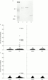
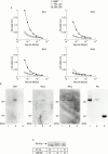
Methods: Autoantibody to YKL-39 was assayed by enzyme linked immunosorbent assay (ELISA) and western blotting in serum samples from patients with RA, systemic lupus erythematosus (SLE), and healthy donors, using recombinant YKL-39 protein. This reactivity was compared with that against a YKL-39 homologue, YKL-40 (human cartilage gp-39/chondrex), which has been reported to be an autoantigen in RA.
Results: Autoantibody to YKL-39 was detected in seven of 87 patients with RA (8%), but not in serum samples from patients with SLE or healthy donors. YKL-40 reactivity was found in only one of 87 RA serum samples (1%), with no cross reactivity to YKL-39.
Conclusion: The existence of anti-YKL-39 antibody in a subset of patients with RA is reported here for the first time. Further, it was shown that the immune response to YKL-39 was independent of that to YKL-40. Clarification of the antibody and T cell responses to autoantigens derived from chondrocyte, cartilage, or other joint components may lead to a better understanding of the pathophysiology of joint destruction in patients with RA.
Figures


Similar articles
-
Autoimmunity against YKL-39, a human cartilage derived protein, in patients with osteoarthritis.J Rheumatol. 2002 Jul;29(7):1459-66. J Rheumatol. 2002. PMID: 12136906
-
The prevalence of autoantibodies against cartilage intermediate layer protein, YKL-39, osteopontin, and cyclic citrullinated peptide in patients with early-stage knee osteoarthritis: evidence of a variety of autoimmune processes.Rheumatol Int. 2005 Nov;26(1):35-41. doi: 10.1007/s00296-004-0497-2. Epub 2004 Sep 18. Rheumatol Int. 2005. PMID: 15378262
-
Proteomic surveillance of autoimmunity in osteoarthritis: identification of triosephosphate isomerase as an autoantigen in patients with osteoarthritis.Arthritis Rheum. 2004 May;50(5):1511-21. doi: 10.1002/art.20189. Arthritis Rheum. 2004. PMID: 15146421
-
[YKL 40: marker of disease activity in rheumatoid arthritis?].Minerva Med. 1999 Nov-Dec;90(11-12):437-41. Minerva Med. 1999. PMID: 10829806 Review. Italian.
-
Neoantigens in osteoarthritic cartilage.Curr Opin Rheumatol. 2004 Sep;16(5):604-8. doi: 10.1097/01.bor.0000133661.52599.bf. Curr Opin Rheumatol. 2004. PMID: 15314502 Review.
Cited by
-
Adenovirus vector-mediated YKL-40 shRNA attenuates eosinophil airway inflammation in a murine asthmatic model.Gene Ther. 2021 Apr;28(3-4):177-185. doi: 10.1038/s41434-020-00202-0. Epub 2020 Oct 12. Gene Ther. 2021. PMID: 33046836
-
Identification of novel citrullinated autoantigens of synovium in rheumatoid arthritis using a proteomic approach.Arthritis Res Ther. 2006;8(6):R175. doi: 10.1186/ar2085. Arthritis Res Ther. 2006. PMID: 17125526 Free PMC article.
-
Chitinase-like proteins are autoantigens in a model of inflammation-promoted incipient neoplasia.Genes Cancer. 2011 Jan;2(1):74-87. doi: 10.1177/1947601911402681. Genes Cancer. 2011. PMID: 21779482 Free PMC article.
-
Structural and thermodynamic insights into chitooligosaccharide binding to human cartilage chitinase 3-like protein 2 (CHI3L2 or YKL-39).J Biol Chem. 2015 Jan 30;290(5):2617-29. doi: 10.1074/jbc.M114.588905. Epub 2014 Dec 4. J Biol Chem. 2015. PMID: 25477513 Free PMC article.
-
Expression of Osteoarthritis Marker YKL-39 is Stimulated by Transforming Growth Factor Beta (TGF-beta) and IL-4 in Differentiating Macrophages.Biomark Insights. 2008 Feb 14;3:39-44. Biomark Insights. 2008. PMID: 19578492 Free PMC article.
References
Publication types
MeSH terms
Substances
LinkOut - more resources
Full Text Sources
Other Literature Sources
Medical
