. 2001 Jan;21(1):209-23.
doi: 10.1128/MCB.21.1.209-223.2001.

Identification and characterization of human orthologues to Saccharomyces cerevisiae Upf2 protein and Upf3 protein (Caenorhabditis elegans SMG-4)

Affiliations

Identification and characterization of human orthologues to Saccharomyces cerevisiae Upf2 protein and Upf3 protein (Caenorhabditis elegans SMG-4)

G Serin et al. Mol Cell Biol. 2001 Jan.

Abstract

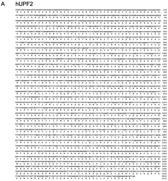
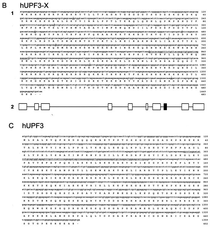
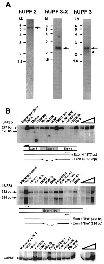
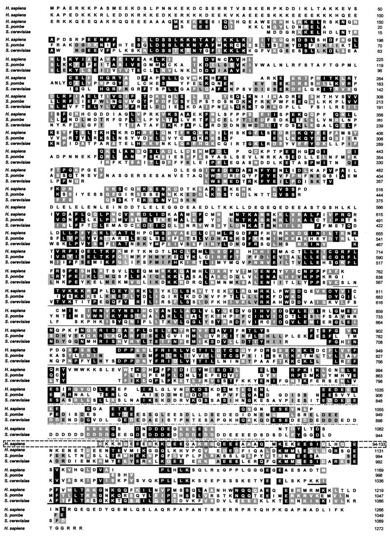
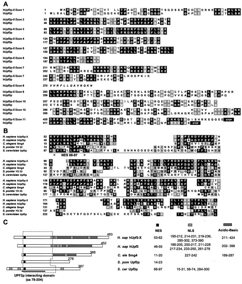
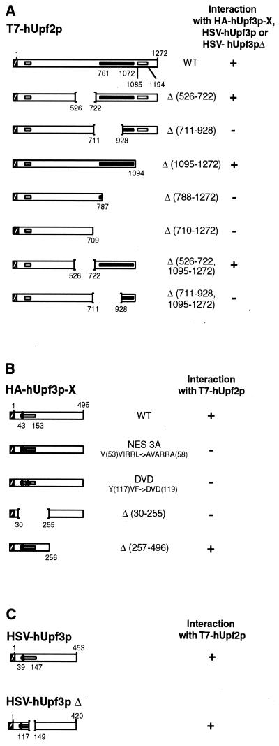
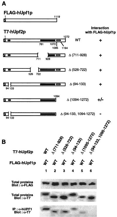
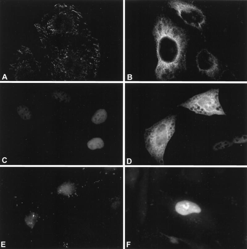
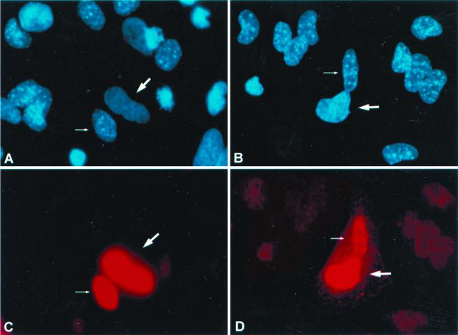
Nonsense-mediated mRNA decay (NMD), also called mRNA surveillance, is an important pathway used by all organisms that have been tested to degrade mRNAs that prematurely terminate translation and, as a consequence, eliminate the production of aberrant proteins that could be potentially harmful. In mammalian cells, NMD appears to involve splicing-dependent alterations to mRNA as well as ribosome-associated components of the translational apparatus. To date, human (h) Upf1 protein (p) (hUpf1p), a group 1 RNA helicase named after its Saccharomyces cerevisiae orthologue that functions in both translation termination and NMD, has been the only factor shown to be required for NMD in mammalian cells. Here, we describe human orthologues to S. cerevisiae Upf2p and S. cerevisiae Upf3p (Caenorhabditis elegans SMG-4) based on limited amino acid similarities. The existence of these orthologues provides evidence for a higher degree of evolutionary conservation of NMD than previously appreciated. Interestingly, human orthologues to S. cerevisiae Upf3p (C. elegans SMG-4) derive from two genes, one of which is X-linked and both of which generate multiple isoforms due to alternative pre-mRNA splicing. We demonstrate using immunoprecipitations of epitope-tagged proteins transiently produced in HeLa cells that hUpf2p interacts with hUpf1p, hUpf3p-X, and hUpf3p, and we define the domains required for the interactions. Furthermore, we find by using indirect immunofluorescence that hUpf1p is detected only in the cytoplasm, hUpf2p is detected primarily in the cytoplasm, and hUpf3p-X localizes primarily to nuclei. The finding that hUpf3p-X is a shuttling protein provides additional indication that NMD has both nuclear and cytoplasmic components.


Figures

FIG. 1
FIG. 1
Characterization of hUPF2 (A), hUPF3-X (B1), and hUPF3 (C) cDNAs. Nucleotide and deduced amino acid sequences of hUPF2, hUPF3-X, and hUPF3 cDNAs are numbered at the right. (B1) Vertical lines in the hUPF3-X nucleotide sequence correspond to exon-exon junctions deduced from the X-chromosome sequence of PAC clone DJ 327A19. Underlined sequences correspond to an exon absent in the fibroblast-derived EST AA071043, suggesting that it is alternatively spliced. (B2) Exon-intron organization of the hUPF3-X gene. Introns are represented at one-tenth the scale of the exons. The black box corresponds to the exon absent from EST AA071043. (C) Underlined sequences in the hUPF3 nucleotide sequence correspond to the alternatively spliced exon evident from the analysis of HeLa cell RNA (see Results).
FIG. 1
FIG. 1
Characterization of hUPF2 (A), hUPF3-X (B1), and hUPF3 (C) cDNAs. Nucleotide and deduced amino acid sequences of hUPF2, hUPF3-X, and hUPF3 cDNAs are numbered at the right. (B1) Vertical lines in the hUPF3-X nucleotide sequence correspond to exon-exon junctions deduced from the X-chromosome sequence of PAC clone DJ 327A19. Underlined sequences correspond to an exon absent in the fibroblast-derived EST AA071043, suggesting that it is alternatively spliced. (B2) Exon-intron organization of the hUPF3-X gene. Introns are represented at one-tenth the scale of the exons. The black box corresponds to the exon absent from EST AA071043. (C) Underlined sequences in the hUPF3 nucleotide sequence correspond to the alternatively spliced exon evident from the analysis of HeLa cell RNA (see Results).
FIG. 2
FIG. 2
Analysis of hUPF2, hUPF3-X, and hUPF3 transcripts. (A) Poly(A)+ RNA from 75 μg of total HeLa-cell RNA was subject to Northern blotting and probed with coding region sequences from hUPF2, hUPF3-X, or hUPF3 cDNAs. hUPF2 mRNA migrates at ∼5.4 kb, hUPF3-X mRNA migrates at ∼2.4 kb, and hUPF3 mRNAs migrate at ∼2.1 and ∼2.4 kb. (B) cDNA was generated using total RNA (2.5 μg) from either the specified human tissue (Clontech) or HeLa cells. hUPF3-X, hUPF3, and, as a control, G3PDH cDNA were PCR amplified. In order to assay for exon skipping, hUPF3-X cDNA was amplified from exon 3 to exon 5, and hUPF3 cDNA was amplified from sequences corresponding to hUPF3-X exon 3 to sequences corresponding to hUPF3-X exon 5. Partial arrows specify the positions of PCR primer annealing. The right-most four lanes contain twofold (hUPF3-X) or threefold (hUPF3 and G3PDH) serial dilutions of HeLa-cell RNA in order to demonstrate a linear relationship between the amounts of input cDNA and RT-PCR products. Results are representative of two independently performed experiments.
FIG. 3
FIG. 3
Comparison of Upf2 proteins from H. sapiens, S. pombe, and S. cerevisiae. Amino acid sequences are numbered at the right. White-letter amino acids in black or grey boxes correspond to S. pombe and S. cerevisiae amino acids that are, respectively, identical or similar to those of H. sapiens. Black-letter amino acids in grey boxes correspond only to those that are identical or similar between S. cerevisiae and S. pombe (rather than to those that are identical or similar to those of H. sapiens). Amino acids underlined with a thin or a thick line represent S. cerevisiae sequences known to interact with S. cerevisiae Upf3p or Upf1p, respectively (18, 19). Notably, hUpf2p sequences that constitute the two putative hUpf1p binding sites derive from the N terminus (broken-line box; amino acids 94 to 133) and the C terminus (thick underline; amino acids 1085 to 1124 and 1167 to 1194). hUpf2p sequences that constitute the putative hUpf3p binding site are specified by the thin line, which signifies the major binding determinant, and the broken line, which signifies sequences that contribute to binding. Amino acids underlined with a double line correspond to the putative NLS in S. cerevisiae (17).
FIG. 4
FIG. 4
Comparison of Upf3-X and hUpf3 proteins from H. sapiens, C. elegans, S. pombe, and S. cerevisiae. (A) Amino acid alignment of hUpf3p-X and hUpf3p according to the coding potential of hUPF3-X gene exons. Amino acid sequences are numbered at the left. White-letter amino acids in black or grey boxes correspond to amino acids that are, respectively, identical or similar between hUpf3p-X and hUpf3p. STOP, end of the coding region. Italicized amino acids specify alternatively spliced exons. (B) Amino acid alignment is provided only for regions that show significant similarity. White-letter amino acids in black or grey boxes correspond to amino acids that are, respectively, identical and similar to those of H. sapiens, and black-letter amino acids in grey boxes correspond only to amino acids that are identical or similar among the three other species. Amino acids underlined with a thin line represent S. cerevisiae sequences shown be required for the interaction with S. cerevisiae Ufp2p (18). The region between arrowheads specifies the S. cerevisiae NES that spans amino acids 88 to 97 (40). (C) Regions corresponding to putative NES, NLS, or acidic-basic domains are specified with black, grey, or lined boxes, respectively. The S. cerevisiae Upf2p interacting domain is underlined, and the broken-line box sets off the region shown in panel B.
FIG. 5
FIG. 5
Characterization of the interaction between hUpf2p and hUpf3p-X or hUpf3p. (A) Diagram of WT and mutated T7-hUpf2p. Striped, black, and grey boxes specify, respectively, the T7 epitope tag, putative hUpf3p binding site, and putative hUpf1p binding sites. Δ, amino acid deletion. (B) Diagram of WT and mutated HA–hUpf3p-X. NES, putative NES, the counterpart of which has function in S. cerevisiae Upf2p (40). Striped, black, and grey boxes specify, respectively, the HA epitope tag, putative NES, and putative hUpf2p binding site. (C) Diagram of WT HSV-hUpf3p and WT HSV-hUpf3pΔ. Striped, black, and grey boxes specify, respectively, the HSV epitope tag, putative NES, and putative hUpf2p binding site. (D) Total proteins (10 μl of lysate) from 104 HeLa cells that had been transiently transfected with the specified T7-hUPF2 and HA–hUPF3-X expression vectors were subjected to Western blot analysis using α-T7 or α-HA antibody either before or after immunoprecipitation (IP) with α-T7 or α-HA antibody as specified. (E and F) Total proteins (10 μl of lysate) from 104 HeLa cells that had been transiently transfected with the specified T7-hUPF2 and HSV-hUPF3 or HSV-hUPF3Δ expression vectors were subjected to Western blot analysis using α-T7 or α-HSV antibody either before or after immunoprecipitation with α-T7 antibody as specified. Results typify three independently performed experiments that, taken as a whole, rule out the possibility that the absence of an interaction is attributable to a low expression level of any particular epitope-tagged protein.
FIG. 5
FIG. 5
Characterization of the interaction between hUpf2p and hUpf3p-X or hUpf3p. (A) Diagram of WT and mutated T7-hUpf2p. Striped, black, and grey boxes specify, respectively, the T7 epitope tag, putative hUpf3p binding site, and putative hUpf1p binding sites. Δ, amino acid deletion. (B) Diagram of WT and mutated HA–hUpf3p-X. NES, putative NES, the counterpart of which has function in S. cerevisiae Upf2p (40). Striped, black, and grey boxes specify, respectively, the HA epitope tag, putative NES, and putative hUpf2p binding site. (C) Diagram of WT HSV-hUpf3p and WT HSV-hUpf3pΔ. Striped, black, and grey boxes specify, respectively, the HSV epitope tag, putative NES, and putative hUpf2p binding site. (D) Total proteins (10 μl of lysate) from 104 HeLa cells that had been transiently transfected with the specified T7-hUPF2 and HA–hUPF3-X expression vectors were subjected to Western blot analysis using α-T7 or α-HA antibody either before or after immunoprecipitation (IP) with α-T7 or α-HA antibody as specified. (E and F) Total proteins (10 μl of lysate) from 104 HeLa cells that had been transiently transfected with the specified T7-hUPF2 and HSV-hUPF3 or HSV-hUPF3Δ expression vectors were subjected to Western blot analysis using α-T7 or α-HSV antibody either before or after immunoprecipitation with α-T7 antibody as specified. Results typify three independently performed experiments that, taken as a whole, rule out the possibility that the absence of an interaction is attributable to a low expression level of any particular epitope-tagged protein.
FIG. 6
FIG. 6
hUpf1p and hUpf2p coimmunoprecipitate. (A) Diagrams of epitope-tagged proteins (here, open inset boxes indicate hUpf1p binding sites), as well as FLAG-hUpf1p. (B) Total proteins (10 μl of lysate) from 104 HeLa cells that had been transiently transfected with the specified combination of FLAG-hUpf1p and either WT or mutated T7-hUpf2p expression vectors were subjected to Western blot analysis using α-FLAG or α-T7 antibody. Immunoprecipitations (IP) with α-hUpf1p antibody were analyzed by Western blotting using α-T7 antibody.
FIG. 7
FIG. 7
hUpf1p and hUpf2p are primarily cytoplasmic, while hUpf3p-X is primarily nuclear. HeLa cells were mock transfected (left) or transiently transfected with FLAG-hUpf1p, T7-hUpf2p, or HA–hUpf3p-X expression vectors (right) and fixed. The subcellular location of each protein was determined by indirect immunofluorescence using antibody against each epitope tag (FLAG [A and B], T7 [C and D], and HA [E and F]) and an appropriate rhodamine-conjugated secondary antibody.
FIG. 8
FIG. 8
hUpf3p-X shuttles between nuclei and cytoplasm. HeLa cells were transfected with either the T7-hnRNP A1 (A and C) or HA-hUpf3p-X (B and D) expression vector. At 24 h posttransfection, cells were incubated with cycloheximide, subsequently fused with mouse NIH 3T3 cells using polyethylene glycol to form heterokaryons, incubated further with cycloheximide, and fixed. Expressed proteins were localized by indirect immunofluorescence (bottom). Cells were simultaneously incubated with Hoechst 33258 for differential staining of human and mouse nuclei (top).

Similar articles

Cited by

References

    1. Applequist S E, Selg M, Raman C, Jäck H M. Cloning and characterization of HUPF1, a human homolog of the Saccharomyces cerevisiae nonsense mRNA-reducing UPF1 protein. Nucleic Acids Res. 1997;25:814–821. - PMC - PubMed
    1. Aravind L, Koonin E V. Eukaryote-specific domains in translation initiation factors: implications for translational regulation and evolution of the translational system. Genome Res. 2000;10:1172–1184. - PMC - PubMed
    1. Atkin A L, Altamura N, Leeds P, Culbertson M R. The majority of yeast UPF1 co-localizes with polyribosomes in the cytoplasm. Mol Biol Cell. 1995;6:611–625. - PMC - PubMed
    1. Atkin A L, Schenkman L R, Eastham M, Dahlseid J N, Lelivelt M J, Culbertson M R. Relationship between yeast polyribosomes and Upf proteins required for nonsense mRNA decay. J Biol Chem. 1997;272:22163–22172. - PubMed
    1. Cáceres J F, Screaton G R, Krainer A R. A specific subset of SR proteins shuttles continuously between the nucleus and the cytoplasm. Genes Dev. 1998;12:55–66. - PMC - PubMed

Publication types

MeSH terms

Associated data