. 2001 Jan;21(1):39-50.
doi: 10.1128/MCB.21.1.39-50.2001.

Analysis of the steroid receptor coactivator 1 (SRC1)-CREB binding protein interaction interface and its importance for the function of SRC1

Affiliations

Analysis of the steroid receptor coactivator 1 (SRC1)-CREB binding protein interaction interface and its importance for the function of SRC1

H M Sheppard et al. Mol Cell Biol. 2001 Jan.

Abstract

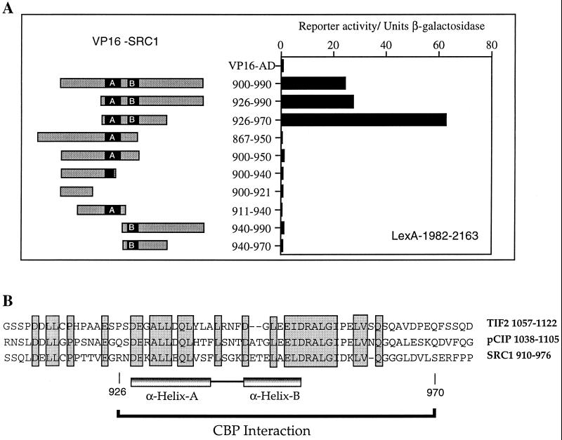
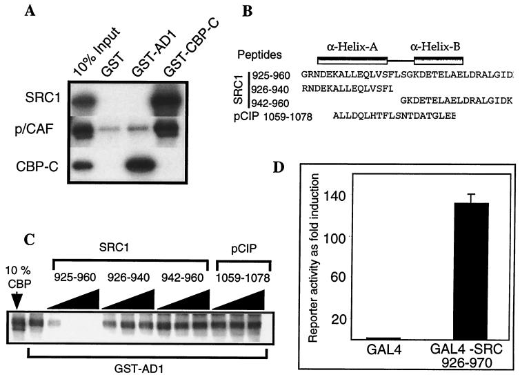
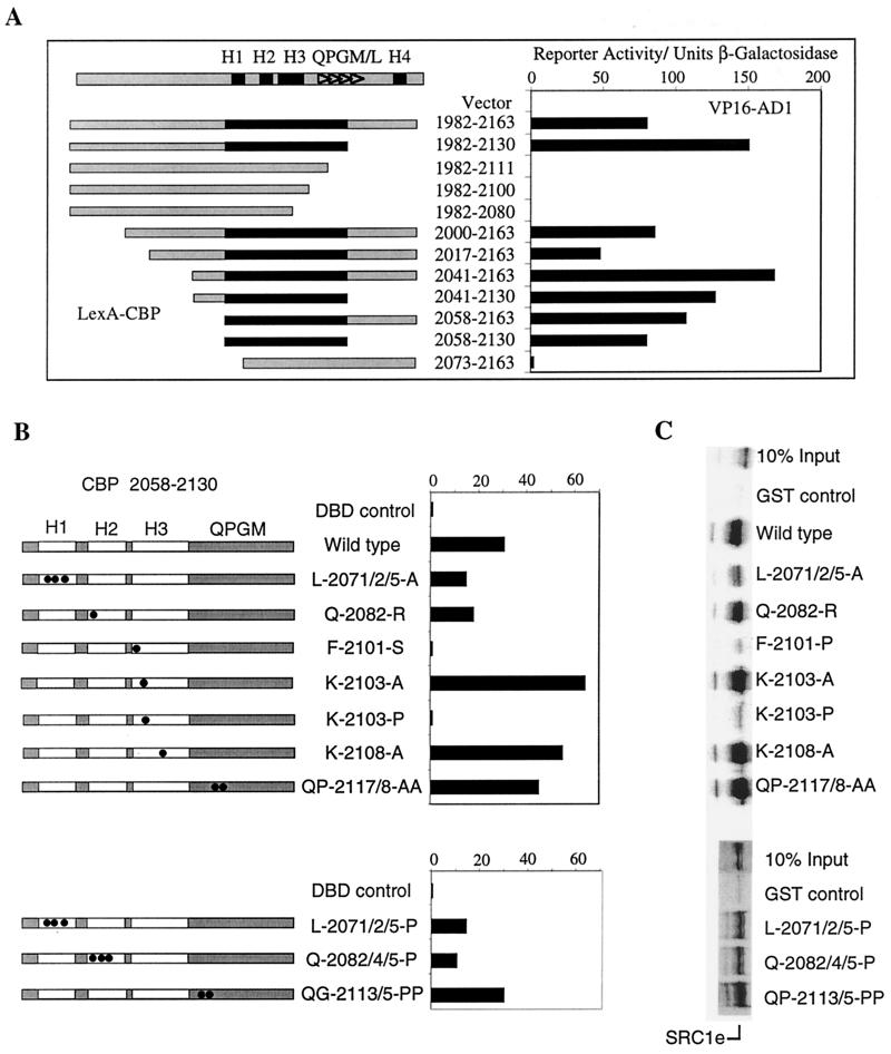
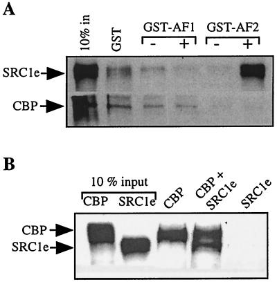
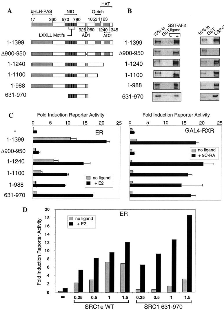
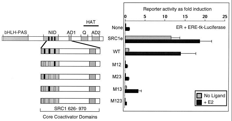
The transcriptional activity of nuclear receptors is mediated by coactivator proteins, including steroid receptor coactivator 1 (SRC1) and its homologues and the general coactivators CREB binding protein (CBP) and p300. SRC1 contains an activation domain (AD1) which functions via recruitment of CBP and and p300. In this study, we have used yeast two-hybrid and in vitro interaction-peptide inhibition experiments to map the AD1 domain of SRC1 to a 35-residue sequence potentially containing two alpha-helices. We also define a 72-amino-acid sequence in CBP necessary for SRC1 binding, designated the SRC1 interaction domain (SID). We show that in contrast to SRC1, direct binding of CBP to the estrogen receptor is weak, suggesting that SRC1 functions primarily as an adaptor to recruit CBP and p300. In support of this, we show that the ability of SRC1 to enhance ligand-dependent nuclear receptor activity in transiently transfected cells is dependent upon the integrity of the AD1 region. In contrast, the putative histone acetyltransferase domain, the Per-Arnt-Sim basic helix-loop-helix domain, the glutamine-rich domain, and AD2 can each be removed without loss of ligand-induced activity. Remarkably, a construct corresponding to residues 631 to 970, which contains only the LXXLL motifs and the AD1 region of SRC1, retained strong coactivator activity in our assays.


Figures

FIG. 1
FIG. 1
The CBP interaction domain in SRC1 maps to amino acids 926 to 970. (A) The interaction between SRC1 and a C-terminal region of CBP was examined in a yeast two-hybrid system. S. cerevisiae L40 was cotransformed with LexA-CBP 1982-2163 and the plasmid pASV3 expressing the acidic activation domain (411 to 490) of VP16 (VP16 AD), or a series of SRC1 constructs were fused in frame with VP16 AD. The SRC1 sequences are represented schematically by grey rectangles, and the putative helices A and B are depicted by black boxes. Reporter activity in cell extracts is expressed in terms of units of β-galactosidase activity. The results from a representative experiment are shown, and similar results were obtained in triplicate experiments. Western blots using anti-LexA and anti-VP16 AD antibodies (Santa Cruz) confirmed that the levels of the bait protein did not vary significantly between different clones and that the levels of VP16 AD1 were similar in all transformants. (B) Sequence alignment of the CBP interaction domain in SRC1 with the corresponding regions in TIF2 and pCIP. The positions of the predicted α-helices and the CBP interaction domain as identified in panel A are indicated. Identical residues present in all three proteins are boxed.
FIG. 2
FIG. 2
Two predicted α-helices in the CBP interaction domain of SRC1 are required in order to maintain interaction with CBP. (A) Glutathione-Sepharose-bound GST, GST-AD1 (781 to 988 of SRC1), and GST–CBP-C (1891 to 2165) were incubated with 35S-labeled full-length SRC1e, pCAF, or CBP-C. Bound proteins were eluted and analyzed by SDS-PAGE and autoradiography. One-tenth of the total labeled protein used in each binding reaction is shown for comparative purposes (10% input). (B) Sequences of synthetic peptides used in competition experiments are shown, with the positions of the predicted helices indicated. (C) Competition experiment showing the effect of increasing concentrations of the competitor peptides on the interaction of GST-AD1 with in vitro-translated 35S-labeled full-length SRC1. (D) Cos-1 cells were transiently transfected as described in Materials and Methods with a GAL4 reporter construct (pGAL4-E16ΔLUC) and 1 μg of vector expressing either GAL4 or the fusion protein GAL4-SRC 926-970. Luciferase activity was measured 48 h later, and the data were normalized to β-galactosidase activity. The activity of GAL4 alone was set at 1, and GAL4-SRC 926-970 activity is expressed relative to it. The values shown represent the average of triplicate samples, and the error bars indicate standard deviation.
FIG. 3
FIG. 3
The SID in CBP maps to amino acids 2058 to 2130. (A) Yeast two-hybrid interactions between VP16 AD1 (SRC1 900-970) and a series of LexA-CBP fusion proteins were assayed as described in the legend to Fig. 1. A schematic representation of the CBP sequence 1982 to 2163 is shown at the top, with the relative positions of four putative α-helices (H1 to H4 [black boxes]) and the QPGM/L repeat sequences (triangles) indicated. CBP sequences are represented schematically below by grey rectangles, whereas the black rectangle denotes the minimal SRC1 interaction domain mapped in these experiments. Reporter activity is expressed in terms of units of β-galactosidase activity, and the results of a representative experiment are shown. Similar results were obtained in triplicate experiments. Western blots confirmed that all LexA constructs were expressed at comparable levels and that VP16 AD1 levels were similar in all cell extracts. (B) Effect of mutations in the SID sequence on its interaction with SRC1 AD1. Yeast two-hybrid interactions between LexA-CBP 2058-2130 mutants and VP16 AD1 were assayed. The LexA-CBP constructs are shown schematically, and the boxed regions represent the relative positions of H1 to H3 and the QPGM/L region. Construct L-2071/2/5-A indicates alteration of leucines at positions 2071, 2072, and 2075 to alanine; similar nomenclature is used for the other constructs. The relative position of each mutation in relation to the predicted α-helices or the QPGM/L motifs is indicated with black circles. Western blots confirmed similar expression of LexA constructs. (C) GST pull-down experiments showing the effects of mutations in the SID on binding of SRC1e. The CBP 2058-2130 fragments identical to those shown in panel B were expressed as GST fusion proteins, and their abilities to bind in vitro-translated 35S-labeled full-length SRC1e were assayed as for Fig. 2A.
FIG. 4
FIG. 4
Direct binding of CBP to the ER is weak. (A) GST pull-down assays were performed as described in the legend to Fig. 2A. Glutathione-Sepharose-bound GST, GST-AF1 (containing the ligand-independent activation function of ER), or GST-AF2 (containing the ligand-dependent activation function of ER) was incubated with 35S-labeled full-length SRC1e or CBP in the presence of 10−6 M E2 or vehicle as indicated. Bound proteins were eluted and analyzed by SDS-PAGE and autoradiography. Ten percent of the total labeled protein used in each binding reaction is shown for comparative purposes (10% in). (B) Immunoprecipitation assays were performed by incubating anti-HA tag antibody and protein A-protein G agarose beads with 35S-labeled full-length CBP, which contains an N-terminal HA tag, and/or SRC1e as indicated. Bound proteins were eluted and analyzed by SDS-PAGE and autoradiography. Ten percent of the total labeled protein used in each reaction is shown for comparative purposes (10% input).
FIG. 5
FIG. 5
AD1 is necessary and, when fused to the NID, sufficient to maintain the coactivator potential of SRC1 in transient-transfection assays. (A) Schematic diagram of the functional domains (indicated by grey boxes) identified in SRC1 and the deletion constructs used in subsequent experiments: bHLH-PAS, sequence similarity with basic helix-loop-helix and Per-Arnt-Sim motifs; NID, with LXXLL motifs indicated by black bars; AD1 and AD2, two autonomous activation domains; Q-rich, a glutamine-rich sequence; and a putative HAT domain. (B) GST pull-down assays performed as described in the legend to Fig. 2A. Glutathione-Sepharose-bound GST, GST-AF2 (containing the ligand-dependent activation function of ER), and GST–CBP-C (1891 to 2165, containing the SID) were incubated with 35S-labeled SRC1, full length or with deletions as indicated. Ligand (10−6 M; E2) or vehicle was added to the binding reaction mixture as appropriate. (C) Cos-1 cells were transiently transfected as described in Materials and Methods with appropriate reporter constructs, a vector expressing either ER or GAL4-RXR, and SRC1e, full-length (1 to 1399) or with deletion mutants, as indicated. Luciferase activity was measured 24 h after the addition of appropriate ligand, and the data were normalized to β-galactosidase activity. The activity of each NR in the presence of ligand was set at 1 for each experiment, and other values are expressed relative to it. The values shown represent the average of triplicate samples, and the error bars indicate standard deviation. These results are representative of experiments performed at least three times. (D) Titration experiment to determine whether luciferase reporter activities induced by SRC1e wild type (WT) or SRC1 631-970 as shown in panel C are at saturating levels. Cos-1 cells were transiently transfected as in panel C using the estrogen-responsive ERE reporter, internal control reporter, ER expression vector, and increasing amounts (expressed in micrograms) of SRC1e wild type or SRC1 631-970 expression vector as indicated. A representative experiment is shown, and similar results were obtained in three replications.
FIG. 6
FIG. 6
Mutation of LXXLL motifs in the context of the SRC1 minimal coactivator abrogates activity. A schematic diagram is shown representing full-length SRC1 (see the legend to Fig. 5A for details of the functional domains indicated by grey boxes) and the deletion constructs spanning the core coactivator domains and containing mutations in which the leucine doublet in two or more LXXLL motifs was mutated to alanines. Cos-1 cells were transfected as described in Materials and Methods with appropriate reporter constructs, vector expressing ER, and the SRC1 constructs as indicated. Luciferase activity was measured 24 h after the addition of 10−8 M E2, and the data were normalized to β-galactosidase activity. The activity of ER in the presence of ligand was set at 1, and the other values are expressed relative to it. The values shown represent the average of triplicate samples, and the error bars indicate standard deviation. The results shown are representative of experiments performed at least three times.

Similar articles

Cited by

References

    1. Anzick S L, Kononen J, Walker R L, Azorsa D O, Tanner M M, Guan X-Y, Sauter G, Kallioniemi O-P, Trent J M, Meltzer P S. AIB1, a steroid receptor coactivator amplified in breast and ovarian cancer. Science. 1997;277:965–968. - PubMed
    1. Bannister A J, Kouzarides T. The CBP co-activator is a histone acetyltransferase. Nature. 1996;384:641–643. - PubMed
    1. Bevan C L, Hoare S, Classens F, Heery D M, Parker M G. The AF1 and AF2 domains of the androgen receptor interact with distinct regions of SRC1. Mol Cell Biol. 1999;19:8383–8392. - PMC - PubMed
    1. Blanco J C, Minucci S, Lu J, Yang X J, Walker K K, Chen H, Evans R M, Nakatani Y, Ozato K. The histone acetylase PCAF is a nuclear receptor coactivator. Genes Dev. 1998;12:1638–1651. - PMC - PubMed
    1. Cavailles V, Dauvois S, Danielian P S, Parker M G. Interaction of proteins with transcriptionally active estrogen-receptors. Proc Natl Acad Sci USA. 1994;91:10009–10013. - PMC - PubMed

Publication types

MeSH terms

LinkOut - more resources