. 2000 Dec 1;19(23):6622-33.
doi: 10.1093/emboj/19.23.6622.

Separate domains in GCN1 for binding protein kinase GCN2 and ribosomes are required for GCN2 activation in amino acid-starved cells

Affiliations

Separate domains in GCN1 for binding protein kinase GCN2 and ribosomes are required for GCN2 activation in amino acid-starved cells

E Sattlegger et al. EMBO J. .

Abstract

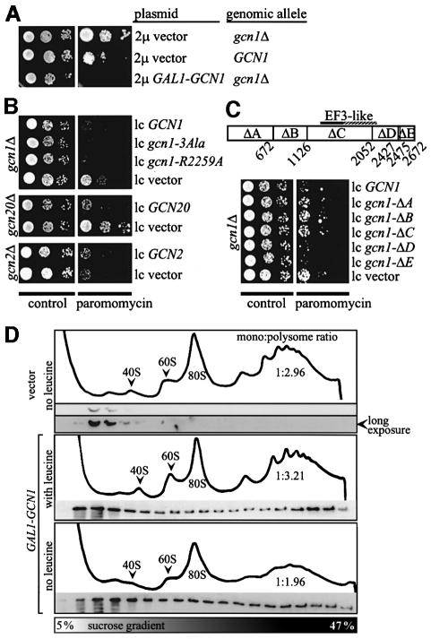
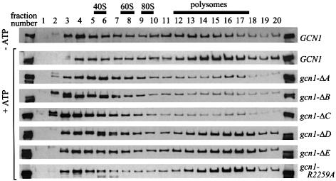
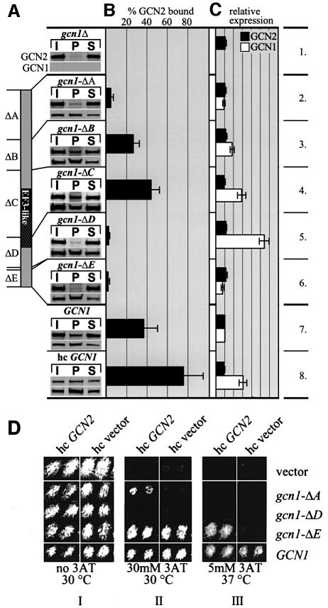
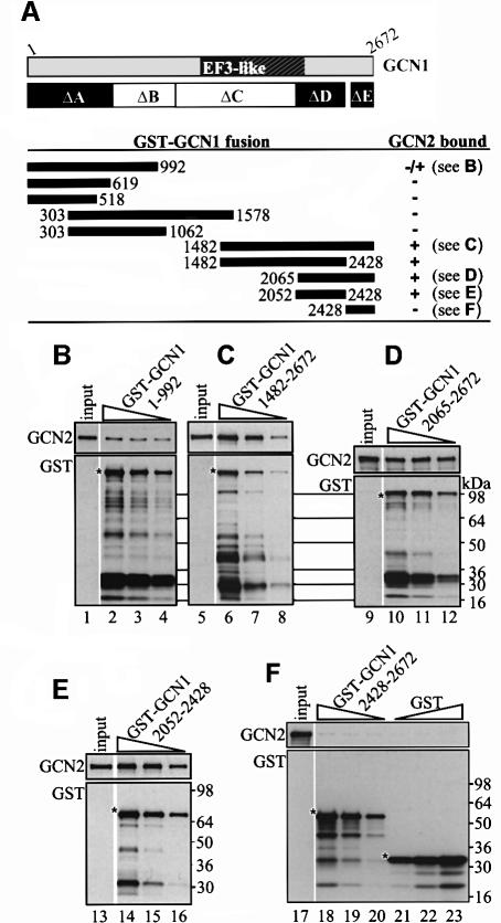
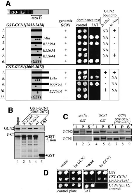
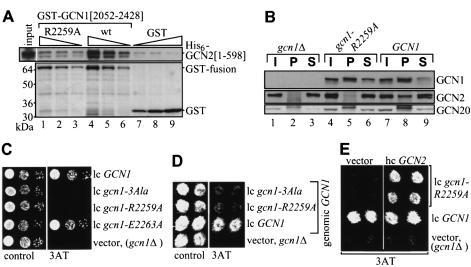
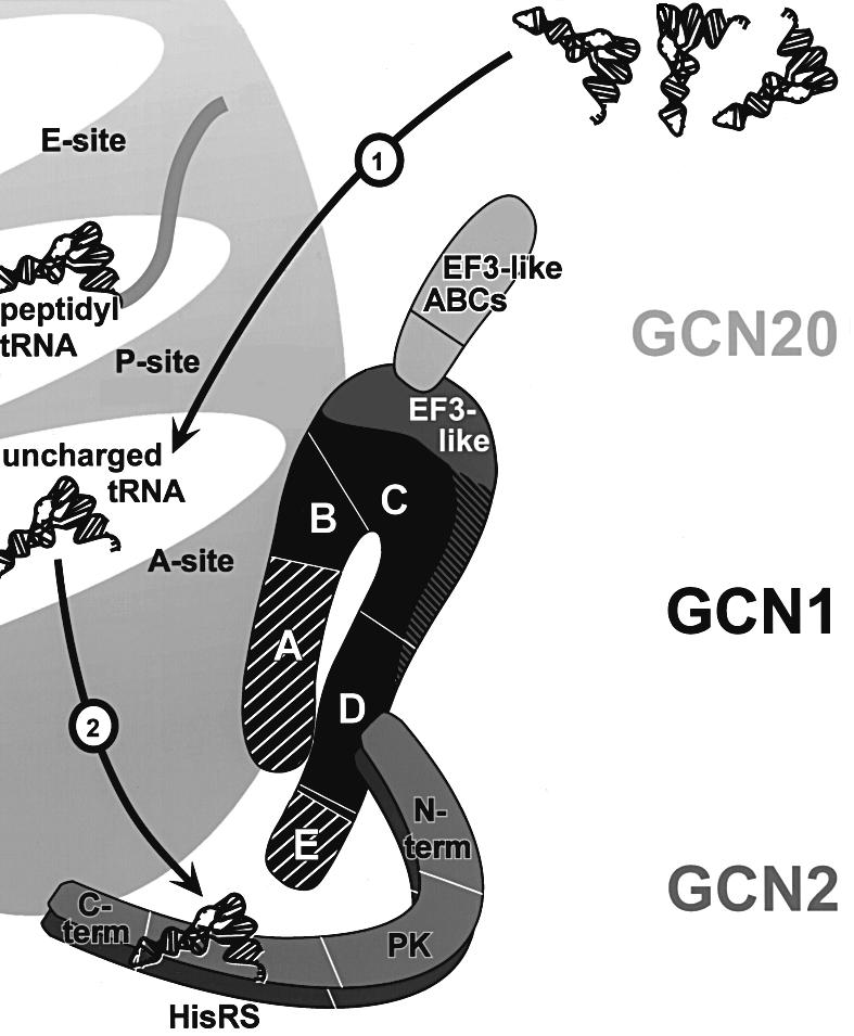
GCN2 stimulates GCN4 translation in amino acid-starved cells by phosphorylating the alpha-subunit of translation initiation factor 2. GCN2 function in vivo requires the GCN1/GCN20 complex, which binds to the N-terminal domain of GCN2. A C-terminal segment of GCN1 (residues 2052-2428) was found to be necessary and sufficient for binding GCN2 in vivo and in vitro. Overexpression of this fragment in wild-type cells impaired association of GCN2 with native GCN1 and had a dominant Gcn(-) phenotype, dependent on Arg2259 in the GCN1 fragment. Substitution of Arg2259 with Ala in full-length GCN1 abolished complex formation with native GCN2 and destroyed GCN1 regulatory function. Consistently, the Gcn(-) phenotype of gcn1-R2259A, but not that of gcn1Delta, was suppressed by overexpressing GCN2. These findings prove that GCN2 binding to the C-terminal domain of GCN1, dependent on Arg2259, is required for high level GCN2 function in vivo. GCN1 expression conferred sensitivity to paromomycin in a manner dependent on its ribosome binding domain, supporting the idea that GCN1 binds near the ribosomal acceptor site to promote GCN2 activation by uncharged tRNA.


Figures

None
Fig. 1. GCN1 expression confers paromomycin sensitivity dependent on the N-terminal three-quarters of the protein. (A) Transformants of gcn1Δ strain H2556 and GCN1 strain H1511 harboring the empty 2 μ vector pEMBLyex4 or the 2µ GAL1-GCN1 plasmid p1827, respectively, were grown to saturation and 5 µl of serial dilutions (of OD600 = 0.15, 0.015 and 0.0015) were spotted on plates containing galactose as carbon source in the presence or absence of 1 mg/ml paromomycin, as indicated, and incubated at 30°C for 3 days. (B) Except for the use of glucose as carbon source, the same analysis as described in (A) was carried out for (top) transformants of gcn1Δ strain H2556 bearing the GCN1 alleles indicated on the right on low copy plasmids (from top to bottom, p2367, pES161-1-2, pES174-3-2 and empty vector pRS316), (middle) transformants of gcn20Δ strain H2558 bearing low copy GCN20 plasmid p1867 or vector pRS316 or (bottom) transformants of gcn2Δ strain H2557 bearing low copy GCN2 plasmid p722 or vector pRS316. (C) (Top) A map of GCN1 showing the amino acid locations of the end-points of the indicated internal deletions, ΔA–ΔE. The EF3-like region in GCN1 is indicated. (Bottom) The same analysis as described in (B) was carried out for transformants of gcn1Δ strain H2556 bearing the indicated GCN1 alleles on low copy plasmids (from top to bottom, p2362, p2354, p2363, p2358 and pES32-1) or the empty vector pRS316. (D) Overexpressed GCN1 co-sediments with polysomes. Transformants of GCN1 (H1511) strains harboring empty vector pEMBLyex4 or the 2µ GAL1-GCN1 plasmid p1827, respectively, were grown on galactose-containing medium and 15 A260 units of each whole-cell extract were resolved by velocity sedimentation in sucrose density gradients in the absence of ATP, as described previously (Marton et al., 1997). Fractions were collected while measuring A254 to identify the positions of polysomes, 80S ribosomes and 40S and 60S subunits. Equivalent proportions of the fractions were subjected to immunoblot analysis using antibodies against GCN1. Based on the A254 tracings, we calculated that the extracts from the transformants overexpressing GCN1 contained 91% (middle) or 51% (lower) of the polysomes present in the transformant expressing GCN1 at wild-type levels (upper). Hence, the amounts of GCN1 bound per ribosome in the polysome fractions of the transformants overexpressing GCN1, compared with the vector transformant, are somewhat greater than they appear in the figure.
None
Fig. 2. The N-terminal three-quarters of GCN1 is required for polysome binding in cell extracts. Whole-cell extracts prepared from the strains described in Figure 1C and from gcn1Δ strain H2556 containing gcn1-R2259A (pES174-3-2) were resolved by velocity sedimentation in sucrose density gradients in the presence or absence of ATP, as described previously (Marton et al., 1997). Fractions (numbered 1–20) were collected while measuring A254 in the gradient lacking ATP. All fractions were subjected to immunoblot analysis using antibodies against the c-myc epitope present at the C-terminus of wild-type and mutant GCN1 proteins. The first and last lanes contain 1% of the extract loaded on the gradient.
None
Fig. 3. Mapping regions in GCN1 necessary for GCN2 binding. (A) Whole-cell extracts prepared from the strains described in Figure 1C and from gcn1Δ strain H2556 containing high copy GCN1 (p1834) were immunoprecipitated with GCN1 antibodies as described previously (Garcia-Barrio et al., 2000) and the immune complexes subjected to immunoblot analysis using GCN2 antibodies or antibodies against the c-myc epitope present at the C-terminus of the GCN1 proteins. P, pellet; I, 10% input; S, 10% supernatant. Data from at least five experiments were averaged to determine (B) the percentage of total GCN2 bound to GCN1 and (C) the amounts of GCN1 and GCN2 in the extracts relative to wild-type GCN1 extract. Standard deviations are indicated by error bars. (D) GCN2 overexpression suppresses the 3ATS phenotype associated with gcn1-ΔE and partially that of gcn1-ΔA. Transformants of the gcn1Δ strain harboring the plasmid-borne gcn1 alleles listed on the right or empty vector, and also bearing high copy (hc) GCN2 plasmid pAH15 or the corresponding empty vector YEp13 were replica printed on medium containing 3AT (Hinnebusch and Fink, 1983) at the indicated concentrations and incubated at the temperature shown. Two independent transformants of each strain were tested side by side.
None
Fig. 4. A C-terminal segment of GCN1 (region D) is sufficient for GCN2 binding in vitro. GCN1 fragments fused to GST were expressed in E.coli, immobilized on glutathione–Sepharose beads and incubated with 1 mg whole-cell extract from yeast gcn1Δ strain H2556 containing the high copy GCN2 plasmid pAH15. After washing, the beads were subjected to immunoblot analysis using GCN2 and GST antibodies to measure the amounts of bound proteins. (A) A schematic of GCN1 is shown, labeled as in Figure 1C, and the regions highlighted in black are necessary for GCN2 binding in vivo (see Figure 3). Below are shown schematically the GCN1 fragments contained in the GST–GCN1 fusions (solid rectangles with numbers indicating amino acid positions) and a qualitative summary of their GCN2 binding activities based on the results in (B–F) and data not shown. The plasmids encoding the GST–GCN1 fusions were (from top to bottom): pES131-2, pES145-1, pES141-2, pES99-3, pES84-2, pES79-1, pES86-1, pES123-B1 and pES136-1. (BF) Results for each of the indicated GST–GCN1 fusions of three binding assays with different concentrations of fusion protein (each one differing by a factor of 2 from the other) and a sample containing 10% of the yeast extract (input) are shown. The upper and lower panels show the results of immunoblot analysis obtained with GCN2 and GST antibodies, respectively. An asterisk indicates the predicted full-length GST fusion proteins.
None
Fig. 5. Overexpression of the C-terminal GCN2-binding domain of GCN1 has a dominant Gcn phenotype that can be suppressed by overexpressing GCN2. (A) Overexpression of GST–GCN1[2052–2428] or GST–GCN1[2065–2672] has a dominant Gcn phenotype, dependent on Arg2259. The indicated GST–GCN1 fusions bearing wild-type or mutant GCN1 segments were expressed in GCN1 strain H1511 from a galactose-inducible promoter from plasmids (listed top to bottom) pES124-B2, pES153-7-2, pES167-2E, pES168-4F, pES169-1g, pES110-32, pES163-6K, pES175-A1, pES180-1-1 and pEG(KT) encoding GST alone, and pEG(KT) expressed in gcn1Δ strain H2556. These strains were tested for growth on 3AT medium containing galactose as carbon source (columns headed dominance test). Two independent transformants of each strain were tested side by side. The next to last column summarizes the binding to endogenous GCN2 of selected fusions expressed in gcn1Δ yeast strain H2256 in assays of the type described below in (B). The last column summarizes the binding of selected fusions expressed in bacteria with the GCN2 present in yeast cell extracts, in assays of the type described in Figure 4. We verified by immunoblot analysis that the gcn1 point mutations introduced into these constructs did not affect the protein levels. NA, not analyzed; ND, not detectable. (B) Binding of GST–GCN1[2065– 2672] to GCN2 in vivo is dependent on Arg2259. Yeast whole-cell extracts prepared from transformants of gcn1Δ strain H2556 expressing the indicated GST fusions from a galactose-inducible promoter were incubated with glutathione–Sepharose beads and proteins bound to the beads were detected by immunoblot analysis. As all GST fusions were expressed at similar levels, only the input amount for the wild-type (wt) extract was analyzed in lane 1. (C) Overexpression of GST–GCN1[2065–2672] titrates GCN2 from endogenous GCN1. Co-immunoprecipitation assays were performed as described in Figure 3A using extracts from gcn1Δ or GCN1 strains expressing GST–GCN1 [2065–2672] or GST alone from a galactose-inducible promoter. (D) The dominant Gcn phenotype of GST–GCN1[2065–2672] can be partially suppressed by GCN2 overexpression. Growth tests were conducted as in (A), but with strains containing high copy (hc) plasmid pAH15 bearing GCN2 or vector YEp13 alone, in addition to the plasmids encoding GST–GCN1[2065–2672] or GST alone (pES125-B2-1 or pES128-9-1). The latter plasmids lacked the leu2-d gene in order to permit selection for the LEU2 marker on pAH15 or YEp13. Two transformants of the GCN1 strain bearing YEp13 or the gcn1Δ strain bearing pAH15 were analyzed as controls.
None
Fig. 6. Arg2259 in GCN1 is essential for GCN2 binding and GCN1 regulatory function. (A) GST–GCN1[2052–2428] directly interacts with His6–GCN2[1–598] in vitro, dependent on Arg2259. The indicated GST–GCN1 fusions or GST alone encoded by plasmids (from left to right) pES164-2A, ES123-B1 or pGEX-6p-1 were expressed in E.coli and immobilized on glutathione–Sepharose beads, then incubated with E.coli extract containing His6–GCN2[1–598] encoded by plasmid pES171-III-1. Proteins bound to the beads were identified by immunoblot analysis, as described in Figure 4 using anti-His6 antibodies. GST proteins were visualized by Coomassie staining. (B) Arg2259 in GCN1 is essential for GCN1–GCN2 interaction in vivo. Co-immunoprecipitation assays were performed as described in Figure 3A using transformants of gcn1Δ strain H2256 harboring plasmid-borne gcn1-R2259A (pES174-3-2), GCN1 (p2367) or vector alone (gcn1Δ,pRS316), as indicated at the top of the panel. Immunoblots were probed for GCN1, GCN2 and GCN20. (C) Arg2259 is essential for GCN1 function. Serial dilutions of gcn1Δ strain H2556 harboring the indicated gcn1 alleles on plasmid (from top to bottom) p2367, pES161-1-2, pES174-3-2, pES179-1-2 or vector pRS316 alone (gcn1Δ) were spotted on plates containing 3AT as indicated and incubated at 30°C. (Dgcn1-R2259A confers a dominant Gcn phenotype. GCN1 strain H1511 containing plasmid-borne gcn1 alleles as shown (plasmids as in C), or gcn1Δ strain H2556 harboring vector alone, were subjected to a dominance test as described in Figure 5A. (E) GCN2 overexpression suppresses the Gcn phenotype of gcn1-R2259A. gcn1Δ strain H2556 harboring gcn1 alleles as indicated (plasmids as in C) and high copy GCN2 plasmid pAH15 or vector YEp13 alone were studied for growth on medium containing 3AT, as in (D).
None
Fig. 7. Model for GCN1 function in activation of GCN2 by uncharged tRNA. GCN1 is shown in black and the positions of regions A–E are indicated, as well as the EF3-like domain located predominantly in region C. Regions A and E (hatched) are largely dispensable for GCN1 function in cells overexpressing GCN2, suggesting that the most crucial domains in GCN1 reside within the core segment comprised of regions B–D. The EF3-like domain of GCN1 interacts with the N-terminus of GCN20. GCN1 has homology to the N-terminus of EF3, whereas the GCN20 C-terminus shows similarity to the C-terminal part of EF3, including the ATP-binding cassettes (ABCs). GCN2 is shown as a dimer. Its subdomains are indicated as a region encompassing a highly conserved N-terminus, a charged region and a degenerate kinase domain (N-term), a protein kinase domain (PK), a histidyl-tRNA synthetase-like domain (HisRS) and a C-terminus with dimerization and ribosome-binding activity (C-term). GCN1 region D directly interacts with the N-terminus of GCN2. In addition, core regions B and C of GCN1 mediate ribosome binding in proximity to the ribosomal A-site. GCN1 binding near the A-site may allow it to stimulate codon-dependent binding of uncharged tRNA to the A-site (1). Alternatively, physical contact between GCN1 and GCN2 may position GCN2 on the ribosome in a way that facilitates its interaction with uncharged tRNAs in the A-site. GCN1 could also have a role in ejecting uncharged tRNAs from the A-site and transferring them to GCN2 (2). For more detail see text.

Similar articles

Cited by

References

    1. Berlanga J.J., Santoyo,J. and De Haro,C. (1999) Characterization of a mammalian homolog of the GCN2 eukaryotic initiation factor 2α kinase. Eur. J. Biochem., 265, 754–762. - PubMed
    1. Botstein D., Falco,S.C., Stewart,S.E., Brennan,M., Scherer,S., Stinchcomb,D.T., Struhl,K. and Davis,R.W. (1979) Sterile host yeasts (SHY): a eukaryotic system of biological containment for recombinant DNA experiments. Gene, 8, 17–24. - PubMed
    1. Broach J.R., Strathern,J.N. and Hicks,J.B. (1979) Transformation in yeast: development of a hybrid cloning vector and isolation of the CAN1 gene. Gene, 8, 121–133. - PubMed
    1. Cashel M. and Rudd,K.E. (1987) The stringent response. In Neidhardt,F.C., Ingraham,J.L., Magasanik,B., Low,K.B., Schaechter,M. and Umbarger,H.E. (eds), Escherichia coli and Salmonella typhimurium: Cellular and Molecular Biology. American Society for Microbiology, Washington, DC, pp. 1410–1438.
    1. Cesareni G. and Murray,J.A.H. (1987) Plasmid vectors carrying the replication origin of filamentous single-stranded phages. In Setlow,J.K. and Hollaender,A. (eds), Genetic Engineering: Principles and Methods. Plenum Press, New York, NY, Vol. 9, pp. 135–154.

Publication types

MeSH terms