. 2000 Dec 1;19(23):6427-39.
doi: 10.1093/emboj/19.23.6427.

Transmembrane transforming growth factor-alpha tethers to the PDZ domain-containing, Golgi membrane-associated protein p59/GRASP55

Affiliations

Transmembrane transforming growth factor-alpha tethers to the PDZ domain-containing, Golgi membrane-associated protein p59/GRASP55

A Kuo et al. EMBO J. .

Abstract

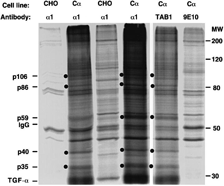
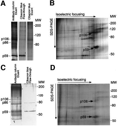
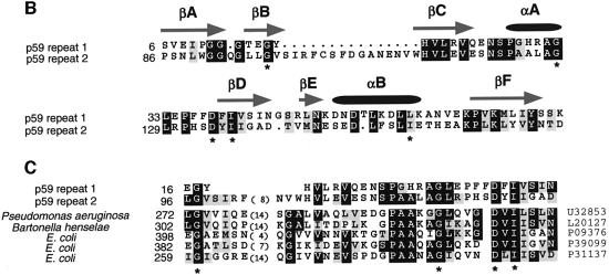
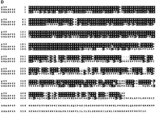
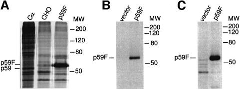
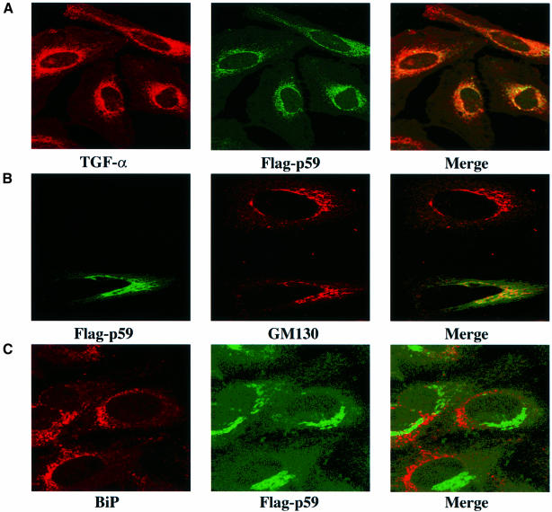
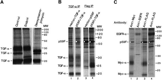
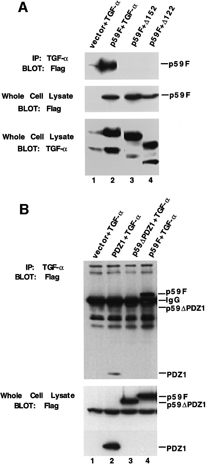
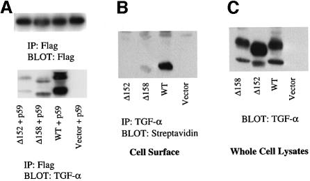
Transforming growth factor-alpha (TGF-alpha) and related proteins represent a family of transmembrane growth factors with representatives in flies and worms. Little is known about the transport of TGF-alpha and other transmembrane growth factors to the cell surface and its regulation. p59 was purified as a cytoplasmic protein, which at endogenous levels associates with transmembrane TGF-alpha. cDNA cloning of p59 revealed a 452 amino acid sequence with two PDZ domains. p59 is myristoylated and palmitoylated, and associates with the Golgi system, where it co-localizes with TGF-alpha. Its first PDZ domain interacts with the C-terminus of transmembrane TGF-alpha and select transmembrane proteins. p59 is the human homolog of GRASP55, which is structurally related to GRASP65. GRASP55 and GRASP65 have been shown to play a role in stacking of the Golgi cisternae in vitro. C-terminal mutations of transmembrane TGF-alpha, which decrease or abolish the interaction with p59, also strongly impair cell surface expression of TGF-alpha. Our observations suggest a role for membrane tethering of p59/GRASP55 to select transmembrane proteins, including TGF-alpha, in maturation and transport to the cell surface.


Figures

None
Fig. 1. Co-immunoprecipitation of associated proteins with transmembrane TGF-α. TGF-α-expressing CHO cells (Cα) or control CHO cells were 35S-labeled, treated with the chemical cross-linker DSP and lysed. Samples were then immunoprecipitated (lanes 1 and 2) or immunoaffinity purified (lanes 3–6) using the monoclonal antibodies α1 or TAB1 against TGF-α, or 9E10 against a myc epitope as control, as indicated. Proteins were then resolved on SDS–PAGE. The positions of the TGF-α-associated proteins are indicated to the left of the panel and with bullets to the left of individual lanes. Note the detection of p59 in lanes 4 and 5, which relied on immunoaffinity purification, and is much less visible in the immunoprecipitation in lane 2. Only the largest, fully glycosylated TGF-α band is apparent on the autoradiogram.
None
Fig. 2. Purification of p59. (A) Analytical Cibacron blue purification of p59. The left lane shows a 35S-labeled Cα cell lysate sample following anti-TGF-α immunoaffinity purification (TGF-α ran off the gel) and prior to Cibacron blue–Sepharose. The next two lanes show p59 in the flow-through fraction but not in the eluant. (B) Analytical two-dimensional gel electrophoresis of p59. A similar immunoaffinity-purified sample to that in (A) was subjected to two-dimensional SDS–PAGE. As in (A), proteins are visualized by autoradiography. (C) Preparative Cibacron blue purification of p59. Unlabeled cell lysate starting from 18 m2 of confluent Cα cells was purified by anti-TGF-α immunoaffinity chromatography and subjected to Cibacron blue– Sepharose. One percent samples of the eluant and flow-through fractions were analyzed by SDS–PAGE, and proteins were visualized by silver staining. (D) Preparative two-dimensional gel electrophoresis of p59. Cell lysate starting from 25 m2 of confluent Cα cells was purified by anti-TGF-α immunoaffinity chromatography and then subjected to preparative two-dimensional gel electrophoresis. A 1% sample was analyzed and proteins are visualized by silver staining. The positions of p59, p86 and p106 are marked.
None
Fig. 3. Polypeptide sequence of human p59. (A) cDNA sequence and corresponding amino acid sequence of human p59. The two PDZ domains are underlined. Peptide sequences obtained from microsequencing are overlined, with asterisks above ambiguous residues. (B) Sequence alignment of the two PDZ domains. Identical (black boxes) and similar (shaded boxes) residues are marked. The asterisks mark highly conserved residues among PDZ domains. The predicted α-helices and β-sheets, characteristic for PDZ domains, are shown by overlining the corresponding amino acids. (C) Alignment of the N-terminal two-thirds of the two PDZ sequences of p59 with the corresponding sequences in other htrA-like PDZ domains. The DDBJ/EMBL/GenBank accession numbers of these sequences are shown to the right. (D) Comparison of the human p59 amino acid sequence with the mouse GRASP55 and GRASP65 sequences. Black boxes show identical residues, while similar residues are shaded.
None
Fig. 4. p59 is myristoylated and palmitoylated. (A) Immunoprecipitation of Flag-tagged p59 (p59F) from transfected cells in comparison with control transfected cells and TGF-α-expressing CHO cells (Cα) in which endogenous p59 was co-immunoprecipitated with TGF-α using an anti-TGF-α antibody (lane 1; as in Figures 1 and 2A). (BIn vitro translation of p59 in the presence of [3H]myristic acid and immunoprecipitation shows that p59 is 3H-myristoylated. (CIn vivo labeling of transfected cells using [3H]palmitic acid, followed by immunoprecipitation, shows that p59 is 3H-palmitoylated.
None
Fig. 5. Subcellular localization of p59, as assessed by immunofluorescence, in transfected HeLa cells. (A) Immunofluorescent localization of TGF-α or Flag-tagged p59 in permeabilized cells, showing that p59 and TGF-α co-localize. (B) Immunofluorescent detection of Flag-tagged p59 or the endogenous Golgi marker GM130. Note that only one of the two cells shown was transfected to express Flag-p59. These results strongly suggest that p59 and GM130 co-localize in the Golgi. (C) Immunofluorescent detection of Flag-tagged p59 or endogenous BiP/GRP78, a resident protein of the endoplasmic reticulum. The data show that p59 does not localize in the endoplasmic reticulum.
None
Fig. 6. p59 associates preferentially with an immaturely glycosylated form of transmembrane TGF-α. (A) TGF-α-expressing CHO cells (Cα) were 35S-labeled and transmembrane TGF-α was immunoprecipitated. The sample in the second lane was treated with endo H after immunoprecipitation, while the sample in the third lane was obtained from cells grown in the presence of deoxymannojirimycin. (B) Co-immunoprecipitation of p59 and transmembrane TGF-α from transfected, 35S-labeled 293 cells. Lanes 1 and 2: anti-TGF-α co-precipitates p59 (lane 2), but p59 is not detected, when cells are not transfected for Flag-tagged p59 expression (lane 1). Note the ratios of the three TGF-α forms. Lanes 3 and 4: immunoprecipitation of Flag-tagged p59 co-precipitates TGF-α. In comparison with the anti-TGF-α immunoprecipitations in lanes 1 and 2, the second TGF-α band, which is a processing intermediate, is highly enriched. No TGF-α was detected in lane 3, in which the cells were not transfected with a TGF-α expression plasmid. (C) Replacement of the ectodomain of transmembrane TGF-α with another ectodomain does not affect p59 association. 293 cells were co-transfected with cDNAs for Flag-tagged p59 and TGF-αΔE, a chimera in which the ectodomain of TGF-α has been replaced by a myc epitope tag (lanes 1–3), or EGFR-α, a chimera in which the ectodomain of TGF-α has been replaced by the ectodomain of EGFR (lane 4). Cells were 35S-labeled, lysed, and samples were immunopurified using the Sepharose-conjugated antibodies, as indicated.
None
Fig. 7. p59 associates through its first PDZ domain with the C-terminal sequence of transmembrane TGF-α. (A) The C-terminal sequence of transmembrane TGF-α interacts with p59. CHO cells were transfected with Flag-tagged p59 (lanes 2–4) and full-size TGF-α (lanes 1 and 2), TGF-αΔ152 (lane 3) or TGF-αΔ122 (lane 4). Immunoprecipitation with anti-TGF-α, followed by anti-p59 western blotting (top panel), showed that only full-size TGF-α, but not TGF-αΔ152 or TGF-αΔ122, associated with p59. The middle and lower panels show expression of Flag-tagged p59 and transmembrane TGF-α, as assessed by western blotting. (B) p59 interacts with transmembrane TGF-α through its first PDZ domain. CHO cells were transfected to express transmembrane TGF-α and PDZ1, a segment of p59 containing the first PDZ domain (lane 2), p59ΔPDZ1, a p59 mutant that lacks the first PDZ domain (lane 3), or full-size p59 (lane 4). Anti-TGF-α immunoprecipitation followed by western blotting for Flag-tagged p59 revealed the association of full-size p59 and PDZ1, but not of p59ΔPDZ1. The lower panel shows the expression levels of p59 and its mutants, as assessed by anti-Flag western blotting.
None
Fig. 8. Specificity of the interaction of p59 with transmembrane TGF-α. (A) Cells were transfected with expression plasmids for TGF-α and HA-tagged EBP50. Immunoprecipitation with anti-TGF-α followed by an anti-HA western blot (top panel) did not reveal TGF-α-associated EBP50, even though EBP50 and TGF-α were expressed, as assessed by western blots (middle and lower panels). (B) p59 interacts with matrix metalloproteinase 1 (MT1-MMP) and kit ligand, but not L-selectin. 293 cells were transfected with the indicated expression plasmids. In the left and middle panels, anti-Flag immunoprecipitation for p59, followed by western blotting for MT1-MMP or kit ligand, respectively, indicated the association of either transmembrane protein with p59. In the right panel, immunoprecipitation for L-selectin, followed by anti-Flag western blotting for p59, did not detect associated p59.
None
Fig. 9. The ability of transmembrane TGF-α mutants to associate with p59 correlates with the levels of transmembrane TGF-α at the cell surface. (A) Wild-type (WT) transmembrane TGF-α, and the TGF-αΔ158 and Δ152 mutants, which lack the C-terminal two and eight amino acids, respectively, were co-expressed with Flag-tagged p59 in CHO cells and their ability to interact was assessed by co-immunoprecipitation analysis. The top panel shows the expression levels of p59 and the lower panel shows the TGF-α that co-precipitates with p59. The control for the total levels of transmembrane TGF-α is essentially as in (C). (B) Cell surface levels of transmembrane TGF-α in transfected CHO cells. Cell surface biotinylation, followed by cell lysis and anti-TGF-α immunoprecipitation, allowed detection of TGF-α at the cell surface, but only of the lower TGF-α band. While wild-type TGF-α is well expressed at the cell surface, the Δ158 mutation resulted in strongly decreased cell surface expression and the Δ152 mutation abolished detectable cell surface expression of TGF-α. (C) Total levels of transmembrane TGF-α in the cell lysates corresponding to (B), as assessed using anti-TGF-α western blotting. This control very closely resembles the total TGF-α levels for (A) (data not shown).

Similar articles

Cited by

References

    1. Alvarez C., Fujita,H., Hubbard,A. and Sztul,E. (1999) ER to Golgi transport: requirement for p115 at a pre-Golgi VTC stage. J. Cell Biol., 147, 1205–1221. - PMC - PubMed
    1. Barr F.A., Puype,M., Vandekerckhove,J. and Warren,G. (1997) GRASP65, a protein involved in the stacking of Golgi cisternae. Cell, 91, 253–262. - PubMed
    1. Barr F.A., Nakamura,N. and Warren,G. (1998) Mapping the interaction between GRASP65 and GM130, components of a protein complex involved in the stacking of Golgi cisternae. EMBO J., 17, 3258–3268. - PMC - PubMed
    1. Baselga J., Mendelsohn,J., Kim,Y.M. and Pandiella,A. (1996) Autocrine regulation of transforming growth factor-α cleavage. J. Biol. Chem., 271, 3279–3284. - PubMed
    1. Bischoff J., Liscum,L. and Kornfeld,R. (1986) The use of deoxy mannojirimycin to evaluate the role of various α-mannosidases in oligosaccharide processing in intact cells. J. Biol. Chem., 261, 4766–4774. - PubMed

Publication types

MeSH terms

LinkOut - more resources