. 2000 Dec 5;97(25):13883-8.
doi: 10.1073/pnas.250471697.

Identification of a multipotent astrocytic stem cell in the immature and adult mouse brain

Affiliations

Identification of a multipotent astrocytic stem cell in the immature and adult mouse brain

E D Laywell et al. Proc Natl Acad Sci U S A. .

Abstract

The mammalian brain contains a population of neural stem cells (NSC) that can both self-renew and generate progeny along the three lineage pathways of the central nervous system (CNS), but the in vivo identification and localization of NSC in the postnatal CNS has proved elusive. Recently, separate studies have implicated ciliated ependymal (CE) cells, and special subependymal zone (SEZ) astrocytes as candidates for NSC in the adult brain. In the present study, we have examined the potential of these two NSC candidates to form multipotent spherical clones-neurospheres-in vitro. We conclude that CE cells are unipotent and give rise only to cells within the glia cell lineage, although they are capable of forming spherical clones when cultured in isolation. In contrast, astrocyte monolayers from the cerebral cortex, cerebellum, spinal cord, and SEZ can form neurospheres that give rise both to neurons and glia. However, the ability to form neurospheres is restricted to astrocyte monolayers derived during the first 2 postnatal wk, except for SEZ astrocytes, which retain this capacity in the mature forebrain. We conclude that environmental factors, simulated by certain in vitro conditions, transiently confer NSC-like attributes on astrocytes during a critical period in CNS development.


Figures
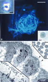
Figure 1
Figure 1
Generation of spherical clones from single, ciliated ependymal cells. (a) GFAP immunofluorescence (blue, AMCA) of a clone (phase microscopy of such a clone is shown in the lower right Inset), derived from a single ciliated ependymal cell (Inset, upper left; notice cilia protruding from the base of the cell). Scale bar in a and lower right Inset = 50 μm; scale bar in upper left Inset = 5 μm. (b) TEM of a clone derived from a single ciliated ependymal cell. Note cilia on the surfaces of cells at the edge of this clone (arrowheads), shown at higher magnification in the lower left Inset. The upper right Inset shows cross-sectioned cilia displaying the characteristic 9 + 2 arrangement of microtubules. Scale bar in b = 5 μm, 0.5 μm in the lower left Inset, and 0.2 μm in the upper right Inset.
Figure 2
Figure 2
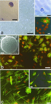
Astrocyte monolayers from P2 mouse cerebral cortex generate spherical clones. (a) Phase micrograph of a confluent layer of astrocytes. Scale bar = 50 μm. Early passage monolayers often contain a small number of microglia, as seen in the lower right Inset using immunofluorescence for Mac-1 (FITC, green), counterstained with propidium iodide (PI, orange). Scale bar in lower right Inset = 10 μm. Astrocyte monolayers from this age also generate spherical clones when grown in suspension culture with EGF and bFGF (upper left Inset, Scale bar = 100 μm; and Inset in b, showing an SEM of such a clone; Scale bar = 50 μm). Similar clones are generated when astrocytes are cultured in isolation (phase micrograph of clone derived from a single astrocyte is shown in the upper right Inset, Scale bar = 30 μm). (b) GFAP immunolabeling (green) and PI counterstaining of most, but not all, astrocytes within the monolayer. Scale bar = 25 μm. (c) Clones are generated from monolayers such as these in which virtually all cells within the monolayer exhibit immunolabeling for the immature astrocyte markers, vimentin (green, PI counterstaining) and S100β (Inset). Scale bar = 25 μm in c and 10 μm in the Inset.
Figure 3
Figure 3
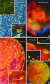
Astrocyte-derived neurospheres are proliferative and express neuronal and glial antigens. (a) BrdUrd (green) and GFAP (blue) immunofluorescence double-labeling of a sphere derived from a P2 cortical astrocyte monolayer, showing the highly proliferative nature of these sphere cells. BrdUrd was present in the culture medium for 24 h before plating. Scale bar = 50 μm. Upper left Inset: Low magnification BrdUrd immunofluorescence labeling of an entire, attached, differentiating neurosphere. Scale bar = 50 μm. Lower right Inset shows double immunolabeling for A2B5 (green) and GFAP (blue), PI counterstained, of differentiating cells from an astrocyte-derived neurosphere. Scale bar = 10 μm. (b) Immunolabeling of a single P7 cerebellar astrocyte-derived neurosphere showing MAP-2 positive cells, indicating the presence of neurons within these clones. The Inset in b shows a higher magnification of a single MAP-2-positive immature neuron within the neurosphere. Scale bars = 30 μm in b, and 10 μm in the Inset. (c) GFAP (blue) and β-III tubulin (green), PI counterstained, double immunolabeling of a neurosphere derived from a P1 cerebral cortex astrocyte monolayer. Note the beta-III tubulin immunopositive neurites arborizing around PI-stained cells, and GFAP astrocytic processes within the lower portion of the neurosphere. Scale bar = 10 μm. (d) β-III tubulin immunolabeling of a neurosphere derived form a P1 spinal cord astrocyte monolayer. Note two β-III tubulin-positive immature neurons at the edge of the neurosphere, compared with apparently more mature immunolabeled neurons (upper right and lower left Insets) that have migrated away from the neurosphere and elaborated long processes. Scale bars = 10 μm in d and both Insets. (e) L1 immunolabeling (green, PI counterstaining) of a neurosphere from a P1 spinal cord astrocyte monolayer, showing pervasive labeling of neurons throughout the neurosphere. Scale bar = 25 μm.
Figure 4
Figure 4
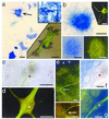
Alkaline phosphatase (AP) enzyme histochemistry and immunocytochemistry of cells and neurospheres derived from Gtv-a transgenic mouse astrocytes infected with RCAS-alkaline phosphatase, showing cells expressing both AP and neuronal phenotype markers. (a) P2 astrocyte monolayer after infection with the avian leukosisvirus expressing the AP reporter gene, showing infected astrocytes (e.g., arrow). Upper right Inset shows AP histochemistry of the DF1 chicken embryo fibroblast line engineered to produce the RCAS-AP leukosisvirus. Lower right Inset shows a single infected astrocyte with both AP histochemical labeling (blue-black punctae) and GFAP immunofluorescence (green, FITC). Scale bars = 25 μm in a, 30 μm in both Insets. (b) A neurosphere derived from a Gtv-a astrocyte monolayer. AP histochemistry reveals cells of this neurosphere expressing the RCAS-AP gene, thus indicating derivation of the clone from a single, infected astrocyte. Upper right Inset shows an example of a neuron derived from such a neurosphere, immunofluorescence for β-III tubulin (green, FITC). Lower pair of Insets show the same neurosphere expressing AP (left) and MAP-2 (right), revealing numerous MAP-2-positive processes emanating from an RCAS-infected neurosphere. Scale bars = 100 μm in b, 50 μm in the lower Insets, and 40 μm in the upper Inset. (c and d) A single RCAS-AP infected neuron that has migrated away from its neurosphere and differentiated. Brightfield labeling of AP reaction product (blue dots) in this neuron (c), colocalized with β-III tubulin immunofluorescence (FITC green) shown in d. Asterisk marks the nucleus of this cell in each figure. Scale bars in c and d = 10 μm. (e) Low (e) and high (lower Inset) magnification of an RCAS-AP-infected neurosphere showing β-III tubulin-positive neurons (FITC, green) within a densely AP-positive neurosphere. Arrow points to double labeled neurons within the neurosphere. Asterisks mark the top edge of the neurosphere. Scale bar = 10 μm. (f and g) A double labeled neuron at the edge of a neurosphere (the neurosphere is in the upper left portion of the field), as seen in single exposures of the same field, brightfield for AP in f and immunofluorescence for β-III tubulin in g. The asterisks are within the nucleus of this double labeled cell in both images, and other nuclei appear as sparsely positive AP ovals. Scale bars = 10 μm.
Figure 5
Figure 5
A theoretical model of roles for glial subtypes in the postnatal generation of neurons and neurospheres. Using our tissue culture paradigm, ependymal cells can generate unipotent spherical clones but do not give rise to neurons or neurospheres. However, the generation of multipotent clones from ependymal cells, under distinct culture conditions, has been described (6). The subependymal zone (SEZ) B cell has been shown to display neural stem cell characteristics (8), and the SEZ B cell may be the cell responsible for neurosphere generation from SEZ-derived astrocyte monolayers. We believe that the neurosphere-generating cell in astrocyte monolayers from non-SEZ regions is another type of astrocyte, possibly a transforming radial glial cell that shares, transiently, the neural stem cell characteristics of the SEZ B cell astrocyte; this cell loses these characteristics after its transformation into a mature astrocyte, as has been shown to occur in the first to second postnatal weeks in rodents (21). All of these putative multipotent cells may be phylogenetically related to the ependymoglial cell of invertebrates (22), which performs all of the functions subserved by mammalian ependymal cells, radial glial cells, and astrocytes.

Similar articles

Cited by

References

    1. Reynolds B A, Weiss S. Science. 1992;255:1707–1710. - PubMed
    1. Weiss S, Dunne C, Hewson J, Wohl C, Wheatly M, Peterson A C, Reynolds B A. J Neurosci. 1996;16:7599–7609. - PMC - PubMed
    1. Altman J. Science. 1962;135:1127–1128. - PubMed
    1. Anderson M J, Waxman S G. Ann N Y Acad Sci. 1985;457:213–233. - PubMed
    1. Molowny A, Nacher J, Lopez-Garcia C. Neuroscience. 1995;68:823–836. - PubMed

Publication types

Substances