. 2000 Dec;74(23):11088-98.
doi: 10.1128/jvi.74.23.11088-11098.2000.

Reconstitution of Marek's disease virus serotype 1 (MDV-1) from DNA cloned as a bacterial artificial chromosome and characterization of a glycoprotein B-negative MDV-1 mutant

Affiliations

Reconstitution of Marek's disease virus serotype 1 (MDV-1) from DNA cloned as a bacterial artificial chromosome and characterization of a glycoprotein B-negative MDV-1 mutant

D Schumacher et al. J Virol. 2000 Dec.

Abstract

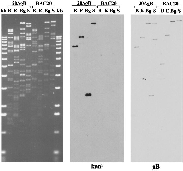
The complete genome of Marek's disease virus serotype 1 (MDV-1) strain 584Ap80C was cloned in Escherichia coli as a bacterial artificial chromosome (BAC). BAC vector sequences were introduced into the U(S)2 locus of the MDV-1 genome by homologous recombination. Viral DNA containing the BAC vector was used to transform Escherichia coli strain DH10B, and several colonies harboring the complete MDV-1 genome as an F plasmid (MDV-1 BACs) were identified. DNA from various MDV-1 BACs was transfected into chicken embryo fibroblasts, and from 3 days after transfection, infectious MDV-1 was obtained. Growth of MDV-1 recovered from BACs was indistinguishable from that of the parental virus, as assessed by plaque formation and determination of growth curves. In one of the MDV-1 BAC clones, sequences encoding glycoprotein B (gB) were deleted by one-step mutagenesis using a linear DNA fragment amplified by PCR. Mutant MDV-1 recovered after transfection of BAC DNA that harbored a 2.0-kbp deletion of the 2.6-kbp gB gene were able to grow and induce MDV-1-specific plaques only on cells providing MDV-1 gB in trans. The gB-negative virus reported here represents the first MDV-1 mutant with a deletion of an essential gene and demonstrates the power and usefulness of BACs to analyze genes and gene products in slowly growing and strictly cell-associated herpesviruses.


Figures

FIG. 1
FIG. 1
Schematic illustration of the cloning procedure of the transfer plasmid to introduce the BAC vector into the MDV-1 genome. The organization of the approximately 180-kbp MDV-1 genome (A) and the BamHI restriction map (B) as determined by Fukuchi et al. (11) are shown. The unique short region (US) and the ORFs located in the US are shown (C and D). A 2.1- and a 3.0-kbp fragment bordering the US2 gene (grey boxes) were amplified by PCR and cloned into plasmid pTZ18R to give rise to recombinant plasmid pDS. The 7.2-kbp BAC vector released from recombinant plasmid pHA1 (15) was inserted into pDS and resulted in plasmid pDS-pHA1 (E). Restriction enzyme sites were determined by Brunovskis and Velicer (7) and are abbreviated as follows: B, BamHI; E, EcoRI; P, PstI; Pa, PacI; S, SalI. The variable lengths of terminal DNA fragments are indicated (var).
FIG. 2
FIG. 2
Digitally scanned image of DNA of 584Ap80C (V) and DNA isolated from chloramphenicol-resistant E. coli DH10B colonies which were named BAC19, BAC20, and BAC24. Viral or BAC DNA was cleaved with BamHI or EcoRI, separated by 0.8% agarose gel electrophoresis, and stained with ethidium bromide (left panel). The restriction enzyme digests are flanked by the 1-kb ladder (Gibco-BRL). Asterisks indicate additional bands or size variations of individual fragments for the three BAC clones (in some cases the additional bands comigrate with other bands). Arrows indicate bands arising by insertion of the BAC vector sequences (left panel). Two subfragments of Bam-HI-A in which an additional BamHI site was introduced by inserting BAC vector sequences and one additional band of 5.8 kbp in EcoRI-digested BAC DNA are indicated by the arrows. After Southern transfer of DNA fragments to nylon membranes, hybridization with digoxigenin-labeled fragments released from plasmid pDS or pHA1 were performed. The sizes (in kilobase pairs) of reactive bands are given to the right.
FIG. 3
FIG. 3
Digitally scanned images of Southern blots to analyze size variations in BAC19, BAC20, and BAC24 DNA. Viral DNA from strain 584Ap80C and individual BACs was cleaved with BamHI or EcoRI and transferred to nylon membranes. Sheets were incubated with digoxigenin-labeled BAC19 DNA, labeled BamHI-C, or BamHI-D. The positions (in kilobase pairs) of size markers (1-kb ladder; Gibco-BRL) are given to the left. The smear-like bands 584Ap80C DNA hybridized with BamHI-D sequences are bracketed. Abbreviations: V, viral DNA from strain 584Ap80C; 19, 20, and 24, DNA from BAC19, BAC20, and BAC24, respectively.
FIG. 4
FIG. 4
(A) IIF analysis of representative MDV-1 plaques after infection with 584Ap80C or recombinant viruses obtained after transfection of BAC19, BAC20, or BAC24 DNA. At 5 days p.i., infected cells were fixed and subjected to IIF using anti-gB MAb 2K11. Detection of bound antibodies was performed with anti-mouse Alexa 488 (Molecular Probes). Approximately 100 plaques induced by each virus were scanned under the fluorescence microscope and no significant differences between virus reconstituted from BAC clones and parental virus or between the reconstituted viruses were observed. Magnification, ×250. (B) Growth curves of MDV-1 strain 584Ap80C and viruses recovered after transfection of various BACs. After infection of CEF cells with 100 PFU of 584Ap80C or transfection progeny of BAC19, BAC20, or BAC24, virus titers were determined at the indicated times p.i. by coseeding with fresh CEF cells. Virus plaques were counted after immunofluorescent staining with MAb 2K11. Each point represents the mean of two independent experiments.
FIG. 5
FIG. 5
Digitally scanned images of Southern blots to analyze the stability of BAC vector sequences in viruses recovered after transfection of BAC19 and BAC20. Transfection progeny was passaged four times, and viral DNA was isolated after each passage. Virus DNA was cleaved with BamHI or EcoRI, separated by 0.8% agarose gel electrophoresis, and transferred to nylon membranes. Southern blot hybridization was performed using digoxigenin-labeled fragments of plasmid pDS or pHA1. Lanes: V, 584Ap80C; 19, BAC19; 20, BAC20; 1 to 4, passages 1 to 4 after transfection of BAC19 DNA, respectively; 4a, DNA isolated after passage 4 after transfection of BAC20 DNA. The sizes (in kilobases) of reactive fragments are given. Asterisks indicate the reactive 1.6-kb band of the marker (1-kb ladder; Gibco-BRL).
FIG. 6
FIG. 6
(A) Schematic illustration of mutagenesis of BAC20 to remove gB-encoding sequences. First, recombinant plasmid pGETrec encoding l-arabinose-inducible recE, recT, and bacteriophage λ gam gene was transformed into BAC20-containing DH10B cells (indicated by step 1). Subsequently, after PCR amplification of the Kanr gene from plasmid pEGFP-N1 (Clontech) with primers that also contained 50-nucleotide homology arms bordering the gB deletion, a 1.6-kbp PCR amplicon was electroporated into DH10B cells harboring both BAC20 and pGETrec (indicated by step 2). Bacterial suspensions were plated on agar containing 30 μg of kanamycin per ml and 30 μg of chloramphenicol per ml. Double-resistant colonies were picked and subjected to further analysis. (B) Schematic illustration of the location of the gB gene in the 180-kbp MDV-1 genome and the replacement of the gB ORF with the Kanr gene by homologous recombination in the recombinant BAC clone 20ΔgB. (C) Schematic representation of the BamHI, BglI, EcoRI, and StuI restriction fragment sizes of 20ΔgB generated by the insertion of the Kanr gene instead of the gB ORF. Also indicated are the restriction fragment sizes for parental BAC20. Fragment sizes are given in kilobase pairs and were calculated according to published sequences (12).
FIG. 7
FIG. 7
Scanned image of an ethidium bromide-stained 0.8% agarose gel containing BAC20 and 20ΔgB DNA which was cleaved with BamHI (B), EcoRI (E), BglI (Bg), or StuI (S) and separated by 0.8% agarose gel electrophoresis (left panel). DNA fragments were transferred to nylon membranes and hybridized with a digoxigenin-labeled Kanr- or gB-specific probe. The Kanr probe was prepared by labeling a 1,015-bp BlnI fragment from pEGFP-N1, and the gB probe was prepared by labeling gB sequences released from plasmid pcMgB.
FIG. 8
FIG. 8
Confocal laser scan analysis of MgB1 cells constitutively expressing MDV-1 gB. MgB1 or QM7 cells were seeded on glass coverslips and incubated with anti-gB MAb 2K11 or convalescent chicken serum anti-MDV (α-MDV). Secondary antibodies were anti-mouse or anti-chicken immunoglobulin G conjugated to Alexa 488 (Molecular Probes). Nuclei were counterstained with propidium iodide. The view shown is 115 by 115 μm.
FIG. 9
FIG. 9
IIF analysis of MgB1, QM7, or CEF cells after transfection with BAC20 (upper panels) or 20ΔgB (lower panels). At 4 days after transfection, cells were fixed with acetone and incubated with anti-pp38 MAb H19. The secondary antibody was anti-mouse immunoglobulin G conjugated to Alexa 488 (Molecular Probes). Whereas MDV-1 plaques were observed on all cell lines after transfection of BAC20 DNA, viral plaques were observed on MgB1 cells only after transfection with 20ΔgB. Only single infected cells were observed on QM7 and CEF cells (arrowheads). Magnification, ×250.

Similar articles

Cited by

References

    1. Anderson A S, Parcells M S, Morgan R W. The glycoprotein D (US6) homolog is not essential for oncogenicity or horizontal transmission of Marek's disease virus. J Virol. 1998;72:2548–2553. - PMC - PubMed
    1. Becker Y, Tabor E, Asher Y, Davidson I, Malkinson M, Witter R L. PCR detection of amplified 132 bp repeats in Marek's disease virus type 1 (MDV-1) DNA can serve as an indicator for critical genomic rearrangement leading to the attenuation of virus virulence. Virus Genes. 1993;7:277–287. - PubMed
    1. Borst E M, Hahn G, Koszinowski U H, Messerle M. Cloning of the human cytomegalovirus (HCMV) genome as an infectious bacterial artificial chromosome in Escherichia coli: a new approach for construction of HCMV mutants. J Virol. 1999;73:8320–8329. - PMC - PubMed
    1. Bradley G, Hayashi M, Lancz G, Tanaka A, Nonoyama M. Structure of the Marek's disease virus BamHI-H gene family: genes of putative importance for tumor induction. J Virol. 1989;63:2534–2542. - PMC - PubMed
    1. Bradley G, Lancz G, Tanaka A, Nonoyama M. Loss of Marek's disease virus tumorigenicity is associated with truncation of RNAs transcribed within BamHI-H. J Virol. 1989;63:4129–4135. - PMC - PubMed

Publication types

LinkOut - more resources