

Synthesis and crystal structure of an octamer RNA r(guguuuac)/r(guaggcac) with G.G/U.U tandem wobble base pairs: comparison with other tandem G.U pairs
- PMID: 11058138
- PMCID: PMC113127
- DOI: 10.1093/nar/28.21.4376
Synthesis and crystal structure of an octamer RNA r(guguuuac)/r(guaggcac) with G.G/U.U tandem wobble base pairs: comparison with other tandem G.U pairs
Abstract
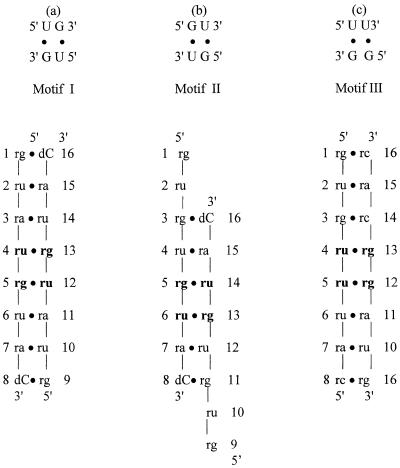
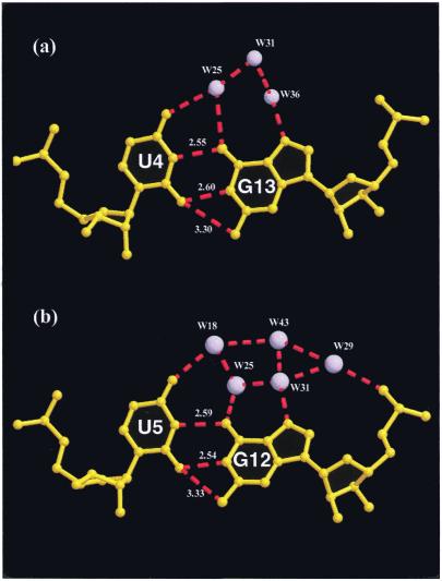
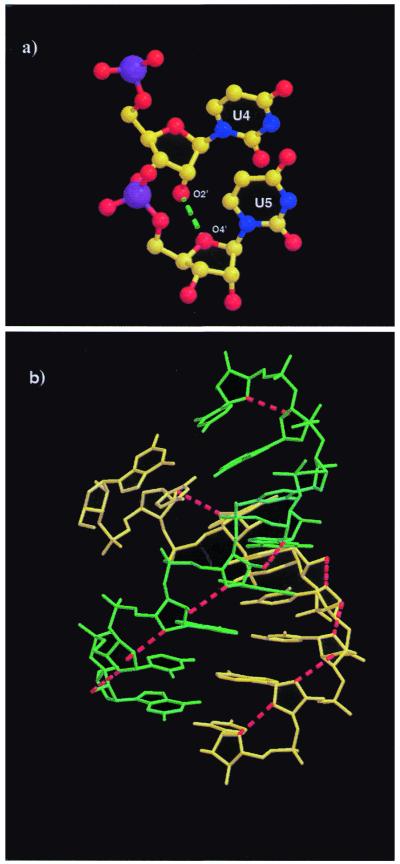
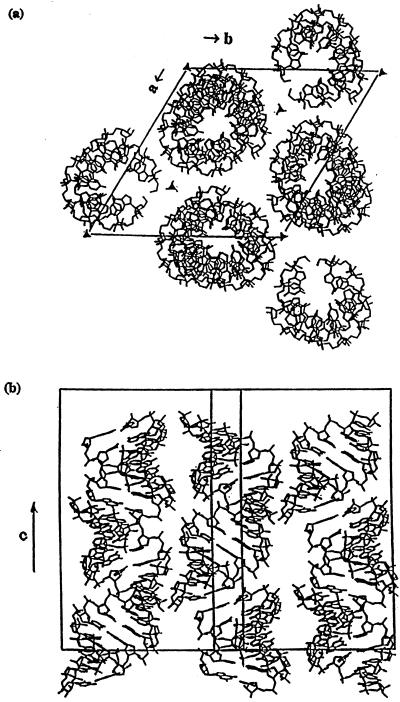
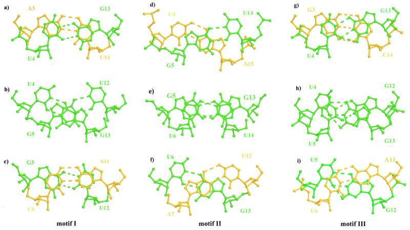
We have determined the crystal structure of the RNA octamer duplex r(guguuuac)/r(guaggcac) with a tandem wobble pair, G.G/U.U (motif III), to compare it with U.G/G.U (motif I) and G.U/U.G (motif II) and to better understand their relative stabilities. The crystal belongs to the rhombohedral space group R3. The hexagonal unit cell dimensions are a = b = 41.92 A, c = 56.41 A, and gamma = 120 degrees, with one duplex in the asymmetric unit. The structure was solved by the molecular replacement method at 1.9 A resolution and refined to a final R: factor of 19.9% and R(free) of 23.3% for 2862 reflections in the resolution range 10.0-1.9 A with F >/= 2sigma(F). The final model contains 335 atoms for the RNA duplex and 30 water molecules. The A-RNA stacks in the familiar head-to-tail fashion forming a pseudo-continuous helix. The uridine bases of the tandem U.G pairs have slipped towards the minor groove relative to the guanine bases and the uridine O2 atoms form bifurcated hydrogen bonds with the N1 and N2 of guanines. The N2 of guanine and O2 of uridine do not bridge the 'locked' water molecule in the minor groove, as in motifs I and II, but are bridged by water molecules in the major groove. A comparison of base stacking stabilities of motif III with motifs I and II confirms the result of thermodynamic studies, motif I > motif III > motif II.
Figures
Similar articles
-
Crystal structure of r(GUGUGUA)dC with tandem G x U/U x G wobble pairs with strand slippage.J Mol Biol. 1997 Jul 18;270(3):511-9. doi: 10.1006/jmbi.1997.1118. J Mol Biol. 1997. PMID: 9237915
-
Crystal structure of an alternating octamer r(GUAUGUA)dC with adjacent G x U wobble pairs.J Mol Biol. 1997 Apr 18;267(5):1149-56. doi: 10.1006/jmbi.1997.0936. J Mol Biol. 1997. PMID: 9150403
-
A single 2'-hydroxyl group converts B-DNA to A-DNA. Crystal structure of the DNA-RNA chimeric decamer duplex d(CCGGC)r(G)d(CCGG) with a novel intermolecular G-C base-paired quadruplet.J Mol Biol. 1994 Feb 11;236(1):275-85. doi: 10.1006/jmbi.1994.1134. J Mol Biol. 1994. PMID: 7508984
-
Hydrogen and hydration of DNA and RNA oligonucleotides.Biophys Chem. 2002 Mar 28;95(3):273-82. doi: 10.1016/s0301-4622(01)00262-9. Biophys Chem. 2002. PMID: 12062385 Review.
-
The G x U wobble base pair. A fundamental building block of RNA structure crucial to RNA function in diverse biological systems.EMBO Rep. 2000 Jul;1(1):18-23. doi: 10.1093/embo-reports/kvd001. EMBO Rep. 2000. PMID: 11256617 Free PMC article. Review.
Cited by
-
Testing the nearest neighbor model for canonical RNA base pairs: revision of GU parameters.Biochemistry. 2012 Apr 24;51(16):3508-22. doi: 10.1021/bi3002709. Epub 2012 Apr 10. Biochemistry. 2012. PMID: 22490167 Free PMC article.
-
Crystal structure of an RNA helix recognized by a zinc-finger protein: an 18-bp duplex at 1.6 A resolution.RNA. 2002 Jul;8(7):924-32. doi: 10.1017/s1355838202028893. RNA. 2002. PMID: 12166647 Free PMC article.
-
An innate twist between Crick's wobble and Watson-Crick base pairs.RNA. 2013 Aug;19(8):1038-53. doi: 10.1261/rna.036905.112. RNA. 2013. PMID: 23861536 Free PMC article. Review.
-
The electrostatic characteristics of G.U wobble base pairs.Nucleic Acids Res. 2007;35(11):3836-47. doi: 10.1093/nar/gkm274. Epub 2007 May 25. Nucleic Acids Res. 2007. PMID: 17526525 Free PMC article.
-
Structural Insights Into the 5'UG/3'GU Wobble Tandem in Complex With Ba2+ Cation.Front Mol Biosci. 2022 Jan 13;8:762786. doi: 10.3389/fmolb.2021.762786. eCollection 2021. Front Mol Biosci. 2022. PMID: 35096964 Free PMC article.
References
-
- Modrich P. (1987) Annu. Rev. Biochem., 56, 435. - PubMed
-
- Pearl L.H. and Savva,R. (1995) Trends Biochem. Sci., 20, 421. - PubMed
-
- Varani G. and Pardi,A. (1994) In Nagai,K. and Mattaj,I.W. (eds), RNA–Protein Interactions. Oxford University Press, Oxford, UK, pp. 1–24.
-
- Varani G., Wimberly,B.and Tinoco,I.,Jr (1989) Biochemistry, 28, 7760. - PubMed
-
- Wimberly B., Varani,G. and Tinoco,I.,Jr (1993) Biochemistry, 32, 1078. - PubMed
Publication types
MeSH terms
Substances
Grants and funding
LinkOut - more resources
Full Text Sources
