. 2000 Nov;20(22):8352-63.
doi: 10.1128/MCB.20.22.8352-8363.2000.

Platelet-derived growth factor receptor association with Na(+)/H(+) exchanger regulatory factor potentiates receptor activity

Affiliations

Platelet-derived growth factor receptor association with Na(+)/H(+) exchanger regulatory factor potentiates receptor activity

S Maudsley et al. Mol Cell Biol. 2000 Nov.

Abstract

Platelet-derived growth factor (PDGF) is a potent mitogen for many cell types. The PDGF receptor (PDGFR) is a receptor tyrosine kinase that mediates the mitogenic effects of PDGF by binding to and/or phosphorylating a variety of intracellular signaling proteins upon PDGF-induced receptor dimerization. We show here that the Na(+)/H(+) exchanger regulatory factor (NHERF; also known as EBP50), a protein not previously known to interact with the PDGFR, binds to the PDGFR carboxyl terminus (PDGFR-CT) with high affinity via a PDZ (PSD-95/Dlg/Z0-1 homology) domain-mediated interaction and potentiates PDGFR autophosphorylation and extracellular signal-regulated kinase (ERK) activation in cells. A point-mutated version of the PDGFR, with the terminal leucine changed to alanine (L1106A), cannot bind NHERF in vitro and is markedly impaired relative to the wild-type receptor with regard to PDGF-induced autophosphorylation and activation of ERK in cells. NHERF potentiation of PDGFR signaling depends on the capacity of NHERF to oligomerize. NHERF oligomerizes in vitro when bound with PDGFR-CT, and a truncated version of the first NHERF PDZ domain that can bind PDGFR-CT but which does not oligomerize reduces PDGFR tyrosine kinase activity when transiently overexpressed in cells. PDGFR activity in cells can also be regulated in a NHERF-dependent fashion by stimulation of the beta(2)-adrenergic receptor, a known cellular binding partner for NHERF. These findings reveal that NHERF can directly bind to the PDGFR and potentiate PDGFR activity, thus elucidating both a novel mechanism by which PDGFR activity can be regulated and a new cellular role for the PDZ domain-containing adapter protein NHERF.


Figures
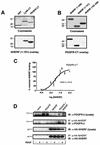
FIG. 1
FIG. 1
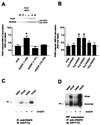
PDGFR-CT binds NHERF. (A) NHERF(1-151) binds to immobilized PDGFR-CT. PDGFR-CT, expressed as a GST fusion protein, was run on an SDS-polyacrylamide gel alongside equal amounts of control GST or the β2AR-CT expressed as a GST fusion protein (top). These samples were blotted to nitrocellulose and overlaid with 25 nM NHERF(1-151), which bound equally well to the PDGFR-CT and β2AR-CT but did not bind to control GST (bottom). (B) PDGFR-CT binds to immobilized NHERF. Equal amounts of NHERF(1-358) (full-length NHERF), NHERF(1-151), and NHERF(152-358) were run on an SDS-polyacrylamide (top), then transferred to nitrocellulose, and overlaid with 25 nM PDGFR-CT. PDGFR-CT bound strongly to full-length NHERF and NHERF(1-151) but only weakly to NHERF(152-358). The numbers adjacent to each panel represent relative molecular masses in kilodaltons. (C) PDGFR-CT binds NHERF(1-151) with high affinity. Increasing concentrations (1 to 1,000 nM) of NHERF(1-151) were overlaid onto immobilized PDGFR-CT, and binding was expressed as a percentage of maximal specific binding. The KD of the interaction was estimated at 26 nM. Points and error bars represent the mean ± SEM for three independent experiments. (D) Full-length PDGFR expressed in CHO cells coimmunoprecipitates with NHERF. Whole-cell lysates and anti-HA immunocomplexes were probed for either the PDGFR (top two panels) or HA-NHERF (lower two panels). Only in CHO cells transiently transfected with cDNAs encoding WT PDGFR and HA-NHERF was PDGFR immunoreactivity detected in anti-HA immunocomplexes. The figures adjacent to each panel represent relative molecular masses in kilodaltons. These experiments were performed in both the absence (−) and presence (+) of stimulation with 200 pM PDGF for 5 min. Stimulation with PDGF had no apparent effect on the association of PDGFR with NHERF. The data shown are representative of six independent experiments. IB, immunoblotting; IP, immunoprecipitation.
FIG. 2
FIG. 2
NHERF potentiates PDGFR signaling. (A) Overexpression of NHERF in CHO cells enhances PDGF-induced ERK1/2 activation. Columns and error bars represent the mean ± SEM for six independent experiments. NS, nonstimulated. (B) Overexpression of NHERF has a biphasic effect on PDGF-induced ERK1/2 activation. The level of NHERF expression is indicated as micrograms of cDNA encoding HA-tagged NHERF transfected per 100-mm-diameter plate of CHO cells. ∗, significant to P < 0.05. (C) Overexpression of NHERF enhances both basal and PDGF-induced PDGFR autophosphorylation. Endogenous PDGFRs from CHO cells that were either unstimulated or stimulated with 200 pM PDGF were immunoprecipitated (IP), resolved by SDS-PAGE, immunoblotted (IB), and probed with an antiphosphotyrosine antibody (anti-P-Tyr). Compared to cells not transfected with NHERF, NHERF overexpression elevated the basal and PDGF-induced tyrosine phosphorylation of the receptor 1.8 ± 0.3-fold (n = 4) and 2.4 ± 0.4-fold (n = 4), respectively. (D) Overexpression of NHERF enhances both basal and PDGF-induced PDGFR dimerization. Endogenous PDGFR in CHO cells was covalently cross-linked with BS3, either under basal conditions or following stimulation with 200 pM PDGF. The receptors were then immunoprecipitated, resolved by SDS-PAGE, blotted, and probed with an antiphosphotyrosine antibody. PDGF stimulation resulted in an increase in tyrosine phosphorylation of both PDGFR monomer and dimer. Compared to cells not transfected with NHERF, NHERF transfection resulted in 2.7 ± 0.1-fold (n = 4) and 2.9 ± 0.3-fold (n = 4), respectively, increases in basal and agonist-stimulated dimer tyrosine phosphorylation. The 205-kDa band is indicated on the left.
FIG. 3
FIG. 3
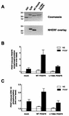
The PDGFR L1106A mutant does not bind to NHERF and is less active than WT PDGFR. (A) L1106A PDGFR-CT does not bind NHERF(1-151). L1106A PDGFR-CT, which is identical to WT PDGFR-CT except that its terminal leucine is mutated to alanine, was expressed as a GST fusion protein and run on an SDS-polyacrylamide gel alongside equal amounts of control GST, β2AR-CT, and WT PDGFR-CT (top). These samples were overlaid with 25 nM NHERF(1-151), which bound well to WT PDGFR-CT but did not bind specifically to either control GST or L1106A PDGFR-CT (bottom). The positions of molecular weight markers are shown on the left in kilodaltons. (B) Full-length L1106A PDGFR exhibits less PDGF-induced autophosphorylation than WT PDGFR in COS-7 cells. WT and L1106A PDGFR were separately transfected into COS-7 cells, and their levels of expression were comparable as assessed by radiolabeled PDGF binding. The differentially transfected cells were then not stimulated (NS) or stimulated with PDGF (40 pM) and assayed for PDGFR autophosphorylation. Bars and error bars represent the mean ± SEM for five independent experiments. (C) L1106A PDGFR exhibits reduced ability to activate ERK relative to WT PDGFR. The low level of endogenous PDGFR in COS-7 cells mediates an approximately 1.4-fold increase in ERK activation following stimulation with 40 pM PDGF (left pair of bars). Overexpression of WT PDGFR enhances PDGF-induced ERK activation (middle pair of bars), whereas overexpression of L1106A PDGFR does not (right pair of bars). The level of ERK activation under all conditions is expressed as fold over the level observed in the absence of transfected PDGFR and the absence of PDGF stimulation. Bars and error bars represent the mean ± SEM for four independent experiments.
FIG. 4
FIG. 4
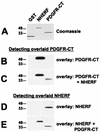
NHERF can facilitate oligomerization of PDGFR-CT, and PDGFR-CT can facilitate oligomerization of NHERF. (A) Equal amounts (2 μg) of GST, NHERF, and PDGFR-CT were run on SDS-polyacrylamide gels. (B) The samples were blotted and overlaid with 25 nM radiolabeled PDGFR-CT, which bound to immobilized NHERF but not to immobilized GST or PDGFR-CT. (C) When the experiment was repeated in the presence of 50 nM NHERF, binding of radiolabeled PDGFR-CT to immobilized PDGFR-CT was detectable. (D) The same samples were overlaid with 25 nM NHERF, which was detected via a far-Western blot approach. The overlaid NHERF bound to immobilized PDGFR-CT but not to immobilized GST or NHERF. (E) When the experiment was repeated in the presence of 250 nM PDGFR-CT, binding of overlaid NHERF to immobilized NHERF was detectable, while binding of overlaid NHERF to immobilized PDGFR-CT was reduced. These data are representative of four to six independent experiments each. The positions of molecular weight standard markers (in kilodaltons) are shown on the left of each panel.
FIG. 5
FIG. 5
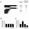
NHERF(1-121) binds PDGFR-CT, does not oligomerize, and inhibits PDGFR autophosphorylation in cells. (A) Truncations of NHERF have differential effects on PDGFR-CT binding versus PDGFR-CT-induced NHERF oligomerization. Equal amounts of various truncated versions of NHERF expressed as fusion proteins were run on SDS-polyacrylamide gels and then blotted to nitrocellulose. The schematic diagram depicts the NHERF truncations and summarizes their abilities to bind overlaid 25 nM PDGFR-CT and to bind overlaid 25 nM NHERF(1-151) in the presence of 250 nM PDGFR-CT. +++, amount of specific binding observed with full-length NHERF; ++, somewhat less binding than observed with full-length NHERF; +, barely detectable binding, −, no specific binding detectable. These data are representative of four to eight independent experiments. (B) EGF-induced EGFR autophosphorylation is unaffected by overexpression of full-length NHERF, NHERF(1-151), or NHERF(1-121) in CHO cells. Human EGFR was transfected into CHO cells and stimulated with 160 pM EGF. The bars and error bars representative the mean ± SEM of three independent experiments. (C) NHERF(1-121) acts as a dominant negative for PDGF-induced PDGFR autophosphorylation in CHO cells. The endogenous PDGFR in CHO cells was stimulated with 100 pM PDGF, and the PDGFR was then immunoprecipitated, run on an SDS-polyacrylamide gel, blotted, and probed with an antiphosphotyrosine antibody. The level of PDGF-induced PDGFR autophosphorylation observed in cells transfected with either full-length NHERF, NHERF(1-151), or NHERF(1-121) is expressed as a percentage of that observed in untransfected cells. Bars and error bars represent the mean ± SEM for four independent experiments. ∗∗, significant to P < 0.01.
FIG. 6
FIG. 6
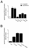
β2AR stimulates ERK phosphorylation via PDGFR transactivation. (A) Isoproterenol-induced ERK activation in CHO cells transfected with WT β2AR is inhibited by PDGFR-specific tyrosine kinase inhibitors and is mediated by a Gi/Gβγ-dependent pathway. Application of 10 μM isoproterenol for 2 min resulted in activation of ERK. This effect was not blocked by preincubation with the EGFR-specific tyrphostin AG1478 (100 nM for 10 min) but was blocked by preincubation with the PDGFR-specific tyrphostin AG1295 (10 μM for 40 min), pertussis toxin (PTX; 100 ng/ml for 16 h), or the PKA inhibitor H89 (10 μM for 10 min) and by coexpression of the Gβγ sequestrant βARK-ct. The data points represent the mean ± SEM for four separate experiments. (B) L413A β2AR exhibits a greater capacity to activate ERK than WT β2AR. Isoproterenol stimulation of the point-mutated L413A β2AR, which is incapable of sequestering cellular NHERF, resulted in more robust activation of ERK than did isoproterenol stimulation of WT β2AR. Overexpression of NHERF with WT β2AR resulted in a potentiation of the ERK activation induced by stimulation of WT β2AR. The data points represent the mean ± SEM for five separate experiments.
FIG. 7
FIG. 7
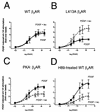
Dissection of G-protein-mediated transactivation from NHERF-mediated inhibition of PDGFR signaling by the β2AR. (A) Prestimulation of WT β2AR stably expressed in AP1-CHO cells does not significantly alter subsequent ERK activation by PDGF. Cells pretreated with isoproterenol (iso) were incubated with 10 μM isoproterenol for 10 min before determination of the levels of ERK activation induced by increasing concentrations of PDGF. (B) Prestimulation of L413A β2AR, stably expressed in AP1-CHO cells, results in an elevation of PDGF-induced ERK activation. This set of experiments reveals that removing the capacity of the β2AR to sequester NHERF relieves an inhibitory action of the β2AR on PDGFR function. (C) Prestimulation of PKA β2AR, stably expressed in AP1-CHO cells, results in an attenuation of PDGF-induced ERK activation. This set of experiments indicates that without the ability to couple to Gi and transactivate the PDGFR, PKA β2AR can still bind NHERF and sequester it from the PDGFR, thus inhibiting PDGFR function. (D) Prestimulation of WT β2AR under conditions of reduced PKA activity results in an attenuation of PDGFR function. AP1-CHO cells stably transfected with WT β2AR were pretreated with the PKA inhibitor H89 and then stimulated with increasing concentrations of PDGF in the absence or presence of isoproterenol pretreatment. Inhibition of PKA prevents β2AR transactivation of the PDGFR and uncovers an inhibitory action of the WT β2AR on PDGFR signaling. This set of experiments reveals the capacity of WT β2AR to inhibit PDGFR function, presumably by sequestering cellular NHERF from the PDGFR.

Similar articles

Cited by

References

    1. Anan K, Morisaki T, Katano M, Ikubo A, Kitsuki H, Uchiyama A, Kuroki S, Tanaka M, Torisu M. Vascular endothelial growth factor and platelet-derived growth factor are potential angiogenic and metastatic factors in human breast cancer. Surgery. 1996;119:333–339. - PubMed
    1. Ariad S, Seymour L, Bezwoda W R. Platelet-derived growth factor (PDGF) in plasma of breast cancer patients: correlation with stage and rate of progression. Breast Cancer Res Treat. 1991;20:11–17. - PubMed
    1. Brenman J E, Chao D S, Gee S H, McGee A W, Craven S E, Santillano D R, Wu Z, Huang F, Xia H, Peters M F, Froehner S C, Bredt D S. Interaction of nitric oxide syntahse with the postsynaptic density protein PSD-95 and α1-syntrophin mediated by PDZ domains. Cell. 1996;84:757–767. - PubMed
    1. Brenman J E, Christopherson K S, Craven S E, McGee A W, Bredt D S. Cloning and characterization of postsynaptic density 93, a nitric oxide synthase interacting protein. J Neurosci. 1996;16:7407–7415. - PMC - PubMed
    1. Buchert M, Schneider S, Meskenaite V, Adams M T, Canaani E, Baechi T, Moelling K, Hovens C M. The junction-associated protein AF-6 interacts and clusters with specific Eph receptor tyrosine kinases at specialized sites of cell-cell contact in the brain. J Cell Biol. 1999;144:361–371. - PMC - PubMed

Publication types

MeSH terms

Substances

LinkOut - more resources