. 2000 Oct;11(10):3341-52.
doi: 10.1091/mbc.11.10.3341.

Regulation of macropinocytosis by p21-activated kinase-1

Affiliations
Free PMC article

Regulation of macropinocytosis by p21-activated kinase-1

S Dharmawardhane et al. Mol Biol Cell. 2000 Oct.
Free PMC article

Abstract

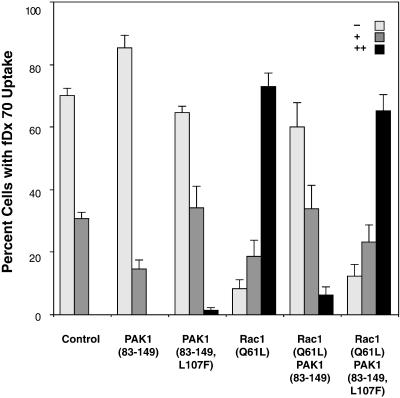
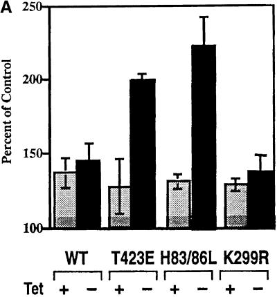
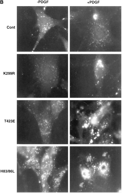
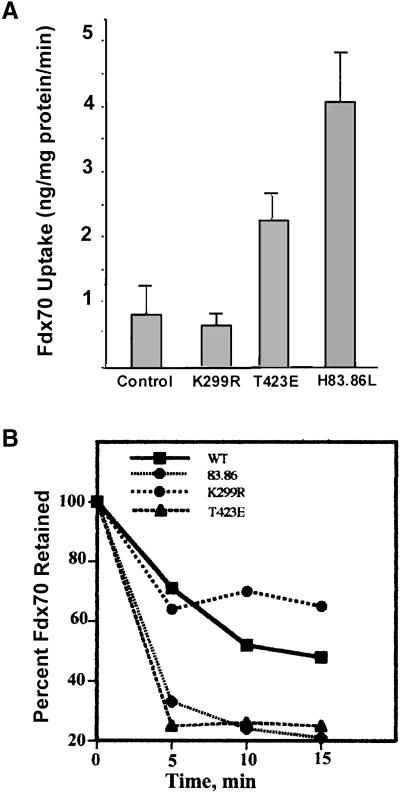
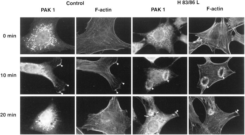
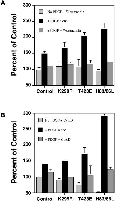
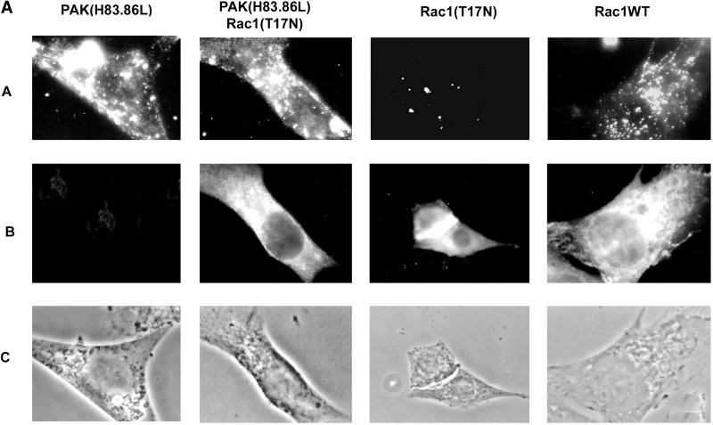
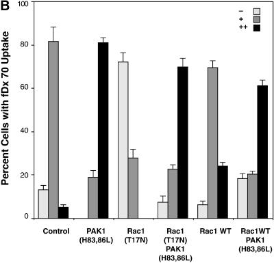
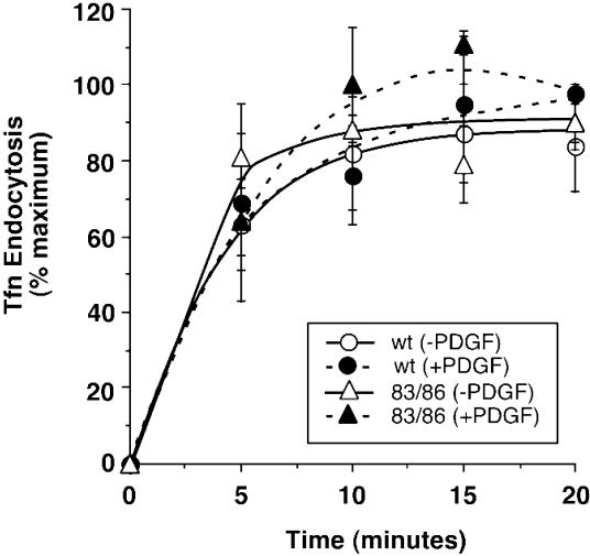
The process of macropinocytosis is an essential aspect of normal cell function, contributing to both growth and motile processes of cells. p21-activated kinases (PAKs) are targets for activated Rac and Cdc42 guanosine 5'-triphosphatases and have been shown to regulate the actin-myosin cytoskeleton. In fibroblasts PAK1 localizes to areas of membrane ruffling, as well as to amiloride-sensitive pinocytic vesicles. Expression of a PAK1 kinase autoinhibitory domain blocked both platelet-derived growth factor- and RacQ61L-stimulated uptake of 70-kDa dextran particles, whereas an inactive version of this domain did not, indicating that PAK kinase activity is required for normal growth factor-induced macropinocytosis. The mechanisms by which PAK modulate macropinocytosis were examined in NIH3T3 cell lines expressing various PAK1 constructs under the control of a tetracycline-responsive transactivator. Cells expressing PAK1 (H83,86L), a mutant that dramatically stimulates formation of dorsal membrane ruffles, exhibited increased macropinocytic uptake of 70-kDa dextran particles in the absence of additional stimulation. This effect was not antagonized by coexpression of dominant-negative Rac1-T17N. In the presence of platelet-derived growth factor, both PAK1 (H83,86L) and a highly kinase active PAK1 (T423E) mutant dramatically enhanced the uptake of 70-kDa dextran. Neither wild-type PAK1 nor vector controls exhibited enhanced macropinocytosis, nor did PAK1 (H83,86L) affect clathrin-dependent endocytic mechanisms. Active versions of PAK1 enhanced both growth factor-stimulated 70-kDa dextran uptake and efflux, suggesting that PAK1 activity modulated pinocytic vesicle cycling. These data indicate that PAK1 plays an important regulatory role in the process of macropinocytosis, perhaps related to the requirement for PAK in directed cell motility.


Figures

Figure 1
Figure 1
Inhibition of Rac Q61L-induced fDx uptake by PAK1 AID. Serum-starved NIH3T3 cells stably transfected with a tetracycline-inducible vector only were infected with Semliki Forest virus containing myc-tagged PAK1 (83–149), PAK1 (83–149, L107F). Rac1 (Q61L), or combinations thereof as indicated (methods as in Edwards et al., 1999; Sanders et al., 1999). After 10 h of infection, the cells were incubated in 5 mg/ml lysine-fixable 70-kDa fDx for 30 min, and then fixed, permeabilized, and stained for the myc epitope by using anti-myc 9E10 antibody, followed by a fluorescein isothiocyanate-tagged secondary antibody. Cells were scored by using a fluorescence microscope with 40× objective as −, +, or ++ according to the level of fDx uptake, as illustrated in Table 1. A total of ∼200 cells was counted per experimental condition by using 10 separate fields per coverslip. Values, given as a percentage of cells in each scoring category, from three separate experiments are shown as the mean ± SE.
Figure 2
Figure 2
PDGF-stimulated pinocytosis in cell lines expressing PAK1 or PAK1 mutants. (A) NIH3T3 cells transformed with wild-type PAK1, PAK1 (T423E), PAK1 (H83,86L), or PAK1 (K299R) were either maintained in tetracycline (+) or grown for 24 h in media without tetracycline (−) to induce PAK protein expression. Expression of PAK proteins was determined to be equal by immunoblot, as in Sells et al. (1999). During the induction period, the cells were serum starved for 18 h, and then cooled on ice for 30 min before the start of the experiment to render them quiescent. The uptake of 70-kDa fDx was measured for 60 min in the presence or absence of 5 ng/ml PDGF as described in MATERIALS AND METHODS. The absorption/emission spectra for fluorescein at 494/520 nm were determined, and data plotted as the percentage of control (i.e,. the amount of fDx70 taken up in the presence of 5 ng/ml PDGF/amount of fDx70 uptake in the absence of PDGF × 100) for each treatment. The values shown are the mean ± SD of the mean for n = 4 determinations in two separate experiments. Tet, tetracycline. (B) Uptake of 70-kDa fDx was performed as above, except that cells were not precooled before the addition of lysine fixable 70-kDa fDx and 5 ng/ml PDGF. The cells were fixed in a periodate–lysine–paraformaldehyde fixative as detailed in MATERIALS AND METHODS, and the fluorescence of fDx was visualized in the various induced PAK1 NIH3T3 cell lines at t = 30 min. Micrographs shown are at 5000×.
Figure 2
Figure 2
PDGF-stimulated pinocytosis in cell lines expressing PAK1 or PAK1 mutants. (A) NIH3T3 cells transformed with wild-type PAK1, PAK1 (T423E), PAK1 (H83,86L), or PAK1 (K299R) were either maintained in tetracycline (+) or grown for 24 h in media without tetracycline (−) to induce PAK protein expression. Expression of PAK proteins was determined to be equal by immunoblot, as in Sells et al. (1999). During the induction period, the cells were serum starved for 18 h, and then cooled on ice for 30 min before the start of the experiment to render them quiescent. The uptake of 70-kDa fDx was measured for 60 min in the presence or absence of 5 ng/ml PDGF as described in MATERIALS AND METHODS. The absorption/emission spectra for fluorescein at 494/520 nm were determined, and data plotted as the percentage of control (i.e,. the amount of fDx70 taken up in the presence of 5 ng/ml PDGF/amount of fDx70 uptake in the absence of PDGF × 100) for each treatment. The values shown are the mean ± SD of the mean for n = 4 determinations in two separate experiments. Tet, tetracycline. (B) Uptake of 70-kDa fDx was performed as above, except that cells were not precooled before the addition of lysine fixable 70-kDa fDx and 5 ng/ml PDGF. The cells were fixed in a periodate–lysine–paraformaldehyde fixative as detailed in MATERIALS AND METHODS, and the fluorescence of fDx was visualized in the various induced PAK1 NIH3T3 cell lines at t = 30 min. Micrographs shown are at 5000×.
Figure 3
Figure 3
Effects of PAK on fDx uptake and efflux kinetics. (A) Effects of preincubation of cells with PDGF on the regulation of fDx uptake by PAK were examined. NIH3T3 cells expressing either vector alone (control), PAK1 wild type, or the indicated point mutants were maintained for 24 h in the absence of tetracycline to allow PAK1 protein expression. Cells were serum starved for 18 h during the expression period, and then PDGF (5 ng/ml) added to each cell line for 45 min. At the end of each incubation period, 5 mg/ml 70-kDa fDx was added and uptake measured at 0, 2.5, 5, 10, and 15 min. Cells were washed free of noninternalized fDx and the amount of fDx in cell lysates quantified by spectrofluorometry. The amount of fDx taken up per minute in the linear phase was plotted (±SE; n = 6). (B) NIH3T3 cell lines were handled as described above. After serum starvation for 18 h, PDGF and 70-kDa fDx were added to the cells for 45 min to allow uptake, and then washed three times in ice cold PBS and incubated in PBS containing 5.5 mM glucose, 1 mM CaCl2, and 10 mM MgCl2 for the indicated times. Cells were lysed and the fDx retained was quantitated by spectrofluorometry. One hundred percent represents the amount of fDx contained within each cell line at t = 0 min. SD for each data point was <18% of the mean (n = 6).
Figure 4
Figure 4
PAK1 and F-actin localization in PAK1 (H83,86L)-expressing cells. Localization of PAK1, determined with a specific affinity purified anti-PAK1 antibody, or of F-actin, determined by rhodamine phalloidin staining, was examined in either vector control (left two columns) or PAK1 (H83,86L) (right two columns)-expressing cell lines at various times after PDGF (5 ng/ml) stimulation, as described in MATERIALS AND METHODS. The arrowheads indicate areas where PAK1 and F-actin colocalize in sites of membrane ruffling. The short arrows point to remnants of macropinocytic vesicles whose outline is visible in the stained cells. Micrographs shown are at 3000×.
Figure 5
Figure 5
Inhibition of PAK1-enhanced macropinocytosis by the PI 3-kinase inhibitor wortmannin and by the F-actin disrupting agent cytochalasin D. Uptake of 70-kDa fDx was measured in cells expressing control vector, PAK1 wild-type, and PAK1 point mutations. As indicated, the cells were pretreated with either 100 nM wortmannin (A) or 10 μg/ml cytochalasin D (B) for 10 min, and then stimulated with 5 ng/ml PDGF and fDx added at t = 0. Uptake was for 45 min. Percentage of control is the amount of fDx70 taken up under each condition compared with the nondrug- or PDGF-treated cells (set to 100%). Results shown are the mean ± SD of 2–3 experiments performed in duplicate.
Figure 6
Figure 6
Macropinocytosis in cells expressing PAK1 and Rac1 mutants. NIH3T3 cell lines expressing PAK1 (H83,86L) were incubated in media with or without (control) tetracycline for protein induction. These cells were then infected with a Semliki Forest virus expression construct containing either myc-tagged dominant-negative Rac1 (T17N) or Rac1 wild type (WT) (Sanders et al., 1999). (A) After 12 h of infection, the cells were incubated in 5 ng/ml PDGF and 5 mg/ml fixable 70-kDa fDX for 45 min, and then fixed, permeabilized, and stained by using the 9E10-myc epitope antibody (Racoosin and Swanson, 1994). Row A, fluorescence micrographs of representative cells demonstrating fDx retained in macropinocytic vesicles. Row B, same cells stained with anti-myc 9E10 to detect Rac1-WT and -T17N expression. Row C, same cells under phase contrast microscopy. (B) After 12 h of infection, the cells were incubated in 5 ng/ml PDGF and 5 mg/ml lysine-fixable 70-kDa fDx for 45 min, and then fixed, permeabilized, and stained for the myc epitope by using anti-myc 9E10 antibody, followed by a fluorescein isothiocyanate-tagged secondary antibody. Cells were scored by using a fluorescence microscope with 40× objective as −, +, or ++ according to the level of fDx uptake, as illustrated in Table 1. A total of ∼200 cells was counted per experimental condition by using 10 separate fields per coverslip. Values, given as a percentage of cells in each scoring category, from three separate experiments are shown as the mean ± SE.
Figure 6
Figure 6
Macropinocytosis in cells expressing PAK1 and Rac1 mutants. NIH3T3 cell lines expressing PAK1 (H83,86L) were incubated in media with or without (control) tetracycline for protein induction. These cells were then infected with a Semliki Forest virus expression construct containing either myc-tagged dominant-negative Rac1 (T17N) or Rac1 wild type (WT) (Sanders et al., 1999). (A) After 12 h of infection, the cells were incubated in 5 ng/ml PDGF and 5 mg/ml fixable 70-kDa fDX for 45 min, and then fixed, permeabilized, and stained by using the 9E10-myc epitope antibody (Racoosin and Swanson, 1994). Row A, fluorescence micrographs of representative cells demonstrating fDx retained in macropinocytic vesicles. Row B, same cells stained with anti-myc 9E10 to detect Rac1-WT and -T17N expression. Row C, same cells under phase contrast microscopy. (B) After 12 h of infection, the cells were incubated in 5 ng/ml PDGF and 5 mg/ml lysine-fixable 70-kDa fDx for 45 min, and then fixed, permeabilized, and stained for the myc epitope by using anti-myc 9E10 antibody, followed by a fluorescein isothiocyanate-tagged secondary antibody. Cells were scored by using a fluorescence microscope with 40× objective as −, +, or ++ according to the level of fDx uptake, as illustrated in Table 1. A total of ∼200 cells was counted per experimental condition by using 10 separate fields per coverslip. Values, given as a percentage of cells in each scoring category, from three separate experiments are shown as the mean ± SE.
Figure 7
Figure 7
PAK1 has no effect on transferrin uptake via clathrin-mediated endocytosis. Uptake of transferrin was determined as described in MATERIALS AND METHODS in the induced vector control (circles) or PAK1 (H83,86L)-expressing (triangles) stable cell lines. The percentage of transferrin uptake is the amount of transferrin internalized as a function of the total amount of transferrin bound for each treatment. The results shown are of one experiment representative of two similar experiments, both of which showed no differences in transferrin uptake stimulated by PDGF (closed symbols) in either the absence or presence of PAK1 (H83,86L) expression. No significant differences in total transferrin binding were observed between the two cell lines.

Similar articles

Cited by

References

    1. Araki N, Johnson MT, Swanson JA. A role for phosphoinositide 3-kinase in the completion of macropinocytosis and phagocytosis by macrophages. J Cell Biol. 1996;135:1–12. - PMC - PubMed
    1. Bagrodia S, Derijard B, Davis RJ, Cerione RA. Cdc42, and PAK-mediated signaling leads to Jun kinase and p38MAP kinase activation. J Biol Chem. 1995;270:27995–27998. - PubMed
    1. Barker SA, Caldwell KK, Hall A, Martinez AM, Pfeiffer JR, Oliver JM, Wilson BS. Wortmannin blocks lipid and protein kinase activities associated with PI 3-kinase and inhibits a subset of responses induced by FcRl cross-linking. Mol Biol Cell. 1995;6:1145–1148. - PMC - PubMed
    1. Bar-Sagi D, Feramisco JR. Induction of membrane ruffling and fluid-phase pinocytosis in quiescent fibroblast by ras proteins. Science. 1986;233:1061–1068. - PubMed
    1. Bretscher MS. Getting membrane flow and the cytoskeleton to cooperate in moving cells. Cell. 1996;87:601–606. - PubMed

Publication types

MeSH terms