A Ufd2/D4Cole1e chimeric protein and overexpression of Rbp7 in the slow Wallerian degeneration (WldS) mouse
- PMID: 11027338
- PMCID: PMC17208
- DOI: 10.1073/pnas.97.21.11377
A Ufd2/D4Cole1e chimeric protein and overexpression of Rbp7 in the slow Wallerian degeneration (WldS) mouse
Abstract
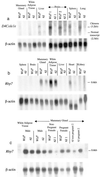
Exons of three genes were identified within the 85-kilobase tandem triplication unit of the slow Wallerian degeneration mutant mouse, C57BL/Wld(S). Ubiquitin fusion degradation protein 2 (Ufd2) and a previously undescribed gene, D4Cole1e, span the proximal and distal boundaries of the repeat unit, respectively. They have the same chromosomal orientation and form a chimeric gene when brought together at the boundaries between adjacent repeat units in Wld(S). The chimeric mRNA is abundantly expressed in the nervous system and encodes an in-frame fusion protein consisting of the N-terminal 70 amino acids of Ufd2, the C-terminal 302 amino acids of D4Cole1e, and an aspartic acid formed at the junction. Antisera raised against synthetic peptides detect the expected 43-kDa protein specifically in Wld(S) brain. This expression pattern, together with the previously established role of ubiquitination in axon degeneration, makes the chimeric gene a promising candidate for Wld. The third gene altered by the triplication, Rbp7, is a novel member of the cellular retinoid-binding protein family and is highly expressed in white adipose tissue and mammary gland. The whole gene lies within the repeat unit leading to overexpression of the normal transcript in Wld(S) mice. However, it is undetectable on Northern blots of Wld(S) brain and seems unlikely to be the Wld gene. These data reveal both a candidate gene for Wld and the potential of the Wld(S) mutant for studies of ubiquitin and retinoid metabolism.
Figures


Similar articles
-
Human homologue of a gene mutated in the slow Wallerian degeneration (C57BL/Wld(s)) mouse.Gene. 2002 Feb 6;284(1-2):23-9. doi: 10.1016/s0378-1119(02)00394-3. Gene. 2002. PMID: 11891043
-
Wallerian degeneration of injured axons and synapses is delayed by a Ube4b/Nmnat chimeric gene.Nat Neurosci. 2001 Dec;4(12):1199-206. doi: 10.1038/nn770. Nat Neurosci. 2001. PMID: 11770485
-
Age-dependent synapse withdrawal at axotomised neuromuscular junctions in Wld(s) mutant and Ube4b/Nmnat transgenic mice.J Physiol. 2002 Sep 15;543(Pt 3):739-55. doi: 10.1113/jphysiol.2002.022343. J Physiol. 2002. PMID: 12231635 Free PMC article.
-
Wallerian degeneration, wld(s), and nmnat.Annu Rev Neurosci. 2010;33:245-67. doi: 10.1146/annurev-neuro-060909-153248. Annu Rev Neurosci. 2010. PMID: 20345246 Free PMC article. Review.
-
Wld(S), Nmnats and axon degeneration--progress in the past two decades.Protein Cell. 2010 Mar;1(3):237-45. doi: 10.1007/s13238-010-0021-2. Epub 2010 Feb 23. Protein Cell. 2010. PMID: 21203970 Free PMC article. Review.
Cited by
-
In vivo nerve-macrophage interactions following peripheral nerve injury.J Neurosci. 2012 Mar 14;32(11):3898-909. doi: 10.1523/JNEUROSCI.5225-11.2012. J Neurosci. 2012. PMID: 22423110 Free PMC article.
-
Molecular chaperones protect against JNK- and Nmnat-regulated axon degeneration in Drosophila.J Cell Sci. 2013 Feb 1;126(Pt 3):838-49. doi: 10.1242/jcs.117259. Epub 2012 Dec 21. J Cell Sci. 2013. PMID: 23264732 Free PMC article.
-
Intracellular signalling pathways in dopamine cell death and axonal degeneration.Prog Brain Res. 2010;183:79-97. doi: 10.1016/S0079-6123(10)83005-5. Prog Brain Res. 2010. PMID: 20696316 Free PMC article.
-
Stimulation of nicotinamide adenine dinucleotide biosynthetic pathways delays axonal degeneration after axotomy.J Neurosci. 2006 Aug 16;26(33):8484-91. doi: 10.1523/JNEUROSCI.2320-06.2006. J Neurosci. 2006. PMID: 16914673 Free PMC article.
-
Differential proteomics analysis of synaptic proteins identifies potential cellular targets and protein mediators of synaptic neuroprotection conferred by the slow Wallerian degeneration (Wlds) gene.Mol Cell Proteomics. 2007 Aug;6(8):1318-30. doi: 10.1074/mcp.M600457-MCP200. Epub 2007 Apr 29. Mol Cell Proteomics. 2007. PMID: 17470424 Free PMC article.
References
-
- Lunn E R, Perry V H, Brown M C, Rosen H, Gordon S. Eur J Neurosci. 1989;1:27–33. - PubMed
-
- Waller A. Philos Trans R Soc London. 1850;140:423–429.
-
- Dal Canto M C, Gurney M E. Brain Res. 1995;676:25–40. - PubMed
-
- Fujimura H, Lacroix C, Said G. Brain. 1991;114:1929–1942. - PubMed
-
- Buckmaster E A, Perry V H, Brown M C. Eur J Neurosci. 1995;7:1596–1602. - PubMed
Publication types
MeSH terms
Substances
Associated data
- Actions
- Actions
- Actions
- Actions
- Actions
LinkOut - more resources
Full Text Sources
Other Literature Sources
Molecular Biology Databases

