. 2000 Oct 2;151(1):107-16.
doi: 10.1083/jcb.151.1.107.

Rapid transport of internalized P-selectin to late endosomes and the TGN: roles in regulating cell surface expression and recycling to secretory granules

Affiliations

Rapid transport of internalized P-selectin to late endosomes and the TGN: roles in regulating cell surface expression and recycling to secretory granules

K S Straley et al. J Cell Biol. .

Abstract

Prior studies on receptor recycling through late endosomes and the TGN have suggested that such traffic may be largely limited to specialized proteins that reside in these organelles. We present evidence that efficient recycling along this pathway is functionally important for nonresident proteins. P-selectin, a transmembrane cell adhesion protein involved in inflammation, is sorted from recycling cell surface receptors (e.g., low density lipoprotein [LDL] receptor) in endosomes, and is transported from the cell surface to the TGN with a half-time of 20-25 min, six to seven times faster than LDL receptor. Native P-selectin colocalizes with LDL, which is efficiently transported to lysosomes, for 20 min after internalization, but a deletion mutant deficient in endosomal sorting activity rapidly separates from the LDL pathway. Thus, P-selectin is sorted from LDL receptor in early endosomes, driving P-selectin rapidly into late endosomes. P-selectin then recycles to the TGN as efficiently as other receptors. Thus, the primary effect of early endosomal sorting of P-selectin is its rapid delivery to the TGN, with rapid turnover in lysosomes a secondary effect of frequent passage through late endosomes. This endosomal sorting event provides a mechanism for efficiently recycling secretory granule membrane proteins and, more generally, for downregulating cell surface receptors.


Figures

Figure 1
Figure 1
Trafficking of internalized LDL receptor in PC12 cells, and models for trafficking of internalized P-selectin. A, Trafficking of LDL receptor. Numbers denote the probabilities that internalized LDL receptors will enter each pathway, and are derived from the measured rates of transport to sorting endosomes (t1/2 = 3–4 min), to the TGN via late endosomes (t1/2 = 2.5–3 h), and to lysosomes (t1/2 = 20 h; Green and Kelly 1992). S.E., sorting endosome; L.E., late endosome; TGN, trans Golgi Network; Lys., lysosome; P.M., plasma membrane (recycling endosomes and endosomal carrier vesicles are not shown). B and C, Models for P-selectin traffic. The observation that P-selectin reaches lysosomes six to seven times faster (t1/2 = 3 h) than LDL receptor in PC12 cells (Green et al. 1994) can be accounted for either by sorting of P-selectin from LDL receptor only in early sorting endosomes (B), or by sorting of P-selectin from LDL receptor only in late endosomes (C). Large arrows indicate the selective step in each model. Early sorting (B) predicts rapid recycling of P-selectin through the TGN, whereas late sorting (C) precludes extensive recycling through the TGN.
Figure 2
Figure 2
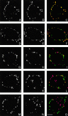
Immunofluorescence localization of organelle markers in PC12 A1-PS cells. A–C, As a control for colocalization using digital deconvolution, PC12 A1-PS cells were fixed and labeled with biotinylated anti–P-selectin COOH-terminal peptide antibody, followed by a mixture of Oregon green-avidin (A) and Texas red-avidin (B). Digital deconvolution was performed on a 25 × 0.2 μm step Z-series for each label and the images were merged (C). D–O, PC12 A1-PS cells were processed for indirect immunofluorescence microscopy and labeled with antibodies recognizing chromogranin A (E and F), synaptophysin (H and I), transferrin receptor (K and L), or CI-MPR (N and O), followed by the appropriate secondary antibody. Cells were then labeled with biotinylated anti–P-selectin COOH-terminal peptide antibody, followed by labeled avidin (D, G, J, and M). For clarity, P-selectin localization (D, G, J, and M) is shown in the green channel, whereas the endogenous proteins (E, H, K, and N) are shown in the red channel of the merged images (F, I, L, and O). Colocalization appears yellow. Bar, 2 μm.
Figure 3
Figure 3
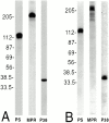
Immunoprecipitation of metabolically labeled proteins from PC12 A1-L14 cells. PC12 A1-PS cells were labeled with [3H]glucosamine (A) or [35S]amino acids (B) for 20 h before detergent lysis. P-selectin (PS), CI-MPR (MPR), and synaptophysin (p38) were sequentially immunoprecipitated from the lysates, resolved by electrophoresis on 7–15% SDS-polyacrylamide gels, and visualized by fluorography. Numbers indicate the position of molecular mass markers in kilodaltons. Virtually all of the radioactivity immunoprecipitated by each antibody migrated as a single band of the appropriate mobility.
Figure 4
Figure 4
Transport of cell surface glycoproteins to the Golgi apparatus. Cell surface glycoproteins on PC12 A1-PS cells were labeled with galactosyltransferase and UDP[3H]galactose at 4°C and recultured for the indicated intervals at 37°C. P-selectin, synaptophysin, and CI-MPR were immunoprecipitated from the cell lysates, and oligosaccharides from the immunoprecipitates were analyzed as described in the text to determine the fraction of galactose that had acquired resistance to β-galactosidase, reflecting addition of terminal sugars in the TGN, during reculture. After a lag period estimated to be ∼25–30 min, the percentage of galactose resistant to β-galactosidase on oligosaccharides from P-selectin increased with a half-time of ∼20–25 min. Each point represents the mean ± SD of three independent experiments for P-selectin and synaptophysin, and the mean of two experiments for CI-MPR, except 20-min chase (n = 1), and 6-h chase (n = 2).
Figure 5
Figure 5
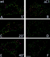
Localization of internalized Alexa488-S12 antibody and DiI-LDL. CHO cells expressing native P-selectin (A, C, and E) or P-selectin-ΔC1 (B, D, and F) were incubated in PBS/BSA containing 10 μg/ml Alexa488-S12 antibody (green channel) and 5 μg/ml DiI-LDL (red channel) for 5 min at 37°C, then in PBS/BSA for 5 min (A and B), 20 min (C and D), or 40 min (E and F). Cells were fixed and mounted for fluorescence microscopy. Stacked images were collected separately using FITC and Cy3 filters. Layers comprising 1.2–1.4-μm beginning at the base of the cells were processed by digital deconvolution and were merged (see Materials and Methods). Vesicles containing both labels appear yellow. Bar, 2 μm.
Figure 6
Figure 6
Quantitation of S12 antibody and LDL colocalization. Images such as those shown in Fig. 5 were used to determine the number of LDL-containing structures that also contained S12 antibody. Images containing a total of 15–21 cells, with an average of 926 LDL-positive vesicles, were analyzed for each data point. Numbers represent the percentage of all LDL-positive vesicles that also contained S12 antibody for the entire sample. Similar results were obtained in two independent experiments.
Figure 7
Figure 7
Selective and nonselective steps in post-Golgi traffic. Post-Golgi trafficking pathways between the TGN, secretory granules (S.G.), plasma membrane (P.M.), sorting endosomes (S.E.), recycling endosomes (R.E.), late endosomes (L.E.), and lysosomes (Lys.) are shown. Solid arrows indicate pathways requiring sorting information for efficient transport; proteins discussed in the text that are recognized in those pathways are named. Dashed arrows indicate nonselective sorting pathways (in nonpolarized cells). MPR appear to be transported selectively from late endosomes to the TGN, whereas this pathway is apparently nonselective for many proteins (see text). Furin and TGN38 are selectively retained in the TGN, accounting in part for their steady state concentration at that site.

Similar articles

Cited by

References

    1. Aniento F., Emans N., Griffiths G., Gruenberg J. Cytoplasmic dynein-dependent vesicular transport from early to late endosomes. J. Cell Biol. 1993;123:1373–1387. - PMC - PubMed
    1. Arvan P., Castle D. Sorting and storage during secretory granule biogenesislooking backward and looking forward. Biochem. J. 1998;332:593–610. - PMC - PubMed
    1. Blagoveshchenskaya A.D., Cutler D.F. Sorting to synaptic-like microvesicles from early and late endosomes requires overlapping but not identical targeting signals. Mol. Biol. Cell. 2000;11:1801–1814. - PMC - PubMed
    1. Blagoveshchenskaya A.D., Norcott J.P., Cutler D.F. Lysosomal targeting of P-selectin is mediated by a novel sequence within its cytoplasmic domain. J. Biol. Chem. 1998;273:2729–2737. - PubMed
    1. Blagoveshchenskaya A.D., Hewitt E.W., Cutler D.F. A complex web of signal-dependent trafficking underlies the triorganellar distribution of P-selectin in neuroendocrine PC12 cells. J. Cell Biol. 1999;145:1419–1433. - PMC - PubMed

Publication types