. 2000 Oct 2;151(1):69-82.
doi: 10.1083/jcb.151.1.69.

Endoplasmic reticulum degradation requires lumen to cytosol signaling. Transmembrane control of Hrd1p by Hrd3p

Affiliations

Endoplasmic reticulum degradation requires lumen to cytosol signaling. Transmembrane control of Hrd1p by Hrd3p

R G Gardner et al. J Cell Biol. .

Abstract

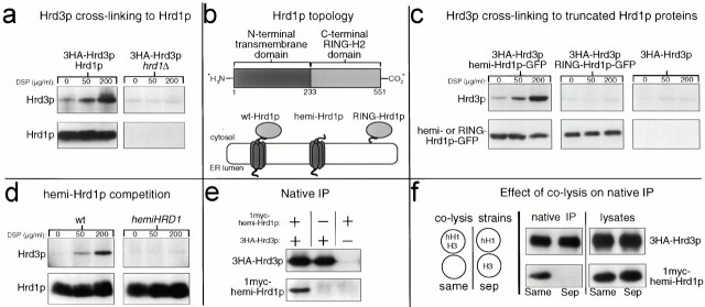
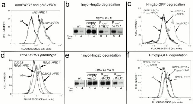
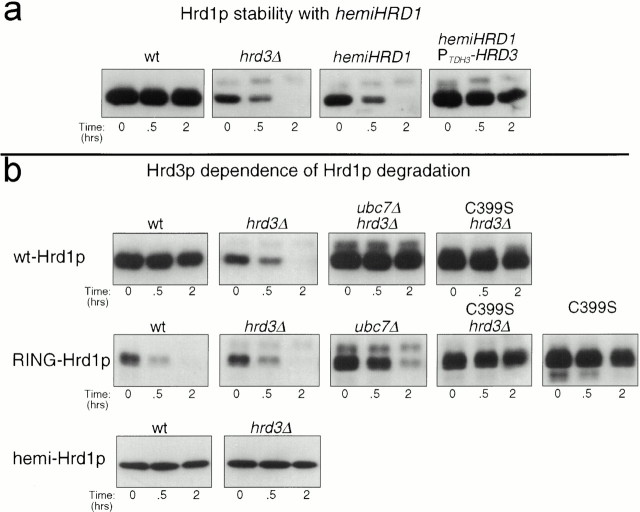
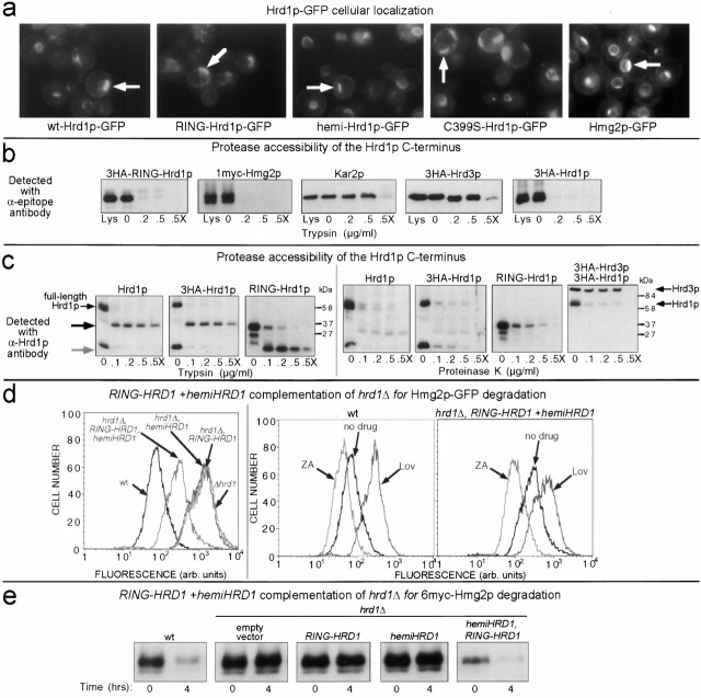
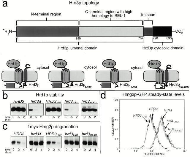
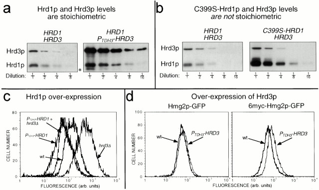
Endoplasmic reticulum (ER)-associated degradation (ERAD) is required for ubiquitin-mediated destruction of numerous proteins. ERAD occurs by processes on both sides of the ER membrane, including lumenal substrate scanning and cytosolic destruction by the proteasome. The ER resident membrane proteins Hrd1p and Hrd3p play central roles in ERAD. We show that these two proteins directly interact through the Hrd1p transmembrane domain, allowing Hrd1p stability by Hrd3p-dependent control of the Hrd1p RING-H2 domain activity. Rigorous reevaluation of Hrd1p topology demonstrated that the Hrd1p RING-H2 domain is located and functions in the cytosol. An engineered, completely lumenal, truncated version of Hrd3p functioned normally in both ERAD and Hrd1p stabilization, indicating that the lumenal domain of Hrd3p regulates the cytosolic Hrd1p RING-H2 domain by signaling through the Hrd1p transmembrane domain. Additionally, we identified a lumenal region of Hrd3p dispensable for regulation of Hrd1p stability, but absolutely required for normal ERAD. Our studies show that Hrd1p and Hrd3p form a stoichiometric complex with ERAD determinants in both the lumen and the cytosol. The HRD complex engages in lumen to cytosol communication required for regulation of Hrd1p stability and the coordination of ERAD events on both sides of the ER membrane.


Figures

Figure 1
Figure 1
Hrd1p and Hrd3p interacted via the Hrd1p NH2-terminal transmembrane domain. (a) Hrd3p cross-linked to Hrd1p. Log phase cells expressing the indicated 3HA epitope–tagged proteins were treated with the indicated concentrations of DSP, lysed, and immunoprecipitated with anti-Hrd1p antisera. Precipitated proteins were next immunoblotted with an anti-HA mAb to detect coimmunoprecipitated 3HA-Hrd3p (top) or with an anti-Hrd1p polyclonal antisera to verify equal amounts of immunoprecipitated Hrd1p (bottom). (b) Cartoon depicting Hrd1p topology. Top row, linear representation of Hrd1p domains. The start and end of each domain are indicated by the number of the corresponding amino acid residue below. Bottom row, cartoon representations of the various Hrd1p constructs including wt-Hrd1p, only the Hrd1p transmembrane domain (hemi-Hrd1p), and only the COOH-terminal RING-H2 domain (termed RING-Hrd1p). (c) Hrd3p cross-linked to hemi-Hrd1p-GFP, but not RING-Hrd1p-GFP. Cells coexpressing 3HA-Hrd3p and the indicated Hrd1p-GFP fusion were subject to the cross-linking assay using anti-GFP antisera to immunoprecipitate the Hrd1p-GFP fusions from the lysates. Precipitated proteins were immunoblotted with an anti-HA mAb to detect coimmunoprecipitated 3HA-Hrd3p (top), or with an anti-GFP mAb to detect immunoprecipitated Hrd1p-GFP fusions (bottom). (d) hemi-Hrd1p expression inhibited Hrd3p cross-linking Hrd1p. The same cross-linking assay in panel a was performed with cells expressing 3HA-Hrd3p with or without the PTDH3-hemi-HRD1 allele. To compensate for lower Hrd1p in the hemi-Hrd1p cells (see Fig. 4 a), four times less lysate was used in the control lanes so that all lanes had identical amounts of immunoprecipitated Hrd1p. (e) Native coimmunoprecipitation of hemi-Hrd1p with Hrd3p. Cells expressing either 1myc-hemi-Hrd1p, 3HA-Hrd3p, or both proteins were lysed under nondenaturing conditions and immunoprecipitated with anti-HA polyclonal antisera. Immunoprecipitates were immunoblotted with the appropriate mAb to detect coimmunoprecipitated 1myc-hemi-Hrd1p (bottom) or immunoprecipitated 3HA-Hrd3p (top). (f) hemi-Hrd1p coimmunoprecipitated with Hrd3p only when expressed in the same cell. Same experiment as in panel d, except 1myc-hemi-Hrd1p and 3HA-Hrd3p were expressed either in the same cell or in separate cells (sep). Cells expressing each protein individually were mixed in equal quantities and lysed. Cells expressing both proteins were mixed with an equal number of empty cells to ensure an equal protein load and lysed (cartoon). Each lysate (right) was immunoblotted with an anti-myc mAb to detect total 1myc-hemi-Hrd1p (bottom) or with an anti-HA mAb to detect total Hrd3p (top).
Figure 2
Figure 2
Dominant negative truncation mutants of Hrd1p. (a) hemi-Hrd1p expression stabilized Hmg2p-GFP similar to a RING-H2 deletion mutant of Hrd1p. Flow cytometry of log phase strains expressing Hmg2p-GFP (wt) or coexpressing hemi-Hrd1p or the RING-H2 motif deletion mutant of Hrd1p as indicated. The stabilizing effect of the hemi-HRD1 allele or the ΔH2-hrd1 allele was seen by the rightward shift in the fluorescence histogram compared with the wild-type strain. (b) Hrd3p overexpression reversed the hemi-HRD1 phenotype for 1myc-Hmg2p degradation. Isogenic strains expressing hemi-Hrd1p only (empty vector) or those coexpressing either the PTDH3-HRD3 or the PTDH3-HRD1 allele were assayed for 1myc-Hmg2p degradation by cycloheximide-chase assay, along with an isogenic wild-type strain (wt) without hemi-Hrd1p. Cell lysates were prepared at the indicated times after cycloheximide addition and immunoblotted using the 9E10 anti-myc mAb. (c) Hrd3p overexpression reversed the PTDH3-hemi-HRD1 phenotype for Hmg2p-GFP degradation. Flow cytometric analysis of strains expressing Hmg2p-GFP and the indicated alleles was performed as above in panel a. (d) RING-Hrd1p expression stabilized Hmg2p-GFP. Isogenic strains expressing Hmg2p-GFP and coexpressing either RING-Hrd1p (RING-HRD1), C399S-RING-Hrd1p (C399S-RING-HRD1), or full-length C399S-Hrd1p (C399S-hrd1) were analyzed by flow cytometry as in panel a. (e) Hrd3p overexpression did not suppress the RING-HRD1 phenotype for 1myc-Hmg2p degradation. Isogenic strains expressing RING-Hrd1p only (empty vector) or those coexpressing either the PTDH3-HRD3 allele or the PTDH3-HRD1 allele, were assayed for 1myc-Hmg2p degradation by cycloheximide chase assay along with a wild-type strain not expressing RING-Hrd1p. Cell lysates were prepared at the indicated times after cycloheximide addition and immunoblotted using the 9E10 anti-myc mAb. (f) Hrd3p overexpression did not reverse the PTDH3-RING-HRD1 phenotype for Hmg2p-GFP degradation. Flow cytometry of strains expressing Hmg2p-GFP and the indicated alleles was performed as in panel a.
Figure 3
Figure 3
Characteristics of Hrd1p stability and degradation. (a) hemi-Hrd1p destabilized Hrd1p by Hrd3p sequestration. Stability of 3HA-Hrd1p was assessed by cycloheximide chase assay in isogenic strains with either the hrd3Δ allele, the PTDH3-hemi-HRD1 allele, or both alleles. Lysates were prepared at the indicated times after cycloheximide addition and immunoblotted for the levels of 3HA-Hrd1p using an anti-HA mAb. (b) The degradation of full-length Hrd1p was programmed by the COOH-terminal RING-H2 domain. Degradation of Hrd1p, C399S-Hrd1p, RING-Hrd1p, C399S-RING-Hrd1p, or hemi-Hrd1p was assessed in otherwise isogenic strains with wild-type (wt), hrd3Δ, or ubc7Δ alleles by cycloheximide chase assay. The presence of the C399S mutation in the degradation substrate is indicated over the appropriate blot. Lysates were prepared at the indicated times after cycloheximide addition and immunoblotted for the levels of 3HA-Hrd1p and 3HA-RING-Hrd1p using an anti-HA mAb or for the levels of 1myc-hemi-hrd1p using the 9E10 anti-myc mAb.
Figure 4
Figure 4
Orientation of Hrd1p proteins and complementation of a hrd1Δ allele by expression of both the RING-HRD1 and hemi-HRD1 alleles. (a) RING-Hrd1p-GFP was localized to the ER. Fluorescence microscopy was performed on otherwise isogenic ubc7Δ strains expressing RING-Hrd1p-GFP, Hrd1p-GFP, hemi-Hrd1p-GFP or C399S-Hrd1p-GFP. Cells were grown to log phase and observed by fluorescence microscopy. Position of the nucleus in each cell was visualized by DAPI staining (data not shown). Arrows indicate the perinuclear, ER localization of the cellular GFP fluorescence, which in all cases is similar to the ER localization of Hmg2p-GFP (right). (b) COOH-terminal RING-H2 domain of Hrd1p was exposed to the cytosol. Protease protection assay using isolated intact microsomes from strains expressing either 3HA-RING-Hrd1p, 3HA-Hrd1p, or 3HA-Hrd3p and 1myc-Hmg2p. Microsomes were treated with the indicated concentrations of trypsin in the absence or presence of Triton X-100 (X). Levels of each protein were assessed by immunoblotting using antibodies specific for the fused epitope tags or for the native protein in the case of Kar2p. For each protein observed, total amount in the cell was comparable to that found only in the crude microsomes (compare Lys lane with 0 lane). (c) Protease protection of untagged Hrd1p. Same experiment as in panel b, using both trypsin (left) or proteinase K (right). Hrd1p was immunoblotted with antisera against the 203 residue COOH-terminal region of Hrd1p. On the left, the black arrow indicates the trypsin-insensitive fragment of full-length Hrd1p, and the gray arrow indicates the trypsin-insensitive fragment for RING-Hrd1p. For the proteinase K experiment, the blot for 3HA-Hrd1p (second from left) was stripped and reprobed with an anti-HA mAb to detect both 3HA-Hrd1p and 3HA-Hrd3p (far right). (d) Coexpression of RING-Hrd1p and hemi-Hrd1p complemented the hrd1Δ allele for mevalonate pathway–regulated Hmg2p-GFP degradation. Cells containing the hrd1Δ allele and expressing Hmg2p-GFP and either RING-Hrd1p, hemi-Hrd1p, or both were grown to log phase and analyzed by flow cytometry (left). To assess proper regulation of Hmg2p-GFP degradation, zaragozic acid (10 μg/ml) or lovastatin (25 μg/ml) was added to cells early in log phase and cells were allowed to grow for 4 h before flow cytometric analysis (middle and right). (e) Coexpression of RING-Hrd1p and hemi-Hrd1p complemented the hrd1Δ allele for constitutive 6myc-Hmg2p degradation. Cells containing the hrd1Δ allele and expressing 6myc-Hmg2p and either RING-Hrd1p, hemi-Hrd1p, or both were subject to a cycloheximide chase assay. Cell lysates were prepared at the indicated times after cycloheximide addition and immunoblotted using the 9E10 anti-myc mAb.
Figure 5
Figure 5
Hrd3p was required for ER degradation, not just Hrd1p stability. (a) Topology of Hrd3p. The start and end of each putative domain are marked with the corresponding amino acid residue number in the Hrd3p coding sequence. (b) Hrd1p stability in the presence of Hrd3p truncation mutants. Stability of 3HA-Hrd1p was assessed by cycloheximide chase assay in isogenic strains coexpressing 3HA-Hrd1p and 1myc-Hmg2p with either the normal HRD3 allele, the hrd3Δ allele, the HRD3 1–767 allele, the hrd3 1–392 allele, or the hrd3 357–833 allele. Lysates were prepared at the indicated times after cycloheximide addition and immunoblotted for the levels of 3HA-Hrd1p using an anti-HA mAb (top) or levels of 1myc-Hmg2p using an anti-myc mAb (bottom). (c) Hmg2p-GFP degradation in the presence of various Hrd3p truncation mutants. Strains expressing Hmg2p-GFP with either the normal HRD3 allele, the hrd3Δ allele, the HRD3 1–767 allele, the hrd3 1–392 allele, or the hrd3 357–833 allele were analyzed by flow cytometry.
Figure 6
Figure 6
Regulation of Hrd1p by Hrd3p results in equal stoichiometry. (a) Hrd1p and Hrd3p are expressed at equivalent levels in cells. Log phase cells coexpressing 3HA-Hrd1p from its native promoter and either 3HA-Hrd3p from its native promoter or the TDH3 promoter (PTDH3HRD3) were lysed and immunoblotted with an anti-HA mAb to determine the levels of 3HA-Hrd1p and 3HA-Hrd3p. Fractions indicate dilutions of original lysates. Asterisk denotes a proteolytic product of Hrd3p as a result of cell lysis. (b) Effect of C399S mutation on Hrd1p levels. Strains that coexpressed 3HA-Hrd3p from its native promoter and either 3HA-Hrd1p or 3HA-C399S-Hrd1p from the native HRD1 promoter were lysed and immunoblotted with an anti-HA mAb as in panel a. (c) Hrd1p overexpression enhanced Hmg2p-GFP degradation and suppressed the stabilizing effect of the hrd3Δ allele. Strains expressing Hmg2p-GFP with either the HRD3 or hrd3Δ allele and carrying the PTDH3-HRD1 allele were analyzed by flow cytometry. (d) Hrd3p overexpression slightly stabilized both Hmg2p-GFP and 6myc-Hmg2p-GFP. Hmg2p-GFP or 6myc-Hmg2p-GFP steady state levels in otherwise isogenic strains were determined by flow cytometric analysis, with or without the PTDH3-HRD3 allele.

Similar articles

Cited by

References

    1. Biederer T., Volkwein C., Sommer T. Degradation of subunits of the Sec61p complex, an integral component of the ER membrane, by the ubiquitin-proteasome pathway. EMBO (Eur. Mol. Biol. Organ.) J. 1996;15:2069–2076. - PMC - PubMed
    1. Bitter G.A., Egan K.M. Expression of heterologous genes in Saccharomyces cerevisiae from vectors utilizing the glyceraldehyde-3-phosphate dehydrogenase gene promoter. Gene. 1984;32:263–274. - PubMed
    1. Bordallo J., Wolf D.H. A RING-H2 finger motif is essential for the function of Der3/Hrd1 in endoplasmic reticulum associated protein degradation in the yeast Saccharomyces cerevisiae . FEBS Lett. 1999;448:244–248. - PubMed
    1. Bordallo J., Plemper R.K., Finger A., Wolf D.H. Der3p-Hrd1p is required for endoplasmic reticulum-associated degradation of misfolded lumenal and integral membrane proteins. Mol. Biol. Cell. 1998;9:209–222. - PMC - PubMed
    1. Chun K.T., Bar-Nun S., Simoni R.D. The regulated degradation of 3-hydroxy-3-methylglutaryl-CoA reductase requires a short-lived protein and occurs in the endoplasmic reticulum. J. Biol. Chem. 1990;265:22004–22010. - PubMed

Publication types