. 2000 Oct;20(20):7685-92.
doi: 10.1128/MCB.20.20.7685-7692.2000.

Endosomal localization and receptor dynamics determine tyrosine phosphorylation of hepatocyte growth factor-regulated tyrosine kinase substrate

Affiliations

Endosomal localization and receptor dynamics determine tyrosine phosphorylation of hepatocyte growth factor-regulated tyrosine kinase substrate

S Urbé et al. Mol Cell Biol. 2000 Oct.

Abstract

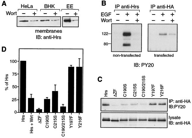
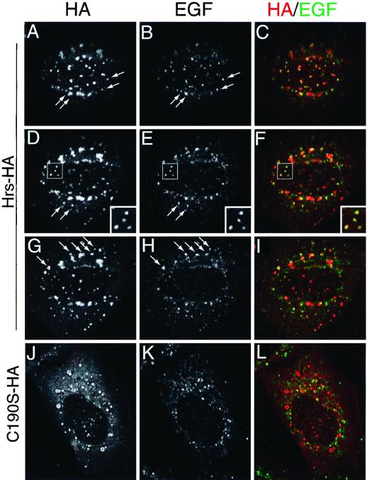
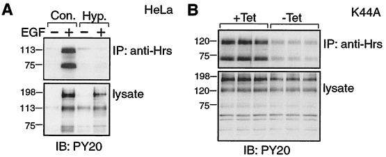
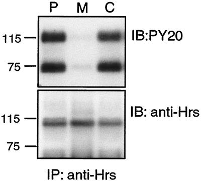
Hepatocyte growth factor-regulated tyrosine kinase substrate (Hrs) is a prominent substrate for activated tyrosine kinase receptors that has been proposed to play a role in endosomal membrane trafficking. The protein contains a FYVE domain, which specifically binds to the lipid phosphatidylinositol (PI) 3-phosphate (PI 3-P). We show that this interaction is required both for correct localization of the protein to endosomes that only partially coincides with early endosomal autoantigen 1 and for efficient tyrosine phosphorylation of the protein in response to epidermal growth factor stimulation. Treatment with wortmannin reveals that Hrs phosphorylation also requires PI 3-kinase activity, which is necessary to generate the PI 3-P required for localization. We have used both hypertonic media and expression of a dominant-negative form of dynamin (K44A) to inhibit endocytosis; under which conditions, receptor stimulation fails to elicit phosphorylation of Hrs. Our results provide a clear example of the coupling of a signal transduction pathway to endocytosis, from which we propose that activated receptor (or associated factor) must be delivered to the appropriate endocytic compartment in order for Hrs phosphorylation to occur.


Figures

FIG. 1
FIG. 1
Immunolocalization of Hrs-HA. HeLa cells were transfected with Hrs-HA and processed for immunofluorescence at 22 (A, C, D, E, and F) or 40 (B) h posttransfection. The cells were either saponin permeabilized before fixation (A, B, D, and F) or fixed immediately and permeabilized with Triton X-100 (C and E) and costained with anti-HA (shown in red) and either anti-EEA1 (A and B), anti-M6PR (D), or anti-CD63 (E) (all shown in green). Cells shown in panel C were incubated for 15 min with biotinylated transferrin (25 μg/ml in Dulbecco's modified Eagle medium) prior to fixation and costained with Oregon Green 488-labeled streptavidin. Cells shown in panel F were also cotransfected with GFP-NAGTI. All panels show a composite of a Z series taken at 260-nm intervals. Large bodies were apparent in cells expressing large amounts of Hrs, which were more frequent at longer times posttransfection (B) but could also be seen in a minority cells at earlier times (E and F). These structures contain EEA1 (B), M6PR, and transferrin receptor (data not shown) but exclude lysosomal and Golgi markers (E and F).
FIG. 2
FIG. 2
FYVE domain mutants of Hrs do not colocalize with EEA1. HeLa cells were transfected with Hrs-HA (A to C), C215S-HA (D to F), ΔZF-HA (G to I), or C190/215S-HA (J to L) and processed for immunofluorescence at 22 h posttransfection. The cells were either saponin permeabilized before fixation to highlight the particulate structures (A to F) or fixed immediately and permeabilized with Triton X-100 (G to L) and costained with anti-HA (shown in red) and anti-EEA1 (shown in green). The areas indicated with a star are shown enlarged in the insets (A through F). All panels show a single confocal section.
FIG. 3
FIG. 3
PI 3-kinase activity together with an intact FYVE domain is required for EGF-dependent tyrosine phosphorylation of Hrs. (A) Wortmannin sensitivity of membrane association. (Left) Membrane fractions were prepared from HeLa and BHK cells that had been preincubated for 15 min with (+) or without (−) 100 nM wortmannin (Wort). (Right) A fraction enriched in early endosomes (EE) by separation on a flotation gradient as described by Gorvel et al. (9) was prepared from BHK cells pretreated for 15 min with (+) or without (−) 100 nM wortmannin. In each case, 10 μg of protein was analyzed by SDS–12% PAGE, followed by transfer to nitrocellulose and blotting with anti-Hrs antibody. (B) Hrs phosphorylation is sensitive to wortmannin. Nontransfected or Hrs-HA-transfected HeLa cells were starved for 16 h in serum-free medium, then preincubated for 22 h posttransfection for 15 min with (+) or without (−) wortmannin, and stimulated for 8 min with EGF (100 ng/ml) with (+) or without (−) wortmannin (100 nM). A lysate was prepared from the nontransfected and transfected cells and subjected to immunoprecipitation with anti-Hrs or anti-HA antibodies, respectively. Phosphorylation was assessed by immunoblotting with PY20 antibody. Molecular weight markers are indicated. (C and D) An intact FYVE domain is required for efficient Hrs phosphorylation. (C) HeLa cells were transfected with either Hrs-HA, ΔZF-HA, Y197F-HA, Y216F-HA, C215S-HA, C190S-HA, or C190/215S-HA. Cells were starved for 12 h before the experiment. The cells were stimulated at 22 h posttransfection with 100 ng of EGF per ml, then lysed, and processed as described in Materials and Methods. The lysates were subjected to immunoprecipitation with anti-HA antibody, and the immunoprecipitated proteins were analyzed by immunoblotting with PY20 antibody (top). Levels of expression of the different mutants were compared by immunoblotting 25 μg of lysate with anti-HA antibody (bottom). Quantitation of collected experiments (n ≥ 3, for each mutant) is shown in panel D. Mutation of the FYVE domain reduces phosphorylation to levels similar to those seen in the presence of wortmannin (Hrs + Wrt, n = 4).
FIG. 4
FIG. 4
Internalized EGF colocalizes with Hrs-HA but not with C190S-HA. HeLa cells were transfected with Hrs-HA (A through I) or C190S-HA (J through L) and starved in serum-free medium for 12 h. Biotinylated EGF (50 ng/ml) was internalized for 8 min, and the cells were fixed with PFA and processed for immunofluorescence. The cells were labeled with anti-HA antibody, followed by a Texas red-coupled secondary antibody (A, D, G, and J; red in overlays). Internalized EGF was labeled with streptavidin coupled to Oregon Green 488 (B, E, H, and K; green in overlays). Panels A to C, D to F, and G to I show three consecutive sections taken at 260-nm intervals. Note the corresponding labeling pattern for HA and EGF in single channels (first and second columns). Arrows have been used to highlight examples of punctae associated with both labels. In panels D to F, a constellation of double-labeled punctae bounded by a box is also shown at higher magnification. Panels J to L show a single confocal section.
FIG. 5
FIG. 5
Clathrin-mediated endocytosis is required for EGF-dependent tyrosine phosphorylation of Hrs. (A) HeLa cells were starved for 16 h in serum-free medium and stimulated for 8 min with (+) or without (−) EGF (100 ng/ml) in the absence (Con.) or presence (Hyp [hypertonic medium]) of 450 mM sucrose. The cells were lysed, and proteins were immunoprecipitated with anti-Hrs antibody. Immunoprecipitated and total proteins (lysate) were analyzed by immunoblotting with PY20 antibody. (B) Stably transfected K44A cells (+Tet) were induced to express a dominant negative dynamin mutant by tetracycline withdrawal (−Tet). Cells were then starved for 16 h in serum-free medium, stimulated for 8 min with EGF (100 ng/ml), and lysed. Tyrosine-phosphorylated proteins immunoprecipitated with anti-Hrs antibody, and total proteins from triplicate experiments were analyzed as described in the legend to panel A. No phosphotyrosine signal was immunoprecipitated in the absence of EGF (data not shown). The tyrosine-phosphorylated band in the lysate just below the 198-kDa marker corresponds to EGF receptor and is decreased in the absence of clathrin-mediated endocytosis in the results shown in panels A and B.
FIG. 6
FIG. 6
Phosphorylated Hrs is predominantly cytosolic. HeLa cells were starved for 16 h in serum-free medium and stimulated for 8 min with 100 ng of EGF per ml. Postnuclear supernatant (P), membranes (M), and cytosol (C) were prepared as described in Materials and Methods. Equal amounts of protein were then subjected to immunoprecipitation with anti-Hrs antibody and analyzed by immunoblotting with PY20 antibody (top) or anti-Hrs (bottom). Molecular weight markers (prestained) are indicated.

Similar articles

Cited by

References

    1. Asao H, Sasaki Y, Arita T, Tanaka N, Endo K, Kasai H, Takeshita T, Endo Y, Fujita T, Sugamura K. Hrs is associated with STAM, a signal-transducing adaptor molecule. J Biol Chem. 1997;272:32785–32791. - PubMed
    1. Bankaitis V A, Johnson L M, Emr S D. Isolation of yeast mutants defective in protein targeting to the vacuole. Proc Natl Acad Sci USA. 1986;83:9075–9079. - PMC - PubMed
    1. Bean A J, Seifert R, Chen Y A, Sacks R, Scheller R H. Hrs-2 is an ATPase implicated in calcium-regulated secretion. Nature. 1997;385:826–829. - PubMed
    1. Burd C G, Babst M, Emr S D. Novel pathways, membrane coats and PI kinase regulation in yeast lysosomal trafficking. Semin Cell Dev Biol. 1998;9:527–533. - PubMed
    1. Burd C G, Emr S D. Phosphatidylinositol(3)-phosphate signaling mediated by specific binding to RING FYVE domains. Mol Cell. 1998;2:157–162. - PubMed

Publication types

MeSH terms

Substances