. 2000 Sep 15;20(18):6898-906.
doi: 10.1523/JNEUROSCI.20-18-06898.2000.

Cathepsin D deficiency induces lysosomal storage with ceroid lipofuscin in mouse CNS neurons

Affiliations

Cathepsin D deficiency induces lysosomal storage with ceroid lipofuscin in mouse CNS neurons

M Koike et al. J Neurosci. .

Abstract

Cathepsin D-deficient (CD-/-) mice have been shown to manifest seizures and become blind near the terminal stage [approximately postnatal day (P) 26]. We therefore examined the morphological, immunocytochemical, and biochemical features of CNS tissues of these mice. By electron microscopy, autophagosome/autolysosome-like bodies containing part of the cytoplasm, granular osmiophilic deposits, and fingerprint profiles were demonstrated in the neuronal perikarya of CD-/- mouse brains after P20. Autophagosomes and granular osmiophilic deposits were detected in neurons at P0 but were few in number, whereas they increased in the neuronal perikarya within days after birth. Some large-sized neurons having autophagosome/autolysosome-like bodies in the perikarya appeared in the CNS tissues, especially in the thalamic region and the cerebral cortex, at P17. These lysosomal bodies occupied the perikarya of almost all neurons in CD-/- mouse brains obtained from P23 until the terminal stage. Because these neurons exhibited autofluorescence, it was considered that ceroid lipofuscin may accumulate in lysosomal structures of CD-/- neurons. Subunit c of mitochondrial ATP synthase was found to accumulate in the lysosomes of neurons, although the activity of tripeptidyl peptidase-I significantly increased in the brain. Moreover, neurons near the terminal stage were often shrunken and possessed irregular nuclei through which small dense chromatin masses were scattered. These results suggest that the CNS neurons in CD-/- mice show a new form of lysosomal accumulation disease with a phenotype resembling neuronal ceroid lipofuscinosis.


Figures

Fig. 1.
Fig. 1.
Extracellular recordings obtained from the CA1 and CA3 regions of hippocampal slices obtained from a cathepsin D-deficient mouse at P18. intact, Control recordings;a, recordings after cutting the connection between the CA1 and the CA3 subfields; a+b, recordings after an additional cutting of the connection between the CA1 and the dentate gyrus (DG). In situations of a anda+b, spontaneous burst discharges were detected in the CA3 region.
Fig. 2.
Fig. 2.
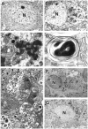
Electron micrographs of autophagosome/autolysosome-like bodies in the neuronal perikarya.A, A cerebral cortex neuron obtained from a control littermate mouse at P23. B, C, A cerebral cortex neuron obtained from a cathepsin D-deficient mouse at P23. A low-power view clearly shows the presence of numerous autophagosome/autolysosome-like bodies in the neuronal perikarya (B), and a high-power view demonstrates the bodies containing granular osmiophilic deposits (GO) and a part of the cytoplasm with or without fingerprint-like myelin figures (AP) (C). Thearrow indicates a body with part of the cytoplasm, which is encircled by double-layered membranes resembling the endoplasmic reticulum. D, A fingerprint profile appears in the neuronal perikaryon of a Purkinje cell (D).E, Neuronal cell bodies in the CA3 region of the hippocampus obtained from a cathepsin D-deficient mouse brain at P25. The neuronal cell perikarya are completely filled with membrane-bounded compartments having dense amorphous materials, part of the cytoplasm, and fingerprint-like myelin figures (MF). A neuronal cell is seen, possessing a shrunken nucleus with small chromatin masses dispersed in the karyoplasm and is encircled with cytoplasmic processes (arrowheads) of a microglial cell (M). F,G, Appearance of dense granular deposits in the perikarya of CA3 pyramidal neurons in the hippocampus obtained from cathepsin D-deficient mice at P1 (F) and P17 (G). The dense granular bodies (arrows) appear in the neuronal perikarya at P1 but are fewer in number, whereas they increase in number at P17. The matrix of the bodies is similar to those seen after P20 (C), except for fingerprint-like figures, which are rarely detectable at P1. N, Nucleus. Scale bars:A, B, E, F,G, 1.5 μm; C, D, 0.25 μm.
Fig. 3.
Fig. 3.
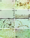
Immunostaining for subunits c and β (Sβ) of mitochondrial F1F0ATPase at P23.A–D, Immunoreactivity for subunit c is large-granular and intensely localized to neuronal perikarya in the CA1 layer (B) and the outer and inner nuclear (granular) layers (ONL and INL) and ganglion cells (GL) of the retina (D) in the CD−/− mouse (−/−), whereas no immunoreactivity is detected in the same regions of the control (+/−) (A,C). E–G, Positive staining of subunit c is large-granular in Kupffer cells (arrows) and fine-granular in hepatocytes (E). Immunodeposits for subunit c are present in epithelial cells of renal tubules (F) and cardiac muscular cells (G). Fine-granular immunoreactivity for subunit β is clearly localized to the CA1 neuronal cell bodies (H). Scale bars, 20 μm.
Fig. 4.
Fig. 4.
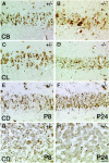
Immunohistochemical demonstration of cathepsin B (CB), cathepsin L (CL), and cathepsin D (CD) in various tissue cells. A–D, Immunostaining of cathepsins B and L in CA1 pyramidal layers of control littermate (+/−) and cathepsin D-deficient (−/−) mice at P26.A, B, Fine-granular immunodeposits for cathepsin B are well localized to the perikarya of +/− pyramidal neurons (A), whereas the immunodeposits are coarse and in some cases large-granular in the perikarya of −/− neurons (B). C, D, No clear-cut difference is detected in the immunoreactivity for cathepsin L between pyramidal neurons obtained from control and knockout mice.E–H, Immunostaining of cathepsin D in CA1 pyramidal layers (E, F) and liver tissues (G, H) obtained from the control CD+/− mice at P8 (E, G) and P24 (F, H). Immunodeposits for cathepsin D are distinct in both tissue cells at each stage, and no clear-cut differences are detected in distribution patterns in these tissues between the two stages, respectively. Scale bar, 20 μm.
Fig. 5.
Fig. 5.
Immunocytochemical staining of cathepsin B and subunit c of mitochondrial F1F0ATPase in neuronal cell bodies of the cerebral cortex from cathepsin D-deficient (D–F) and control littermate (A–C) mice at P23, using the cryothin section immunogold method. A, D, Cathepsin B. Gold labeling is clearly localized to electron-lucent lysosomes in the control (A), whereas it is detected in membrane-bound compartments with electron-dense materials in a deficient mouse (D). B,E, Subunit c. Gold particles label only the mitochondrial inner membrane in the control mouse (B), whereas they are associated with both the inner membrane of intact mitochondria and the membrane-bound compartments with dense materials in the knockout mouse (E). C, F, Double immunostaining of cathepsin B (gold particles, 5 nm in diameter) and subunit c (gold particles, 15 nm in diameter). In the control, small gold particles for cathepsin B clearly label an electron-lucent lysosome, whereas large particles for subunit c are localized to the mitochondrial inner membrane (C). In the deficient mouse, small and large gold particles are colocalized in electron-dense compartments (F). Scale bars, 0.25 μm.
Fig. 6.
Fig. 6.
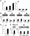
Biochemical analyses of lysosomal proteinases and storage proteins in CD−/− mouse brains. A, Proteolytic activity of cathep-sins B (left) and cathepsin L (right) in brain extracts from cathepsin D-deficient (−/−) and control littermate (+/−) mice at P17, P21, and P23. The cathepsin B activity is significantly increased in knockout mouse brains at P23, compared with that in the control, whereas the differences in the activity are not significant at P17 and P21 between the two groups, respectively. No clear-cut difference is detected in the cathepsin L activity between the −/− and +/− brains on P17, P21, and P23. The activity was expressed as nanomole per minute per milligram of protein (n = 3 in each case).B, Immunoblot analyses of cathepsins B (CB) and L (CL) and subunits c (Sc) and β (Sβ) from cathepsin D-deficient (−/−) and control littermate (+/−) mouse brains. Immunoreactive bands for cathepsin B (single chain form) are distinctly augmented in the −/− mouse brain at P23, whereas no difference is seen between the two groups at P17. Protein bands immunostained for cathepsin L (single chain form) show similar densities between −/− and +/− mouse brains at P17 and P23. Immunoreactive bands for subunit c are much more increased in the −/− mouse brain at P23 than in the +/− mouse brain, whereas no difference is detected in immunoreactive bands for subunit β between the two groups at both P17 and P23. InB, protein makers are on the left side. In each lane, 20 μg of protein was applied. C, Quantification of B. The blotted densities of each protein were measured with a Scanning Imager. D, The activity of TPP-I in CD−/− and control littermate brains at P23. The activity was expressed as nanomole per minute per milligram of protein (n = 3 for each case). E, Immunoblot analysis of TPP-I in CD−/− and control littermate mouse brains at P23. Protein maker is on the left side.F, Quantification of E. G, Immunoblot analysis of subunit c in extracts of liver, kidney, and heart tissues of CD−/− and littermate control mice at P23. H, Quantification of G. The protein amounts applied inE and G and the measurement of protein bands in F and H followedB and C, respectively. The density of each protein band in control brain extracts was estimated as 100%.

Similar articles

Cited by

References

    1. Banay-Schwartz M, Bracco F, DeGuzman T, Lajtha A. Developmental changes in the breakdown of brain tubulin by cerebral cathepsin D. Neurochem Res. 1983;8:51–61. - PubMed
    1. Banay-Schwartz M, Dahl D, Hui KS, Lajtha A. The breakdown of the individual neurofilament proteins by cathepsin D. Neurochem Res. 1987;12:361–367. - PubMed
    1. Bando Y, Kominami E, Katunuma N. Purification and tissue distribution of rat cathepsin L. J Biochem (Tokyo) 1986;100:35–42. - PubMed
    1. Barrett AJ, Kirsche H. Cathepsin B, cathepsin H, cathepsin L. Methods Enzymol. 1981;80:535–561. - PubMed
    1. Bednarski E, Ribak CE, Lynch G. Suppression of cathepsins B and L causes a proliferation of lysosomes and the formation of meganeurites in hippocampus. J Neurosci. 1997;17:4006–4021. - PMC - PubMed

Publication types

MeSH terms

LinkOut - more resources