. 2000 Sep 15;19(18):4885-94.
doi: 10.1093/emboj/19.18.4885.

Ric1p and Rgp1p form a complex that catalyses nucleotide exchange on Ypt6p

Affiliations

Ric1p and Rgp1p form a complex that catalyses nucleotide exchange on Ypt6p

S Siniossoglou et al. EMBO J. .

Abstract

Cells lacking the GTPase Ypt6p have defects in intracellular traffic and are temperature sensitive. Their growth is severely impaired by additional mutation of IMH1, which encodes a non-essential Golgi-associated coiled-coil protein. A screen for mutants that, like ypt6, specifically impair the growth of imh1 cells led to the identification of RIC1. Ric1p forms a tight complex with a previously uncharacterized protein, Rgp1p. The Ric1p-Rgp1p complex binds Ypt6p in a nucleotide-dependent manner, and purified Ric1p-Rgp1 stimulates guanine nucleotide exchange on Ypt6p in vitro. Deletion of RIC1 or RGP1, like that of YPT6, blocks the recycling of the exocytic SNARE Snc1p from early endosomes to the Golgi and causes temperature-sensitive growth, but this defect can be relieved by overexpression of YPT6. Ric1p largely colocalizes with the late Golgi marker Sec7p. Ypt6p shows a similar distribution, but this is altered when RIC1 or RGP1 is mutated. We infer that the Ric1p-Rgp1p complex serves to activate Ypt6p on Golgi membranes by nucleotide exchange, and that this is required for efficient fusion of endosome-derived vesicles with the Golgi.


Figures

None
Fig. 1. Functional interaction between IMH1 and RIC1. (A) Synthetic lethal interaction between imh1Δ and ric1 mutants. The sl233 mutant was transformed with the indicated Ycplac111 plasmid (pLEU), either with no insert, or carrying IMH1, RIC1 or a RIC1–GFP fusion. Transformants were grown on plates containing 5-FOA for 3 days at 30°C. (B) Growth properties of the ric1Δ deletion mutant (ric1). Cells from a ric1Δ strain transformed either with an empty LEU2 plasmid or with the same plasmid carrying the wild-type RIC1 gene were diluted in YEPD, spotted on to selective (–Leu) plates and grown at 30 and 37°C for 2 days.
None
Fig. 2. Ric1p forms a complex with Rgp1p in vivo. (Left) Affinity purification of Rgp1p–PtA and Ric1p–PtA from rgp1Δ and ric1Δ strains, respectively. The purified proteins eluted from the IgG–Sepharose column with low pH were analysed by SDS–PAGE and Coomassie staining. The position of the protein A fusions and the copurifying proteins, identified as Ric1p and Rgp1p by mass spectrometry, are indicated. Molecular mass standards are indicated on the left side of the gel. (Right) The IgG–Sepharose beads carrying the Ric1p–PtA and Rgp1p–PtA fusions were digested by TEV protease and the released fractions containing the cleaved Ric1p and Rgp1p were analysed by SDS–PAGE and Coomassie staining. Molecular mass standards were resolved on the left side of the gel. The position of TEV protease is indicated; the asterisk indicates immunoglobulin heavy chains derived from the affinity column.
None
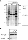
Fig. 3. The Ric1p–Rgp1p complex binds to Ypt6p in vivo in a nucleotide-dependent manner. (A) Affinity purification of a PtA–Ypt6p fusion and identification of proteins that bind to it. A detergent-solubilized supernatant from a ypt6Δ strain expressing a PtAYPT6 fusion, in the absence (–) or presence of GTP-γS, was loaded onto an IgG–Sepharose column and the purified fusion protein eluted at low pH. Samples from the supernatant loaded on to the column (S) and the eluted PtA–Ypt6p (E), (3700-fold concentrated as compared with the supernatant), were analysed by SDS–PAGE followed by Coomassie staining. The position of PtA–Ypt6p and the copurifying Ric1p and Rgp1p, identified by mass spectrometry, are indicated by asterisks. (B) PtA–Ypt6p was affinity purified from a ypt6Δ ric1Δ double deletion strain co-expressing a Ric1p–GFP fusion from a Ycplac33-URA3 plasmid, in the absence of nucleotide, or in the presence of GDP or GTP-γS. The eluates from the IgG–Sepharose column were analysed by SDS–PAGE followed by western blotting using anti-protein A or anti-GFP antibodies.
None
Fig. 4. The Ric1p–Rgp1p complex stimulates GDP exchange on Ypt6p. (A) Ypt6p and the Ric1p–Rgp1p complex were purified from yeast as described in Materials and methods. Ninety picomoles of Ypt6p pre-incubated with [3H]GDP were incubated with the indicated amounts of Ric1p–Rgp1p at 24°C, and protein-bound radioactivity was determined at intervals by filtration of aliquots through nitrocellulose. Background binding (with bovine serum albumin in place of Ypt6p) was equivalent to ∼10% of the total and has not been subtracted. (B) Ypt1p preloaded with [3H]GDP was incubated with the Ric1p–Rgp1p complex at 24°C and nucleotide exchange was determined as in (A). (C) Ypt6p preloaded with [3H]GDP was incubated independently with either Ric1p or Rgp1p or the Ric1p–Rgp1p complex at 24°C. Nucleotide exchange was determined as in (A).
None
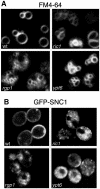
Fig. 5. Defects in vacuolar morphology and GFP–Snc1p recycling in ric1Δ, rgp1Δ and ypt6Δ mutants. (A) The ric1Δ, rgp1Δ and ypt6Δ deletion mutants and a wild-type strain were grown at 30°C to exponential phase, labelled with the endocytic tracer dye FM4-64 for 15 min and chased for 1 h. Note that only vacuoles are visible under these conditions. (B) The deletion mutants were transformed with a plasmid expressing GFP–Snc1p. Transformants were grown to exponential phase at 30°C and inspected by confocal microscopy.
None
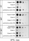
Fig. 6. YPT6 suppresses the temperature-sensitive growth defect of the ric1, rgp1 and ric1 rgp1 deletion mutants. The ric1, rgp1 and ric1 rgp1 deletion mutants were transformed with an empty centromeric plasmid (YCplac111), YCp plasmid(s) carrying the complementing gene(s) as indicated, or with a centromeric plasmid over-expressing PtA–YPT6 [pUN100-(PNOP1)PtA-YPT6]. Transformants were spotted onto selective (–Leu) plates and incubated for 2 days at 37°C.
None
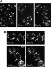
Fig. 7. Localization of Ric1p–GFP, Rgp1p–GFP and GFP–Ypt6p fusions. (ARIC1–GFP, RGP1–GFP and GFP–YPT6 were expressed from centromeric plasmids under the control of their own promoters, in the corresponding deletion mutants. (B) The Ric1p–GFP and GFP–Ypt6p strains were mated with a strain bearing a chromosomal copy of YFP–SEC7, under the control of the TPI1 promoter. YFP and GFP images of diploid cells were obtained as described in Materials and methods. Arrows indicate identical positions in the two images.
None
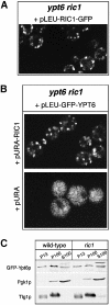
Fig. 8. Subcellular distribution of GFP–Ypt6p is drastically altered in ric1 deletion mutants. (A) The ypt6 ric1 double deletion mutant was transformed with a centromeric plasmid expressing Ric1p–GFP and examined under the confocal microscope. (B) A ypt6 ric1 double deletion mutant transformed with the indicated plasmids was examined under the MRC-600 confocal microscope. (C) The GFP–YPT6 plasmid was transformed into ypt6 cells, thus complementing them (‘wild-type’) or into the double ypt6 ric1 deletion mutant (‘ric1’) and fractionated into 13 000 and 100 000 g pellets (P13 and P100, respectively) and 100 000 g supernatant (S100). Equivalent amounts of each fraction were analysed by SDS–PAGE followed by western blotting with anti-GFP, anti-Pgk1p and anti-Tlg1p antibodies. The upper band visible in the S100 fraction probed with anti-GFP is a cross-reacting endogenous protein.
None
Fig. 9. Multiple sequence alignment of the C-terminal domain of Ric1p with related sequences found in proteins from other species: Drosophila melanogaster (DDBJ/EMBL/GenBank accession No. AF181626.1); Homo sapiens (AB037853.1; brackets in the residue number indicate that it is a partial sequence); Caenorhabditis elegans (Q09417); S.pombe (AL133361.1). Sequences were aligned with Clustal W1.8 (Thompson et al., 1994) and displayed using the program Boxshade (http://www.ch.embnet.org/software/BOX_form.html).

Similar articles

Cited by

References

    1. Aguilera A., Moskowitz,P. and Klein,H.L. (1990) Molecular analysis of RGP1, a new yeast gene required for proper mitotic growth. Nucleic Acids Res., 18, 1064. - PMC - PubMed
    1. Barr F.A. (1999) A novel Rab6-interacting domain defines a family of Golgi-targeted coiled-coil proteins. Curr. Biol., 9, 381–384. - PubMed
    1. Bensen E.S., Costaguta,G. and Payne,G.S. (2000) Synthetic genetic interactions with temperature-sensitive clathrin in Saccharomyces cerevisiae. Roles for synaptojanin-like Inp53p and dynamin-related Vps1p in clathrin-dependent protein sorting at the trans-Golgi network. Genetics, 154, 83–97. - PMC - PubMed
    1. Cao X. and Barlowe,C. (2000) Asymmetric requirements for a Rab GTPase and SNARE proteins in fusion of COPII vesicles with acceptor membranes. J. Cell Biol., 149, 55–66. - PMC - PubMed
    1. Cao X., Ballew,N. and Barlowe,C. (1998) Initial docking of ER-derived vesicles requires Uso1p and Ypt1p but is independent of SNARE proteins. EMBO J., 17, 2156–2165. - PMC - PubMed

Publication types

MeSH terms