. 2000 Sep;124(1):71-84.
doi: 10.1104/pp.124.1.71.

A maize vacuolar invertase, IVR2, is induced by water stress. Organ/tissue specificity and diurnal modulation of expression

Affiliations

A maize vacuolar invertase, IVR2, is induced by water stress. Organ/tissue specificity and diurnal modulation of expression

J Y Kim et al. Plant Physiol. 2000 Sep.

Abstract

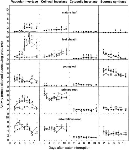
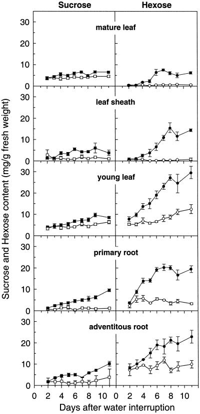
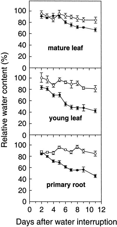
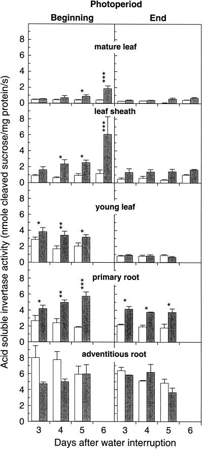
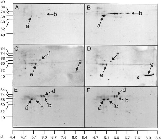
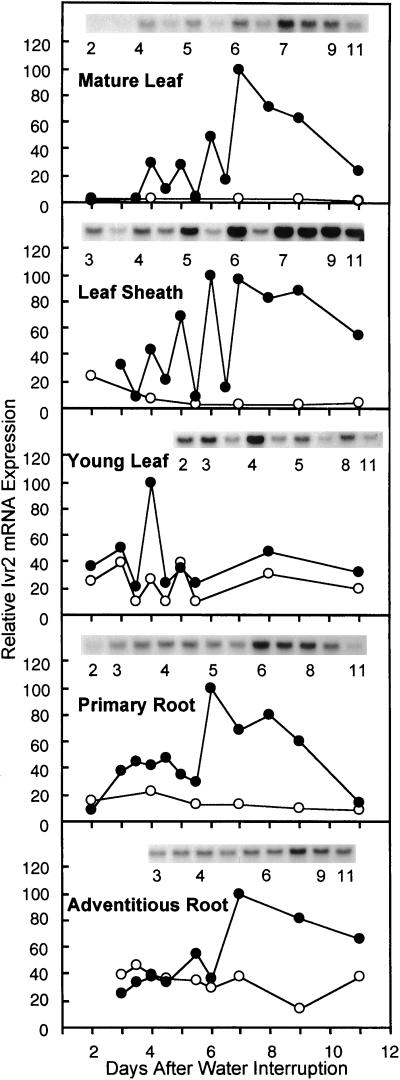
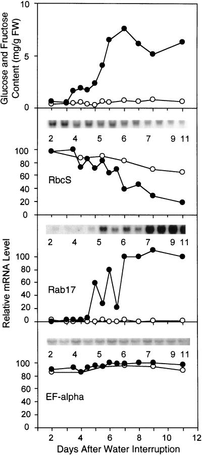
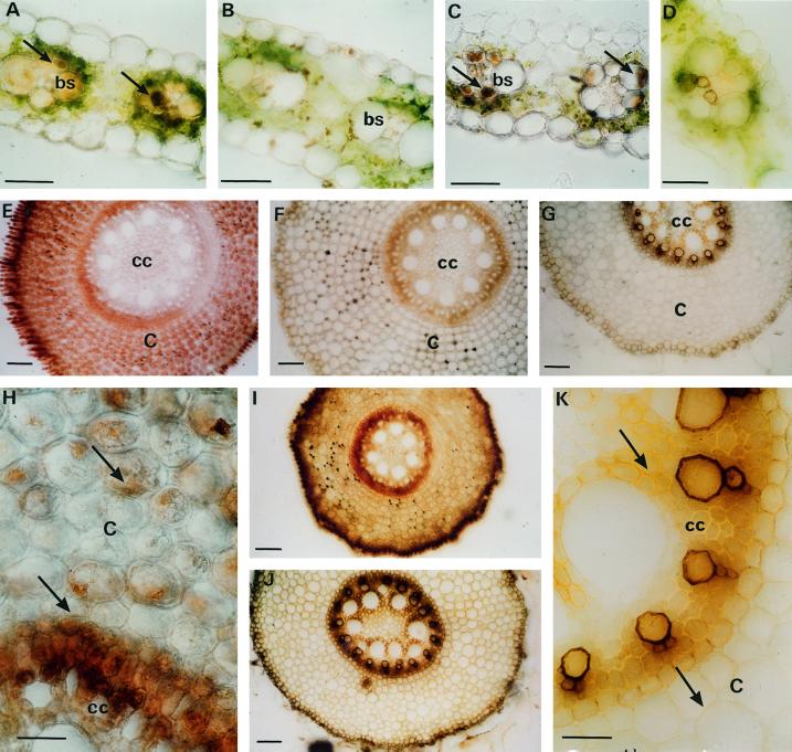
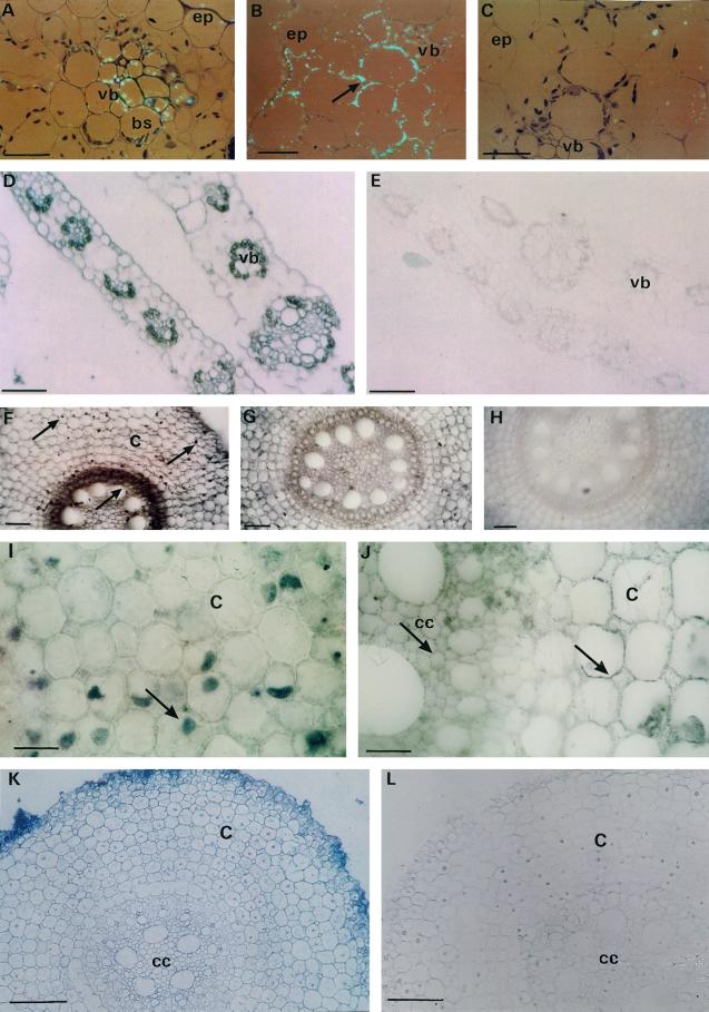
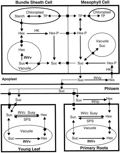
The expression of invertases was analyzed in vegetative organs of well-watered and water-stressed maize (Zea mays) plants. Early changes in sucrose metabolism and in acid soluble invertase expression were observed in vegetative sink and source organs under mild water stress. The organ-specific induction of acid invertase activity was correlated with an increase in the Ivr2 gene transcripts and in the vacuolar invertase proteins. In addition diurnal changes in activity and Ivr2 transcripts for vacuolar invertase were noted in shoots. Hexoses (glucose and fructose) accumulated in all organs examined from water-stressed plants. In situ localization studies showed that glucose accumulation, vacuolar invertase activity, invertase protein, and the Ivr2 transcripts colocalized specifically in bundle sheath and vascular tissue cells of mature stressed leaf; in primary roots the stress-induced increase of Ivr2 transcripts was detected only in root tips. Based on these results different regulatory roles are proposed in sink and source organs for the stress induced Ivr2 vacuolar invertase.


Figures

Figure 1
Figure 1
Effect of drought on acid soluble (vacuolar), acid insoluble (cell wall), and neutral (cytosolic) invertase activity, and Susy activity in five vegetative organs from well-watered and water-stressed maize plants. Samplings were carried out at 9 am (3 h after light on) from d 2 to 11 after water interruption. White symbols, Control plants; black symbols, water-stressed plants. Mean ± se from four independent determinations. Control/stress Student's comparison: *, P < 0.05 significant; **, P < 0.01 very significant; ***, P < 0.001 highly significant.
Figure 2
Figure 2
Effect of drought on Suc and hexoses (Glc and Fru) concentrations on a fresh weight basis in five vegetative organs from well-watered and water-stressed maize plants. Same samples as in Figure 1.
Figure 3
Figure 3
Variation of RWC in mature leaf, young leaf, and root after water deprivation.
Figure 4
Figure 4
Diurnal variation of acid soluble invertase (vacuolar) in five vegetative organs of watered (white bars) and water-stressed plants (gray bars). Samplings were carried out at the beginning, 9 am (3 h after light on), and at the end, 9 pm, of the photoperiod (1 h before light off). Symbols for statistical comparisons between stressed and control plants as in Figure 1.
Figure 5
Figure 5
Immunodetection from two-dimensional gels of vacuolar and cell wall invertase in mature leaf (A–D) and primary root (E and F) from well-watered (left column: A, C, and E) or water-stressed plants (right column: B, D, and F) for 7 d . Homologous groups of spots were designated a, b, c, and d for vacuolar invertase antibodies and e, f, and g for cell wall invertase antibodies. Antiserums raised against an IVR2 oligopeptide (A and B for mature leaf; E and F for primary root) and a cell wall invertase peptide (C and D for mature leaf) were used for invertase immunodetection from crude protein extracts (50 μg) in mature leaf and root. All sampling was done at 9 am. Comparison among loaded protein quantities was carried out from Coomassie Blue gel staining (data not shown). To measure pI, four gels were cut into 15 fragments and four gel fragments were incubated together in 1 mL of distilled water overnight; pH was measured from these eluted solutions.
Figure 6
Figure 6
Time course of Ivr2 transcript accumulation in water-stressed (black symbols) and watered plants (white symbols) in five vegetative organs. Total RNA samples (20 μg/lane) extracted from organs were hybridized with an Ivr2 cDNA probe, radioactivity was stripped, and the membrane was rehybridized with EF-α cDNA. The hybridization signal intensity was measured with an image analyzer and the relative Ivr2 mRNA expression was normalized from the average between the intensity of EF-α and of 18S rRNA staining.
Figure 7
Figure 7
Time course of hexose accumulation in mature leaf in comparison with the expression of a hexose repressed gene (RbcS), an ABA-responsive gene (Rab17) and a constitutively expressed gene (EF-α), under watered and water-stressed conditions. The membrane for mature leaf used in Figure 6 was rehybridized with 32P-labeled Rab17, RbcS, and EF-α cDNA probes.
Figure 8
Figure 8
In situ histochemical staining for Glc and invertase activity in hand-sliced fresh mature leaf (A–D) and primary root (E–K) sampled on d 7 from well-watered and water-stressed plants. A, Stressed mature leaf; Glc localized within bundle sheath and vascular bundle cells (arrows). B, Watered mature leaf; acid invertase activity only detected in cell walls. C, Stressed mature leaf; acid invertase activity localized within bundle sheath and vascular bundle cells (arrows). D, Stressed mature leaf; histochemical control (tissue flushed, no sugar addition). E, Stressed root; strong signal for Glc in cytosol of cortex and outer central cylinder. F, Watered root; weak signal for Glc in both cell walls and cytosol of cortex and central cylinder. G, Stressed root; negative histochemical control showing artifactual xylem staining. H and I, Stressed root; strong intracellular acid invertase activity in epidermis and central cylinder (arrow), weaker signal in cortex (arrow). J and K, Watered root; moderate cell wall signal and weak intracellular signal for acid invertase activity in epidermis, cortex and central cylinder (arrow). bs, Bundle sheath; C, cortex; cc, central cylinder. Bars in A through D, 50 μm; E through G and I and J, 100 μm; H and K, 25 μm.
Figure 9
Figure 9
In situ immunolocalization of invertase protein (A–C and F–J) and in situ hybridization of Ivr2 mRNA transcripts (D–E and K–L) in mature leaves (A–E) and roots (F–L) sampled on d 7 from water-stressed plants. Epipolarization optics of water-stressed mature leaf section exposed to A, Vacuolar invertase antibodies showing intracellular labeling in the vascular bundle; B, cell wall invertase antibodies showing cell wall labeling (arrow); and C, nonimmune serum. D, leaf section of water-stressed mature leaf hybridized to Ivr2 probe, in antisense orientation; E, leaf hybridized to Ivr2 probe in sense orientation. F and I, Root section of water-stressed plants incubated with vacuolar invertase antibodies showing intracellular localization in cortex and central cylinder (arrows); G and J, water-stressed roots exposed to anti-cell wall invertase serum showing immunopositive cell walls in all root tissue (arrows in J); H, root section incubated with nonimmune serum yielded no labeling; K, root section of water-stressed plants hybridized to Ivr2 antisense probe; L, root hybridized to sense probe. c, Cortex cells; cc, central cylinder; ep, epidermis; bs, bundle sheath cells; vb, vascular bundle cells. Bars in A through C, I, and J, 25 μm; D through H, K, and L, 100 μm.
Figure 10
Figure 10
Flowchart of Suc and hexose mobilization among different organs, cells, and compartments in maize. Suc is synthesized in leaf mesophyll cells during the daytime from current photosynthates and in bundle sheath cells at night from starch hydrolysis products. Suc is loaded into phloem or temporally stored in the vacuole and remobilized. Induction of invertase activity in bundle sheath cells during the night in water-stressed maize slows down the Suc export and produces a Suc degradation/synthesis futile cycle. In young leaves Suc is imported without hydrolysis, whereas it is imported with or without hydrolysis in roots. Black squares represent plasmodesmata and black circles represent membrane translocation. Hex, hexoses (Glc or Fru); Hex-P, hexose phosphates (Glc- or Fru-Phosphate); TP, triose-P; INVv, INVc, and INVcw: vacuolar, cytosolic, and cell wall-bound invertases respectively; SPS, Suc-P synthase.

Similar articles

Cited by

References

    1. Bohnert HJ, Nelson DE, Jensen RG. Adaptations to environmental stresses. Plant Cell. 1995;7:1099–1111. - PMC - PubMed
    1. Broglie R, Corruzi G, Keith B, Chua N. Molecular biology of C4 photosynthesis in Zea mays: differential localization of proteins and mRNAs in two cell types. Plant Mol Biol. 1984;3:431–444. - PubMed
    1. Büssis D, Heineke D, Sonnewald U, Willmitzer L, Raschke K, Heldt HW. Solute accumulation and decreased photosynthesis in leaves of potato plants expressing yeast-derived invertase either in the apoplast, vacuole or cytosol. Planta. 1997;202:126–136. - PubMed
    1. Chen MH, Liu LF, Chen YR, Wu HK, Yu SM. Expression of a-amylases, carbohydrate metabolism, and autophagy in cultured rice cells is coordinately regulated by sugar nutrient. Plant J. 1994;6:625–636. - PubMed
    1. Cheng WH, Taliercio EW, Chourey PS. The Miniature 1 seed locus of maize encodes a cell-wall invertase required for normal development of endosperm and maternal cells in the pedicel. Plant Cell. 1996;8:971–973. - PMC - PubMed

Publication types

MeSH terms

LinkOut - more resources