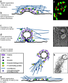
Molecular links between endocytosis and the actin cytoskeleton
- PMID: 10974009
- PMCID: PMC2175242
- DOI: 10.1083/jcb.150.5.f111
Molecular links between endocytosis and the actin cytoskeleton
Figures

Similar articles
-
Pathways linking endocytosis and actin cytoskeleton in mammalian cells.Exp Cell Res. 2001 Nov 15;271(1):45-56. doi: 10.1006/excr.2001.5369. Exp Cell Res. 2001. PMID: 11697881 Review. No abstract available.
-
Mammalian Abp1, a signal-responsive F-actin-binding protein, links the actin cytoskeleton to endocytosis via the GTPase dynamin.J Cell Biol. 2001 Apr 16;153(2):351-66. doi: 10.1083/jcb.153.2.351. J Cell Biol. 2001. PMID: 11309416 Free PMC article.
-
Endocytosis, actin cytoskeleton, and signaling.Plant Physiol. 2004 Jul;135(3):1150-61. doi: 10.1104/pp.104.040683. Plant Physiol. 2004. PMID: 15266049 Free PMC article. Review. No abstract available.
-
Regulation of the actin cytoskeleton-plasma membrane interplay by phosphoinositides.Physiol Rev. 2010 Jan;90(1):259-89. doi: 10.1152/physrev.00036.2009. Physiol Rev. 2010. PMID: 20086078 Review.
-
Synaptic vesicle endocytosis: calcium works overtime in the nerve terminal.Mol Neurobiol. 2000 Aug-Dec;22(1-3):115-28. doi: 10.1385/MN:22:1-3:115. Mol Neurobiol. 2000. PMID: 11414275 Review.
Cited by
-
Transient cell stiffening triggered by magnetic nanoparticle exposure.J Nanobiotechnology. 2021 Apr 26;19(1):117. doi: 10.1186/s12951-021-00790-y. J Nanobiotechnology. 2021. PMID: 33902616 Free PMC article.
-
Cloning, overexpression, purification and crystallization of the CRN12 coiled-coil domain from Leishmania donovani.Acta Crystallogr Sect F Struct Biol Cryst Commun. 2013 May 1;69(Pt 5):535-9. doi: 10.1107/S1744309113007811. Epub 2013 Apr 30. Acta Crystallogr Sect F Struct Biol Cryst Commun. 2013. PMID: 23695571 Free PMC article.
-
Cytoskeletal Components Define Protein Location to Membrane Microdomains.Mol Cell Proteomics. 2015 Sep;14(9):2493-509. doi: 10.1074/mcp.M114.046904. Epub 2015 Jun 19. Mol Cell Proteomics. 2015. PMID: 26091700 Free PMC article.
-
Coordination between the actin cytoskeleton and membrane deformation by a novel membrane tubulation domain of PCH proteins is involved in endocytosis.J Cell Biol. 2006 Jan 16;172(2):269-79. doi: 10.1083/jcb.200508091. J Cell Biol. 2006. PMID: 16418535 Free PMC article.
-
Bidirectional signaling links the Abelson kinases to the platelet-derived growth factor receptor.Mol Cell Biol. 2004 Mar;24(6):2573-83. doi: 10.1128/MCB.24.6.2573-2583.2004. Mol Cell Biol. 2004. PMID: 14993293 Free PMC article.
References
-
- Cremona O., Di Paolo G., Wenk M.R., Lüthi A., Kim W.T., Takei K., Daniell L., Nemoto Y., Shears S.B., Flavell R.A. Essential role of phosphoinositide metabolism in synaptic vesicle recycling. Cell. 1999;99:179–188. - PubMed
-
- De Camilli P., Emr S.D., McPherson P.S., Novick P. Phosphoinositides as regulators in membrane traffic. Science. 1996;271:1533–1539. - PubMed
Publication types
MeSH terms
Substances
Grants and funding
LinkOut - more resources
Full Text Sources
Other Literature Sources
Molecular Biology Databases

