Subcellular localization of wild-type and Parkinson's disease-associated mutant alpha -synuclein in human and transgenic mouse brain
- PMID: 10964942
- PMCID: PMC6772969
- DOI: 10.1523/JNEUROSCI.20-17-06365.2000
Subcellular localization of wild-type and Parkinson's disease-associated mutant alpha -synuclein in human and transgenic mouse brain
Abstract
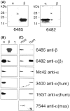
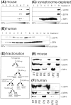
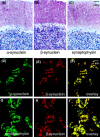
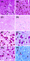
Mutations in the alpha-synuclein (alphaSYN) gene are associated with rare cases of familial Parkinson's disease, and alphaSYN is a major component of Lewy bodies and Lewy neurites. Here we have investigated the localization of wild-type and mutant [A30P]alphaSYN as well as betaSYN at the cellular and subcellular level. Our direct comparative study demonstrates extensive synaptic colocalization of alphaSYN and betaSYN in human and mouse brain. In a sucrose gradient equilibrium centrifugation assay, a portion of betaSYN floated into lower density fractions, which also contained the synaptic vesicle marker synaptophysin. Likewise, wild-type and [A30P]alphaSYN were found in floating fractions. Subcellular fractionation of mouse brain revealed that both alphaSYN and betaSYN were present in synaptosomes. In contrast to synaptophysin, betaSYN and alphaSYN were recovered from the soluble fraction upon lysis of the synaptosomes. Synaptic colocalization of alphaSYN and betaSYN was directly visualized by confocal microscopy of double-stained human brain sections. The Parkinson's disease-associated human mutant [A30P]alphaSYN was found to colocalize with betaSYN and synaptophysin in synapses of transgenic mouse brain. However, in addition to their normal presynaptic localization, transgenic wild-type and [A30P]alphaSYN abnormally accumulated in neuronal cell bodies and neurites throughout the brain. Thus, mutant [A30P]alphaSYN does not fail to be transported to synapses, but its transgenic overexpression apparently leads to abnormal cellular accumulations.
Figures


Similar articles
-
Sensitivity to MPTP is not increased in Parkinson's disease-associated mutant alpha-synuclein transgenic mice.J Neurochem. 2001 May;77(4):1181-4. doi: 10.1046/j.1471-4159.2001.00366.x. J Neurochem. 2001. PMID: 11359883
-
Effect of familial Parkinson's disease point mutations A30P and A53T on the structural properties, aggregation, and fibrillation of human alpha-synuclein.Biochemistry. 2001 Sep 25;40(38):11604-13. doi: 10.1021/bi010616g. Biochemistry. 2001. PMID: 11560511
-
Nuclear and neuritic distribution of serine-129 phosphorylated alpha-synuclein in transgenic mice.Neuroscience. 2009 Jun 2;160(4):796-804. doi: 10.1016/j.neuroscience.2009.03.002. Epub 2009 Mar 9. Neuroscience. 2009. PMID: 19272424
-
Physiology and pathophysiology of alpha-synuclein. Cell culture and transgenic animal models based on a Parkinson's disease-associated protein.Ann N Y Acad Sci. 2000;920:33-41. doi: 10.1111/j.1749-6632.2000.tb06902.x. Ann N Y Acad Sci. 2000. PMID: 11193173 Review.
-
Properties of NACP/alpha-synuclein and its role in Alzheimer's disease.Biochim Biophys Acta. 2000 Jul 26;1502(1):95-109. doi: 10.1016/s0925-4439(00)00036-3. Biochim Biophys Acta. 2000. PMID: 10899435 Review.
Cited by
-
LRRK2 and α-Synuclein: Distinct or Synergistic Players in Parkinson's Disease?Front Neurosci. 2020 Jun 17;14:577. doi: 10.3389/fnins.2020.00577. eCollection 2020. Front Neurosci. 2020. PMID: 32625052 Free PMC article. Review.
-
Familial Parkinson's Disease Mutant E46K α-Synuclein Localizes to Membranous Structures, Forms Aggregates, and Induces Toxicity in Yeast Models.ISRN Neurol. 2011;2011:521847. doi: 10.5402/2011/521847. Epub 2011 Jul 9. ISRN Neurol. 2011. PMID: 22389823 Free PMC article.
-
Adding hydrophobicity or positive charges to the cytosolic half of the α-synuclein 3-11 helix increases membrane association and S129 phosphorylation.FEBS Lett. 2024 Jan;598(2):210-219. doi: 10.1002/1873-3468.14773. Epub 2023 Nov 21. FEBS Lett. 2024. PMID: 37989349 Free PMC article.
-
Synuclein activates microglia in a model of Parkinson's disease.Neurobiol Aging. 2008 Nov;29(11):1690-701. doi: 10.1016/j.neurobiolaging.2007.04.006. Epub 2007 May 29. Neurobiol Aging. 2008. PMID: 17537546 Free PMC article.
-
Model fusion, the next phase in developing animal models for Parkinson's disease.Neurotox Res. 2007 Apr;11(3-4):219-40. doi: 10.1007/BF03033569. Neurotox Res. 2007. PMID: 17449461 Review.
References
-
- Abeliovich A, Schmitz Y, Fariñas I, Choi-Lundberg D, Ho W-H, Castillo PE, Shinsky N, Garcia Verdugo JM, Armanini M, Ryan A, Hynes M, Phillips H, Sulzer D, Rosenthal A. Mice lacking α-synuclein display functional deficits in the nigrostriatal dopamine system. Neuron. 2000;25:239–252. - PubMed
-
- Arawaka S, Saito Y, Murayama S, Mori H. Lewy body in neurodegeneration with brain iron accumulation type 1 is immunoreactive for α-synuclein. Neurology. 1998;51:887–889. - PubMed
-
- Arima K, Uéda K, Sunohara N, Hirai S, Izumiyama Y, Tonozuka-Uehara H, Kawai M. Immunoelectron-microscopic demonstration of NACP/α-synuclein-epitopes on the filamentous component of Lewy bodies in Parkinson's disease and in dementia with Lewy bodies. Brain Res. 1998;808:93–100. - PubMed
-
- Conway KA, Harper JD, Lansbury PT. Accelerated in vitro fibril formation by a mutant α-synuclein linked to early-onset Parkinson disease. Nat Med. 1998;4:1318–1320. - PubMed
Publication types
MeSH terms
Substances
LinkOut - more resources
Full Text Sources
Other Literature Sources
Medical
Molecular Biology Databases
Miscellaneous
