. 2000 Aug;11(8):2719-31.
doi: 10.1091/mbc.11.8.2719.

Syntaxin 17 is abundant in steroidogenic cells and implicated in smooth endoplasmic reticulum membrane dynamics

Affiliations
Free PMC article

Syntaxin 17 is abundant in steroidogenic cells and implicated in smooth endoplasmic reticulum membrane dynamics

M Steegmaier et al. Mol Biol Cell. 2000 Aug.
Free PMC article

Abstract

The endoplasmic reticulum (ER) consists of subcompartments that have distinct protein constituents, morphological appearances, and functions. To understand the mechanisms that regulate the intricate and dynamic organization of the endoplasmic reticulum, it is important to identify and characterize the molecular machinery involved in the assembly and maintenance of the different subcompartments. Here we report that syntaxin 17 is abundantly expressed in steroidogenic cell types and specifically localizes to smooth membranes of the ER. By immunoprecipitation analyses, syntaxin 17 exists in complexes with a syntaxin regulatory protein, rsly1, and/or two intermediate compartment SNARE proteins, rsec22b and rbet1. Furthermore, we found that syntaxin 17 is anchored to the smooth endoplasmic reticulum through an unusual mechanism, requiring two adjacent hydrophobic domains near its carboxyl terminus. Converging lines of evidence indicate that syntaxin 17 functions in a vesicle-trafficking step to the smooth-surfaced tubular ER membranes that are abundant in steroidogenic cells.


Figures

Figure 1
Figure 1
Syntaxin 17 is abundantly expressed in steroidogenic tissues. Postnuclear supernatants from rat heart (H), brain (B), spleen (Sp), lung (Ln), liver (Lv), skeletal muscle (Sk), kidney (K), testis (Ts), adrenal gland (Ad), and placenta (Pl) (30 μg/lane) were analyzed by Western blotting with the use of the mouse monoclonal anti-syntaxin 17 antibody clone 14C10-2. A major band is detected at 38 kDa, with high expression in liver, testis, adrenal gland, and placenta.
Figure 2
Figure 2
Syntaxin 17 is highly expressed in testosterone-synthesizing Leydig cells. Sections of rat testis were labeled with antibodies directed against syntaxin 17 (A and D) and VAMP4 (B and E). (C and F) Overlays of images in A and B and in D and E, respectively. Note that anti-syntaxin 17 antibodies strongly labeled the steroid hormone–secreting Leydig cells (Lc), whereas syntaxin 17 label was drastically reduced in cells of the seminiferous tubules (st) and in the lumen (lu) of the tubules. Bars, 400 μm for A, B, and C (in A) and 100 μm for D, E, and F (in D).
Figure 3
Figure 3
Syntaxin 17 is abundantly expressed in steroid hormone–synthesizing cells of the adrenal cortex. (A and B) Sections of rat adrenal gland were double labeled with antibodies directed against syntaxin 17 (Texas Red–conjugated) and VAMP4 (FITC-conjugated). Syntaxin 17 is highly expressed in steroid hormone–synthesizing cells of the adrenal cortex (cx), whereas syntaxin 17 label is drastically reduced in the catecholamine-secreting cells of the adrenal medulla (md). Bars, 400 μm in A and 100 μm in B. (C) Bovine adrenal gland was dissected into cortex and medulla, postnuclear supernatant was prepared, and the expression of different SNARE proteins was analyzed by Western blotting. Labels on the right indicate the antibodies used for immunoblotting.
Figure 4
Figure 4
Cell lines derived from Leydig cell and adrenal cortical cell tumors express high levels of syntaxin 17. Postnuclear supernatants (30 μg/lane) derived from the cell lines indicated on the top of the panel were separated by SDS-PAGE and immunoblotted with affinity-purified goat anti-syntaxin 17 antibodies. LC540 and R2C are cell lines derived from rat Leydig cell tumor, and H295R and Y-1 are cell lines derived from human and mouse adrenal tumors, respectively. The antibodies recognize a major band of 38 kDa in cell lines of rat or human origin and an ∼40-kDa band in the cell line of mouse origin. Note that only cell lines derived from steroidogenic tissues express high levels of syntaxin 17.
Figure 5
Figure 5
Syntaxin 17 does not colocalize with markers of lysosomes, mitochondria, or RER. H295R adrenal cortical cells were fixed with 4% paraformaldehyde, permeabilized with saponin, and stained with affinity-purified goat anti-syntaxin 17 antibodies (A, D, and G). The cells were costained with mouse mAbs against LAMP1 (B) and cytochrome C (E) and rabbit antibodies against calnexin (H). Texas Red–labeled anti-goat IgG and FITC-labeled anti-mouse and anti-rabbit IgG were used as secondary antibodies. Cells were visualized with confocal microscopy. C, F, and I show merged images. Bars, 2 μm.
Figure 6
Figure 6
Ultrathin cryosection of an adrenal cortical cell. Immunogold labeling of syntaxin 17 (10-nm gold). Syntaxin 17 is present on smooth-surfaced membranes typical of the SER. The appearance of holes in the mitochondria (M) is due to tubular cristae in cross-section. The clustering of gold particles that is seen at some points is due to the labeling method (see MATERIALS AND METHODS) and does not represent local concentrations of syntaxin 17. Bar, 200 nm.
Figure 7
Figure 7
Syntaxin 17 localization in hepatocytes. Ultrathin cryosection (A) and Epon section (B) of rat hepatocytes, both showing the SER-rich region adjacent to a bile capillary (BC). (A) Immunogold label for syntaxin 17 (10-nm gold) is predominantly associated with the SER, whereas RER cisternae (ER) show little or no labeling. (B) The highly convoluted SER membranes are more clearly delineated in Epon sections. M, mitochondrion; P, peroxisome. Bars, 200 nm (A) and 500 nm (B).
Figure 8
Figure 8
The ER-to-SER transition. Ultrathin Epon section (A) and cryosection (B) of rat hepatocytes. (A) Arrows point to continuities between the ER and SER subdomains. (B) Syntaxin 17 (10-nm gold) is predominantly associated with the SER membranes. Bars, 200 nm.
Figure 9
Figure 9
Comparison of SER and VTC membranes in rat hepatocytes. (A) Epon section showing a transitional element (TE) of the ER facing a cluster of VTCs (arrows) that extends up to the Golgi (G). The VTC membranes are morphologically clearly different from the SER membranes shown in the same panel. (B) Cryosection double immunogold labeled for albumin (10-nm gold) and KDELr (15-nm gold) showing the presence of KDELr on VTC membranes between the ER and Golgi (arrows). The Golgi is outlined by the albumin labeling. (C) Cryosection double labeled for syntaxin 17 (10-nm gold) and COPII (15-nm gold). COPII labels some of the VTC membranes between the ER and Golgi (arrows). Syntaxin 17 is only occasionally found on VTC membranes (arrowhead) but strongly labels membranes of the SER. Note that syntaxin 17 is absent from the Golgi. P, plasma membrane. Bars, 200 nm.
Figure 10
Figure 10
Syntaxin 17 is extractable from membranes with high pH. LC540 Leydig tumor cells were homogenized and divided into postnuclear (PNS), cytosolic (CS), and postnuclear membrane fractions. Membrane fractions were extracted with either control buffer (Co), 1.5 M NaCl (NaCl), 0.2 M sodium bicarbonate at pH 11 (pH), or 2% Triton X-100 (TX-100) and centrifuged at 100,000 × g. The resulting supernatants (S) and pellets (P) were analyzed by SDS-PAGE and immunoblotted with the antibodies indicated on the right. Note that only syntaxin 17 and not the other SNARE proteins were extracted from membranes with high pH.
Figure 11
Figure 11
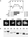
Two carboxyl-terminal hydrophobic domains are required to anchor syntaxin 17 to membranes. (A) Kyte-Doolittle hydrophobicity plot of the carboxyl-terminal part of syntaxin 17 generated with the DNA-Star program. Shown below are the individual carboxyl-terminal deletion constructs. The putative transmembrane domains (in black) are separated by a single positively charged amino acid (K253, *). (B) NRK cells were transfected with amino-terminally myc-tagged syntaxin 17 full-length protein (amino acids [aa] 1–301; wt), myc–syntaxin 17 deleted of its carboxyl-terminal hydrophilic tail (aa 1–272; Δtail), myc–syntaxin 17 deleted of its carboxyl-terminal hydrophilic tail and its second hydrophobic domain (aa 1–253; ΔTM2nd), or myc–syntaxin 17 deleted of its carboxyl-terminal hydrophilic tail and both hydrophobic domains (aa 1–227; ΔTM). Cells were then fixed, permeabilized, and stained with anti-myc mAb. (C) COS-7 cells were transfected with the same syntaxin 17 deletion constructs as in B. Transfected cells were then fractionated into cytosolic (CS) and postnuclear membrane (M) fractions. Ten micrograms of protein was loaded onto each lane, separated by SDS-PAGE, and immunoblotted with anti-myc antibody. Note that the ratio of cytosolic to membrane-bound syntaxin17ΔTM2nd is dramatically increased compared with the full-length construct.
Figure 12
Figure 12
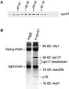
The syntaxin 17 complex(es) contains five vesicle-trafficking proteins. (A) Rat testis membrane extract was fractionated by centrifugation through an 11–35% glycerol velocity gradient, and sequential fractions were analyzed by electrophoresis and immunoblotting with antibodies against syntaxin 17. Note that the entire pool of syntaxin 17 is present in an oligomeric complex(es). (B) Rat testis membranes were isolated and solubilized with 1% Triton X-100. The solubilized membranes were incubated with protein G–Sepharose beads loaded with either mouse monoclonal anti-syntaxin 17 antibody or with nonspecific mouse IgG. After the binding step, the beads were washed, eluted with SDS protein sample dye, resolved by SDS-PAGE, and visualized with Coomassie blue. The indicated bands were excised from the gel, digested with trypsin, and analyzed by microsequencing and mass spectrometry. The labels to the right indicate the identities of the precipitated proteins.

Similar articles

Cited by

References

    1. Bennett MK, Garcia-Arraras JE, Elferink LA, Peterson K, Fleming AM, Hazuka CD, Scheller RH. The syntaxin family of vesicular transport receptors. Cell. 1993;74:863–873. - PubMed
    1. Bock JB, Klumperman J, Davanger S, Scheller RH. Syntaxin 6 functions in trans-Golgi network vesicle trafficking. Mol Biol Cell. 1997;8:1261–1271. - PMC - PubMed
    1. Bock JB, Scheller RH. SNARE proteins mediate lipid bilayer fusion. Proc Natl Acad Sci USA. 1999;96:12227–12229. - PMC - PubMed
    1. Brands R, Slot JW, Geuze HJ. Albumin localization in rat liver parenchymal cells. Eur J Cell Biol. 1983;32:99–107. - PubMed
    1. Dascher C, Balch WE. Mammalian Sly1 regulates syntaxin 5 function in endoplasmic reticulum to Golgi transport. J Biol Chem. 1996;271:15866–15869. - PubMed

MeSH terms