. 2000 Aug;11(8):2691-704.
doi: 10.1091/mbc.11.8.2691.

Specific sequence motif of 8-Cys repeats of TGF-beta binding proteins, LTBPs, creates a hydrophobic interaction surface for binding of small latent TGF-beta

Affiliations
Free PMC article

Specific sequence motif of 8-Cys repeats of TGF-beta binding proteins, LTBPs, creates a hydrophobic interaction surface for binding of small latent TGF-beta

J Saharinen et al. Mol Biol Cell. 2000 Aug.
Free PMC article

Abstract

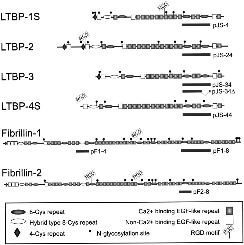
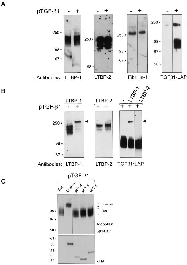
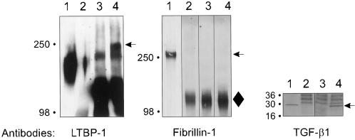
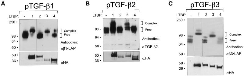
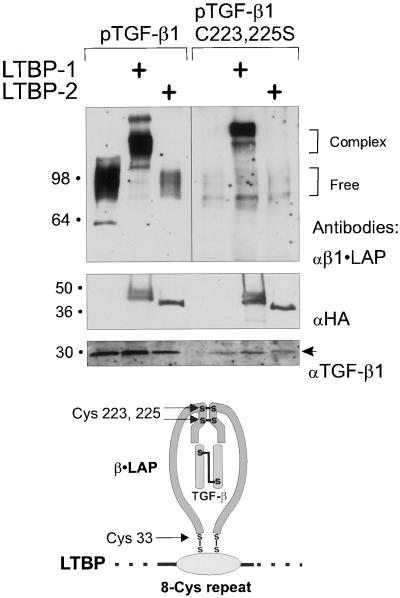
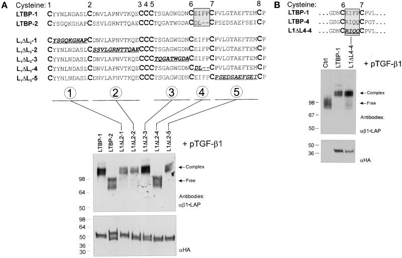
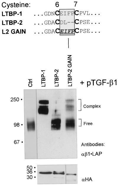
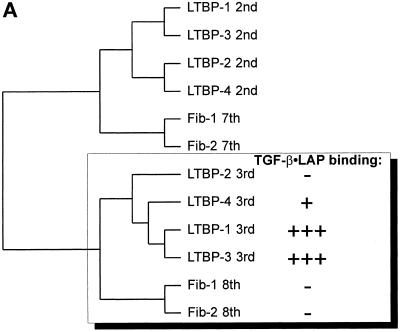
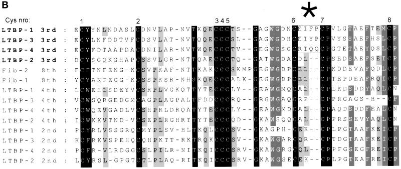
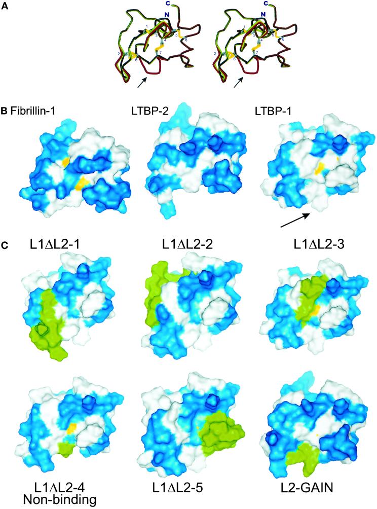
Transforming growth factor (TGF)-betas are secreted in large latent complexes consisting of TGF-beta, its N-terminal latency-associated peptide (LAP) propeptide, and latent TGF-beta binding protein (LTBP). LTBPs are required for secretion and subsequent deposition of TGF-beta into the extracellular matrix. TGF-beta1 associates with the 3(rd) 8-Cys repeat of LTBP-1 by LAP. All LTBPs, as well as fibrillins, contain multiple 8-Cys repeats. We analyzed the abilities of fibrillins and LTBPs to bind latent TGF-beta by their 8-Cys repeats. 8-Cys repeat was found to interact with TGF-beta1*LAP by direct cysteine bridging. LTBP-1 and LTBP-3 bound efficiently all TGF-beta isoforms, LTBP-4 had a much weaker binding capacity, whereas LTBP-2 as well as fibrillins -1 and -2 were negative. A short, specific TGF-beta binding motif was identified in the TGF-beta binding 8-Cys repeats. Deletion of this motif in the 3(rd) 8-Cys repeat of LTBP-1 resulted in loss of TGF-beta*LAP binding ability, while its inclusion in non-TGF-beta binding 3(rd) 8-Cys repeat of LTBP-2 resulted in TGF-beta binding. Molecular modeling of the 8-Cys repeats revealed a hydrophobic interaction surface and lack of three stabilizing hydrogen bonds introduced by the TGF-beta binding motif necessary for the formation of the TGF-beta*LAP - 8-Cys repeat complex inside the cells.


Figures

Figure 1
Figure 1
Schematic representation of the human LTBPs 1–4 and human fibrillins -1 and -2. The small (S) isoforms of LTBPs -1 and -4 are illustrated. The solid bars below the proteins describe the area coded by the respective constructs.
Figure 2
Figure 2
A) Overexpressed TGF-β1•LAP forms covalent complexes with endogenous LTBP-1, but not with LTBP-2 or fibrillin-1 in human fibroblast culture. Confluent human embryonic lung fibroblast cultures were transfected with pTGFβ1. Proteins secreted into fibroblast conditioned medium were separated in SDS-PAGEs. The concentrations of acrylamide in SDS PAGES were the following: 7,5% for LTBP-1 blot, 5% for LTBP-2 blot, 7,5% for fibrillin-1 blot, 4–15% gradient for β1•LAP blot. The formation of β1•LAP complexes with endogenous LTBPs -1 or -2 or fibrillin-1 was detected by immunoblotting. Transfection of TGF-β1 cDNA is indicated above the immunoblots, and the antibodies used are below the immunoblot. Brackets on the right indicate the complexed β1•LAP forms. B) Overexpressed LTBP-1 binds covalently β1•LAP, whereas LTBP-2 does not. pLTBP-1 and pLTBP-2 were transfected together with pTGFβ1 cDNA to 293T cells, and proteins secreted into the conditioned medium were separated by 7.5% PAGEs. The secreted proteins were detected by immunoblotting with antibodies for LTBP-1, LTBP-2, or β1•LAP as indicated, in order to detect the covalent LTBP - β1•LAP complexes. The transfected cDNAs are indicated above the immunoblots, and the antibodies used are below them. Arrows indicate the large latent β1•LAP - LTBP-1 complexes. C) β1•LAP does not associate with 8-Cys repeats of fibrillin-1. Expression constructs containing certain 8-Cys repeats of fibrillins were transfected together with pTGFβ1 to 293T cells, as indicated on the top of the figure. LTBP-1 construct pJS-4 (Figure 1) was used as positive control for a protein capable of forming covalent complexes with β1•LAP. Secreted proteins were separated by 7.5% PAGE and analyzed for β1•LAP by immunoblotting (upper panel). Brackets on the right indicate the free and complexed β1•LAP forms. Expression levels of the proteins coded by all HA-tagged fibrillin and LTBP-1 constructs were verified by 4–15% PAGE followed by immunoblotting with HA-antibody from reduced samples (lower panel).
Figure 3
Figure 3
Endogenous fibrillin does not coprecipitate with endogenous β1•LAP from human fibroblasts. β1•LAP was immunoprecipitated from the conditioned medium lung fibroblasts. The immunoprecipitates were separated by 5% PAGE (LTBP-1 and fibrillin-1) or 4–15% gradient PAGE (TGF-β1) and analyzed for β1•LAP complexed proteins by immunoblotting. Lanes in each panel are: 1) fibroblast conditioned medium; 2) conditioned medium mock-precipitated with nonimmune serum; 3) d-MEM immunoprecipitated with β1•LAP antibodies; 4) fibroblast conditioned medium immunoprecipitated with β1•LAP antibodies. Arrows on the right indicate the latent β1•LAP - LTBP-1 complexes, fibrillin-1 and TGF-β1 dimer. A diamond (♦) on the right of fibrillin-1 immunoblot indicates the migration of a background band.
Figure 4
Figure 4
Association of TGF-β•LAP isoforms 1, 2, and 3 with LTBPs 1–4. Expression constructs covering the 3rd 8-Cys repeats of LTBPs (LTBP-1: pJS-4, LTBP-2: pJS-24, LTBP-3: pJS-34Δ and LTBP-4: pJS-44, illustrated in Figure 1), were transfected together with the three human TGF-β isoforms to 293T cells. The secreted proteins were separated by 7.5% PAGE and detected by immunoblotting with antibodies against β1•LAP (A), TGF-β2 (B), or β3•LAP (C) in order to detect covalent β•LAP - LTBP-fragment complexes (upper panels). The secretion of proteins coded by LTBP constructs was verified by 4–15% gradient PAGE followed by immunoblotting with anti-HA antibody (lower panels). Brackets on the right indicate the free and complexed β•LAP forms. Note the higher molecular weight bands in Figure 4C (lanes 1–3), which most likely represent the unprocessed TGF-β3•LAP, where the TGF-β and LAP parts are not proteolytically processed.
Figure 5
Figure 5
Binding between the 8-Cys repeat of LTBP-1 and Cys-33 of TGF-β1•LAP is mediated by a direct disulfide exchange. Constructs pJS-4 or pJS-24 were transfected with wild-type pTGFβ1 or pTGFβ1 C223,225S to COS-7 cells. The secreted proteins were separated by 7.5% PAGE and detected by immunoblotting with β1•LAP antibodies (top panel). Brackets on the right indicate the free or complexed β1•LAP forms. The expression of proteins coded by LTBP-constructs was verified by 4–15% gradient PAGE followed by anti-HA immunoblot (middle panel). Secretion of TGF-β1 was detected by immunoblotting for TGF-β1 (bottom panel, an arrow). Below the immunoblots is a model illustrating the proposed β1•LAP - 8-Cys repeat interaction.
Figure 6
Figure 6
The region between 6th and 7th Cys-residues of the 8-Cys repeat determines the β•LAP binding ability. A) Loss of function of LTBP-1. The substituted amino acids in chimeric constructs L1ΔL2 1–5 between the 3rd 8-Cys repeats of LTBP-1 and LTBP-2 are underlined. These constructs as well as the wild-type LTBP-1 and LTBP-2 constructs (pJS-4 and pJS-24, respectively) were transfected together with pTGFβ1 cDNA to 293T cells, as indicated above the immunoblot. The secreted proteins were separated by 7.5% PAGE and immunodetected with β1•LAP antibodies. The proteins coded by all LTBP constructs were separated by 4–15% gradient PAGE, and their expression levels were detected by immunoblotting with anti-HA antibody (lower panel). B) The corresponding chimeric construct between LTBP-1 and LTBP-4 retains TGF-β•LAP binding function. Construct L1ΔL4–4, analogous to construct L1ΔL2–4, was transfected with pTGFβ1 cDNA to 293T cells. The conditioned medium was separated by 7.5% PAGE and analyzed for the presence of covalent protein complexes between β1•LAP and L1ΔL4–4 encoded protein. Arrows on the right indicate the free or complexed β1•LAP forms. The secretion of proteins coded by all LTBP constructs were detected by 4–15% gradient PAGE followed by immunoblotting with anti-HA antibody (lower panel).
Figure 7
Figure 7
Replacement of the region between the 6th and 7th cysteine residues in the 3rd 8-Cys repeat of LTBP-2 with that of LTBP-1 results in gain-of-function of TGF-β-β1•LAP binding. Wild-type LTBP -1 and -2 constructs, pJS-4 and pJS-24, respectively, as well as construct L2GAIN were transfected together with TGF-β1 cDNA to 293T cells. The proteins from conditioned medium were separated by 7.5% PAGEs and analyzed for covalent complex formation between the proteins coded by LTBP constructs and β1•LAP. Brackets on the right indicate the free or complexed β1•LAP forms. Secretion of the proteins coded by LTBP constructs was verified by 4–15% gradient PAGEs followed by immunoblotting with anti-HA antibody (lower panel).
Figure 8
Figure 8
A) Relationships between the 8-Cys repeats of human LTBPs 1–4 and fibrillins -1 and -2. The amino acid sequences for all known 8-Cys as well as the hybrid domains of human LTBPs and fibrillins were used to make the dendrogram, and the ones most similar to the 3rd 8-Cys repeats of LTBPs are shown. The TGF-β•LAP binding abilities of the 3rd 8-Cys repeats of LTBPs and the 8th 8-Cys repeats of fibrillins are illustrated. B) Multiple sequence alignment of the 8-Cys repeats of human LTBPs and the 8th 8-Cys repeats of fibrillins. The amino acid sequences for all the 8-Cys repeats of human LTBPs and the 8th 8-Cys repeats of fibrillins were aligned. The sequences covering all eight cysteine residues as well as one amino acid on both N- and C-terminal sides were included. The alignment indicates several conserved residues in the 8-Cys repeats. The three TGF-β binding type 8-Cys repeats (3rd ones of LTBPs -1, -3, and -4) are clearly distinguished by the two amino acids insertion between the 6th and 7th cysteine residues (indicated by an asterisk above the alignment).
Figure 8
Figure 8
A) Relationships between the 8-Cys repeats of human LTBPs 1–4 and fibrillins -1 and -2. The amino acid sequences for all known 8-Cys as well as the hybrid domains of human LTBPs and fibrillins were used to make the dendrogram, and the ones most similar to the 3rd 8-Cys repeats of LTBPs are shown. The TGF-β•LAP binding abilities of the 3rd 8-Cys repeats of LTBPs and the 8th 8-Cys repeats of fibrillins are illustrated. B) Multiple sequence alignment of the 8-Cys repeats of human LTBPs and the 8th 8-Cys repeats of fibrillins. The amino acid sequences for all the 8-Cys repeats of human LTBPs and the 8th 8-Cys repeats of fibrillins were aligned. The sequences covering all eight cysteine residues as well as one amino acid on both N- and C-terminal sides were included. The alignment indicates several conserved residues in the 8-Cys repeats. The three TGF-β binding type 8-Cys repeats (3rd ones of LTBPs -1, -3, and -4) are clearly distinguished by the two amino acids insertion between the 6th and 7th cysteine residues (indicated by an asterisk above the alignment).
Figure 9
Figure 9
Molecular modeling of 8-Cys repeats. A) Stereoimage of the superpositioned structure of the 8th 8-Cys repeat of fibrillin-1 and the models for the 3rd 8-Cys repeat of LTBPs -1 and -2. The backbone of the previously published structure of the 8th 8-Cys repeat of fibrillin (Yuan et al., 1997) is illustrated in black, the molecular models for the 3rd 8-Cys repeat of LTBP-1 and LTBP-2 in red and green, respectively. The side chains of cysteine residues are shown with the numbering referring to the ordinals of the cysteine residues. At the bottom of the illustration is the region between the 6th and 7th Cys-residues, where the backbone alignment between the TGF-β binding and nonbinding type 8-Cys repeats is lost (indicated by an arrow). B) The solvent accessible surfaces for the models of the 3rd 8-Cys repeats of LTBPs. The structures of TGF-β binding (LTBP-1) and nonbinding type (Fibrillin-1, LTBP-2) 8-Cys repeats are shown. Blue: hydrophilic amino acids; white: hydrophobic amino acids; yellow: SH groups of cysteine residues exposed to solvent. The hydrophobic area caused by the insertion of two amino acid between the 6th and 7th Cys-residues of the TGF-β binding type 8-Cys repeat of LTBP-1 is marked by an arrow. C) Solvent accessible surfaces of the models coded by L1ΔL2 1–5 and L2GAIN constructs. The structures of the 8-Cys repeat chimeras, described in Figure 6A, 6B, and 7, were modeled. The modified amino acids in these models are shown in green, hydrophobic in white, hydrophilic in blue and the SH-groups of Cys-residues in yellow.

Similar articles

Cited by

References

    1. Bashir MM, Han MD, Abrams WR, Tucker T, Ma RI, Gibson M, Ritty T, Mecham R, Rosenbloom J. Analysis of the human gene encoding latent transforming growth factor-β-binding protein-2. Int J Biochem Cell Biol. 1996;28:531–542. - PubMed
    1. Dallas SL, Park-Snyder S, Miyazono K, Twardzik D, Mundy GR, Bonewald LF. Characterization and autoregulation of latent transforming growth factor-β (TGF-β) complexes in osteoblast-like cell lines: production of a latent complex lacking the latent TGF-β-binding protein. J Biol Chem. 1994;269:6815–6821. - PubMed
    1. D'Arrigo C, Burl S, Withers AP, Dobson H, Black C, Boxer M. TGF-β1 binding protein-like modules of fibrillin-1 and -2 mediate integrin-dependent cell adhesion. Connect Tissue Res. 1998;37:29–51. - PubMed
    1. Davis CG. The many faces of epidermal growth factor repeats. New Biol. 1990;2:410–419. - PubMed
    1. Dubois CM, Laprise MH, Blanchette F, Gentry LE, Leduc R. Processing of transforming growth factor-β 1 precursor by human furin convertase. J Biol Chem. 1995;270:10618–10624. - PubMed

Publication types

MeSH terms