. 2000 Aug;11(8):2591-604.
doi: 10.1091/mbc.11.8.2591.

The AP-3 complex required for endosomal synaptic vesicle biogenesis is associated with a casein kinase Ialpha-like isoform

Affiliations
Free PMC article

The AP-3 complex required for endosomal synaptic vesicle biogenesis is associated with a casein kinase Ialpha-like isoform

V V Faundez et al. Mol Biol Cell. 2000 Aug.
Free PMC article

Abstract

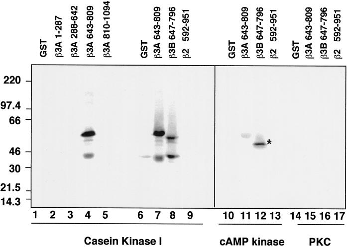
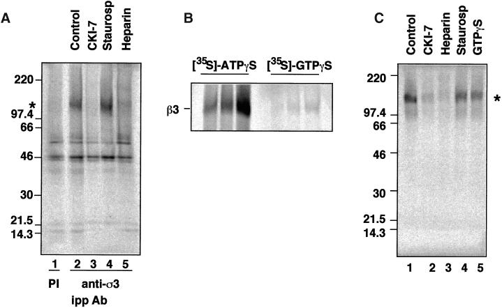
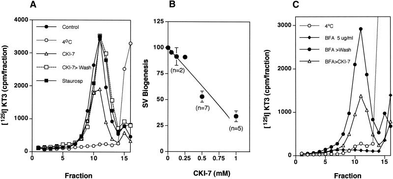
The formation of small vesicles is mediated by cytoplasmic coats the assembly of which is regulated by the activity of GTPases, kinases, and phosphatases. A heterotetrameric AP-3 adaptor complex has been implicated in the formation of synaptic vesicles from PC12 endosomes (). When the small GTPase ARF1 is prevented from hydrolyzing GTP, we can reconstitute AP-3 recruitment to synaptic vesicle membranes in an assembly reaction that requires temperatures above 15 degrees C and the presence of ATP suggesting that an enzymatic step is involved in the coat assembly. We have now found an enzymatic reaction, the phosphorylation of the AP-3 adaptor complex, that is linked with synaptic vesicle coating. Phosphorylation occurs in the beta3 subunit of the complex by a kinase similar to casein kinase 1alpha. The kinase copurifies with neuronal-specific AP-3. In vitro, purified casein kinase I selectively phosphorylates the beta3A and beta3B subunit at its hinge domain. Inhibiting the kinase hinders the recruitment of AP-3 to synaptic vesicles. The same inhibitors that prevent coat assembly in vitro also inhibit the formation of synaptic vesicles in PC12 cells. The data suggest, therefore, that the mechanism of AP-3-mediated vesiculation from neuroendocrine endosomes requires the phosphorylation of the adaptor complex at a step during or after AP-3 recruitment to membranes.


Figures

Figure 1
Figure 1
Nucleotide requirements for the ARF-dependent protein recruitment onto SV. Synaptic vesicle coating reactions were performed with synaptic vesicles isolated from 125I-KT3-labeled PC12N49A cells. Synaptic vesicles were incubated in the presence of 3 mg/ml normal or dialyzed cytosol plus 30 μM Q71L ARF1 mutant. Dialyzed cytosol was supplemented with either an ATP-regenerating system (O'Grady et al. 1998) or with different nucleotides at 200 μM concentration, except for GTP, which was at a final concentration of 1 mM. Samples were warmed to 37°C for 30 min, and reactions were stopped at 0°C. Vesicle sedimentation was analyzed in 10–45% continuous sucrose gradients, and the sucrose concentration was measured at the peak of radioactivity. Only the poorly hydrolyzable ATP analog ATPγS could fully replace the ATP requirements of the coating reaction and shifts the density from 22 to 32% sucrose.
Figure 2
Figure 2
Recruitment of AP-3 to immobilized synaptic vesicles. (A) AP-3 is recruited to SVs in the presence of ATPγS. Coating assays were performed using unlabeled PC12 N49A synaptic vesicles attached to an antisynaptophysin-protein G-Sepharose affinity column. Reactions were performed with rat brain cytosol, supplemented (lanes 1 and 2) or not (lanes 3 and 4) with an ATP-regenerating system. AP-3 recruitment to SV was induced at 37°C for 30 min either by GTPγS (20 μM, lane 2) or solely by ATPγS (200 μM, lane 4). AP-3 recruitment to synaptic vesicles was assessed by immunoblot with antibodies against either β3 or ς3 subunits of the complex. Similarly, ARF1 binding to membranes was determined with an anti-ARF1 antibody. (B) The β3 subunit of the synaptic vesicle-bound AP-3 complex is thiophosphorylated by acasein kinase activity. Immunoimmobilized PC12 N49A synaptic vesicles were coated in vitro in the presence of dialyzed rat brain cytosol. Coating was triggered by adding [35S]-ATPγS (100 μCi) and warming the reactions at 37°C for 40 min. Reactions were stopped at 4°C, and bound complexes were washed extensively in intracellular buffer. Coated SVs were solubilized from the column by either sample buffer to directly resolve the proteins in SDS-PAGE (lanes 1–4) or in buffer B (lanes 5–8), from which AP-3 β subunits were immunoprecipitated and immunocomplexes were analyzed by SDS-PAGE. The intensity of the β3 thiophosphorylated band was reduced by the casein kinase inhibitors CKI-7 (500 μM, lanes 2 and 6) and heparin (10 μg/ml, lanes 4 and 9) but not by a generic serine–threonine kinase inhibitor staurosporine (10 μM, lanes 4 and 8).
Figure 3
Figure 3
The hinge domain of β3A and β3B are selectively thiophosphorylated by casein kinase I. GST and GST fusion proteins encompassing the trunk of β3A (residues 1–287 and 288–642; lanes 2 and 3, respectively), the hinge domain of β3A (residues 643–809; lanes 4, 7, 11, and 15) and β3B (residues 647–796; lanes 8, 12, and 16), the ear domain of β3A (residues 810–1094, lane 5), or the hinge–COOH terminal region of β2 (residues 592–951; lanes 9, 13, and 17) were thiophosphorylated as described with purified casein kinase I (10 U/assay; lanes 1–9), catalytic subunit of the cAMP-dependent kinase (10U/assay; lanes 10–13), or the catalytic tryptic fragment of brain protein kinase C (20 ng/assay; lanes 14–17). Recombinant proteins were TCA-precipitated from the reaction mixture and were analyzed by SDS-PAGE. Only the hinge domains of β3A and β3B were thiophosphorylated by casein kinase I. The asterisk in lane 12 corresponds to a bacterial contaminant band.
Figure 4
Figure 4
The AP-3 coat complex is tightly associated with casein kinase I. (A) Casein kinase I activity coimmunoprecipitates with the AP-3 complex. Native AP-3 complex was immunoprecipitated from rat brain cytosol with antibodies against ς3 subunits (lanes 2–5) with preimmune sera (lane 1) as a control. Immunocomplexes were reconstituted at 4°C for 15 min in intracellular buffer in the absence (lanes 1 and 2) or the presence of CKI-7 (500 μM, lane 3), staurosporine (10 μM, lane 4), or heparin (10 μg/ml, lane 5). Thiophosphorylation was started at 24°C by adding 20 μCi [35S]-ATPγS. Reactions were stopped in ice by adding buffer A plus 20 mM EDTA. Immunocomplexes were extensively washed in buffer A before SDS-PAGE. An asterisk indicates the position of phosphorylated β chain. (B) Immunoprecipitated brain AP-3 complex was thiophosphorylated for 5, 10, and 20 min, using either 20 μCi [35S]-ATPγS or 20 μCi [35S]-GTPγS, as previously described. Only [35S]-ATPγS was effective in thiophosphorylating the β3 subunit of the AP-3 complex. (C) Purified brain AP-3 complex is tightly associated with the casein kinase I activity. MonoS-purified bovine brain AP-3 complex in intracellular buffer was reconstituted at 4°C for 15 min in the absence (lane 1) or presence of CKI-7 (500 μM, lane 2), heparin (10 μg/ml, lane 3), staurosporine (300 nM, lane 4), or GTPγS (20 μM, lane 5). Thiophosphorylation reactions were performed as described in the presence of 20 μCi [35S]-ATPγS. Thiophosphorylated AP-3 complex was TCA-precipitated from the reaction mixture and was analyzed by SDS-PAGE. Both the immunoprecipitated and purified AP-3 complex displayed a similar inhibition profile. (D) A Iα-like casein kinase is associated with the AP-3 complex. Either 5.6 μg of liver-purified casein kinase I (lanes 1, 3, and 5) or 6 μg of bovine brain-purified AP-3 complex were resolved in SDS-PAGE gels and immunoblotted with affinity-purified anti-casein kinase Iα antibodies (CKI-α RA and CKI-α SC) or anti-δ/ε casein kinase I isoforms. Only the antibodies directed against the Iα isoform, RA and SC, cross-reacted with a 45-kDa band present in the AP-3 complex.
Figure 4
Figure 4
The AP-3 coat complex is tightly associated with casein kinase I. (A) Casein kinase I activity coimmunoprecipitates with the AP-3 complex. Native AP-3 complex was immunoprecipitated from rat brain cytosol with antibodies against ς3 subunits (lanes 2–5) with preimmune sera (lane 1) as a control. Immunocomplexes were reconstituted at 4°C for 15 min in intracellular buffer in the absence (lanes 1 and 2) or the presence of CKI-7 (500 μM, lane 3), staurosporine (10 μM, lane 4), or heparin (10 μg/ml, lane 5). Thiophosphorylation was started at 24°C by adding 20 μCi [35S]-ATPγS. Reactions were stopped in ice by adding buffer A plus 20 mM EDTA. Immunocomplexes were extensively washed in buffer A before SDS-PAGE. An asterisk indicates the position of phosphorylated β chain. (B) Immunoprecipitated brain AP-3 complex was thiophosphorylated for 5, 10, and 20 min, using either 20 μCi [35S]-ATPγS or 20 μCi [35S]-GTPγS, as previously described. Only [35S]-ATPγS was effective in thiophosphorylating the β3 subunit of the AP-3 complex. (C) Purified brain AP-3 complex is tightly associated with the casein kinase I activity. MonoS-purified bovine brain AP-3 complex in intracellular buffer was reconstituted at 4°C for 15 min in the absence (lane 1) or presence of CKI-7 (500 μM, lane 2), heparin (10 μg/ml, lane 3), staurosporine (300 nM, lane 4), or GTPγS (20 μM, lane 5). Thiophosphorylation reactions were performed as described in the presence of 20 μCi [35S]-ATPγS. Thiophosphorylated AP-3 complex was TCA-precipitated from the reaction mixture and was analyzed by SDS-PAGE. Both the immunoprecipitated and purified AP-3 complex displayed a similar inhibition profile. (D) A Iα-like casein kinase is associated with the AP-3 complex. Either 5.6 μg of liver-purified casein kinase I (lanes 1, 3, and 5) or 6 μg of bovine brain-purified AP-3 complex were resolved in SDS-PAGE gels and immunoblotted with affinity-purified anti-casein kinase Iα antibodies (CKI-α RA and CKI-α SC) or anti-δ/ε casein kinase I isoforms. Only the antibodies directed against the Iα isoform, RA and SC, cross-reacted with a 45-kDa band present in the AP-3 complex.
Figure 5
Figure 5
Casein kinase inhibitors hinder β3 thiophosphorylation in a dose-dependent manner. (A) Native AP-3 complex was immunoprecipitated from rat brain cytosol with antibodies against ς3 subunits. Thiophosphorylation reactions were performed as described. Before adding 20 μCi [35S]-ATPγS, the reaction mixtures were preincubated at 4°C for 15 min in intracellular buffer in the absence or presence of increasing concentrations of either CKI-7 (closed circles) or DRB (5,6-Dichloro-1-β-d-rybofuranosylbenzimidazole; open circles). Thiophosphorylation was stopped with ice-cold buffer A plus 20 mM EDTA. Immunocomplexes were extensively washed in buffer A to then be resolved in SDS-PAGE gels. Bands were quantified by PhosphorImager. The IC50 for CKI-7 and DRB were both ∼10 μM (n = 2). (B) Depicted is the quantitation of the effects of 300 nM staurosporine (diagonally striped bar) or 10 μg/ml heparin (n = 3) on the kinase coimmunoprecipitated with the AP-3 complex.
Figure 6
Figure 6
The neuronal AP-3 complex is associated with casein kinase I. AP-3 complexes from mouse brain cytosol (250 μg/assay) prepared from C57BL6 mice (lanes 1–3), or the Hermansky–Pudlak-like mice mutants mocha (lanes 4–6), pearl (lanes 7–9), and pale ear (lanes 10–12) were immunoprecipitated with preimmune sera (lanes 1, 4, 7, and 10) or antibodies against ς3 subunits. Immunocomplexes were reconstituted at 4°C for 15 min in intracellular buffer in the absence or presence of CKI-7 (250 μM, lanes 3, 6, 9, and 12). Thiophosphorylation was started at 24°C by adding 20 μCi [35S]-ATPγS. Reactions were stopped in ice by adding buffer A plus 20 mM EDTA, were extensively washed, and the complexes were resolved in SDS–PAGE gels. AP-3 complexes immunoprecipitated from pearl mutants that only have the neuronal form of AP-3 were thiophosphorylated. No β3-phosphorylated bands were detected in mocha mice brain cytosol that lacks both neuronal and nonneuronal AP-3 complexes.
Figure 7
Figure 7
Characterization of inhibition by CK1–7. (A) Casein kinase I activity is required for the coat recruitment. Synaptic vesicle coating reactions were performed with radioactive SVs from 125I-KT3 endocytically labeled PC12N49A cells in the presence of 3 mg/ml dialyzed cytosol plus 150 μM ATPγS. Before adding the nucleotide, reaction mixtures were incubated for 15 min at 37°C in the absence or presence of CKI-7 (500 μM, n = 4) or staurosporine (10 μM, n = 4). CKI-7 concentrations < 200 μM were ineffective. Coating reactions were triggered by ATPγS at 37°C for 30 min then were stopped at 0°C. Radioactive vesicle sedimentation was analyzed as before in 10–45% continuous sucrose gradients. CKI-7 partially inhibited the coating reaction. (B) Coating assays were performed using unlabeled PC12 N49A synaptic vesicles bound to an antisynaptophysin-protein G-Sepharose affinity column. Reactions were performed at 37°C with dialyzed rat brain cytosol. Before the addition (even lanes) or not (odd lanes) of ATPγS (150 μM), the coating mixtures were preincubated at 37°C for 15 min in the absence (lanes 1 and 2) or in the presence of either CKI-7 (500 μM, lanes 3 and 4) or staurosporine (10 μM, lanes 5 and 6). Coating was stopped at 4°C, and the adsorbed coated SV was extensively washed in intracellular buffer. Complexes were resolved in SDS–PAGE gels, and the AP-3 content was assessed by immunoblot with antibodies against the β3 subunit. CKI-7 reduced the AP-3 recruited to SV to 40 ± 17% of the control reactions (n = 7). (C) ATPγS-dependent AP-3 recruitment to 50K-g membrane fractions is reduced by casein kinase inhibition. A donor membrane-enriched fraction was incubated in the absence or presence of ATPγS (150 μM). ATPγS-treated assays either were added or not to CKI-7. Reaction mixtures were incubated as described in panel B. Reactions were stopped by chilling down and sedimentation through a sucrose cushion. Blot analysis was performed as in panel B. CKI-7 reduced AP-3 recruitment to donor-enriched membranes by 58 ± 16% (n = 5) and 32 ± 21 (n = 6) at 0.5 and 1 mM, respectively. All data represent average ± SE.
Figure 8
Figure 8
CKI-7 inhibits in vivo SV budding from PC12 cell endosomes. PC12 N49A cells were labeled with 125I-KT3 at 15°C as described. Unbound radiolabeled antibodies were extensively washed at 4°C, and cells were incubated for 30 min at 15°C in the absence or presence of CKI-7 (500 μM, panel A, or staurosporine (300 nM). In (B) cells were incubated in increasing concentrations of CK1–7. Cells were warmed at 37°C for 20 min to allow SV formation, and the reactions were stopped by chilling down the cells. Recovery experiments (n = 2) were performed after extensively washing the cells with DME media at 4°C before returning them for 20 min to 37°C. Cells were homogenized and fractionated as previously described, and the SV production was assessed in glycerol velocity gradients. CKI-7 inhibited SV budding with an IC50 of 500 μM (B). No inhibition was detected by staurosporine (n = 5). (C) CKI-7 inhibits in vivo endosomal SV budding at a step close to coat recruitment. PC12 N49A cells were labeled with 125I-KT3 at 15°C, unbound label was washed away at 4°C to then incubate the cells either at 4°C (open circles) or for 15 min at 37°C in DME H21 media supplemented with 5 μg/ml brefeldin A to release coat from the membranes. Brefeldin A was kept (closed diamonds) or washed away at 4°C, and the cells were incubated at 15°C for 30 min in the absence (closed circles) or presence of 500 μM CKI-7 (open triangles). Without removing the inhibitor cells, they were warmed at 37°C for 20 min to allow SV formation, and the reactions were stopped by chilling down the cells. Cells were homogenized and fractionated as previously described, and the SV content was assessed in glycerol velocity gradients. CKI-7 hindered SV formation from brefeldin A-treated endosomes as effectively as it inhibited SV formation for endosomes labeled at 15°C. Depicted is a representative experiment of two.

Similar articles

Cited by

References

    1. Anderson HA, Roche PA. Phosphorylation regulates the delivery of MHC class II invariant chain complexes to antigen processing compartments. J Immunol. 1998;160:4850–4858. - PubMed
    1. Bauerfeind R, Takei K, De Camilli P. Amphiphysin I is associated with coated endocytic intermediates and undergoes stimulation-dependent dephosphorylation in nerve terminals. J Biol Chem. 1997;272:30984–30992. - PubMed
    1. Beyaert R, Vanhaesebroeck B, Declercq W, Van Lint J, Vandenabele P, Agostinis P, Vandenheede JR, Fiers W. Casein kinase-1 phosphorylates the p75 tumor necrosis factor receptor and negatively regulates tumor necrosis factor signaling for apoptosis. J Biol Chem. 1995;270:23293–23299. - PubMed
    1. Bioukar EB, Marricco NC, Zuo D, Larose L. Serine phosphorylation of the ligand-activated beta-platelet-derived growth factor receptor by casein kinase I-gamma2 inhibits the receptor's autophosphorylating activity. J Biol Chem. 1999;274:21457–21463. - PubMed
    1. Bonifacino JS, Dell'Angelica EC. Molecular bases for the recognition of tyrosine-based sorting signals. J Cell Biol. 1999;145:923–926. - PMC - PubMed

Publication types

MeSH terms

Substances

LinkOut - more resources