

Intratumoral injection of adenoviral vectors encoding tumor-targeted immunoconjugates for cancer immunotherapy
- PMID: 10922073
- PMCID: PMC16849
- DOI: 10.1073/pnas.97.16.9221
Intratumoral injection of adenoviral vectors encoding tumor-targeted immunoconjugates for cancer immunotherapy
Abstract
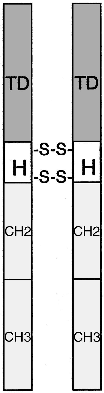
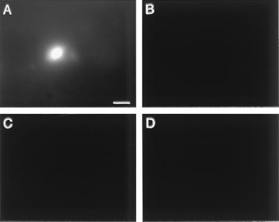
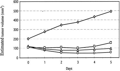
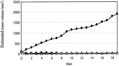
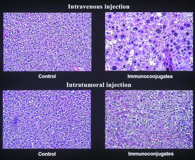
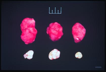
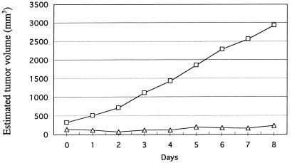
The efficacy and safety of a new immunotherapy protocol for cancer were tested in a severe combined immunodeficient mouse model of human skin and metastatic lung melanoma. The protocol involves intratumoral injections of replication-incompetent adenoviral vectors encoding immunoconjugates composed of the Fc region of an IgG1 immunoglobulin conjugated to a tumor-targeting domain. One targeting domain is factor VII that binds to tissue factor expressed on endothelial cells lining the tumor neovasculature and on tumor cells; the active site of factor VII was mutated to inhibit the initiation of blood coagulation. Another targeting domain is a single-chain Fv antibody that binds to a cognate antigen expressed on human melanoma cells. The adenoviral vectors infect mainly the cells of the injected tumor, which synthesize and secrete the immunoconjugates. The bloodborne immunoconjugates induce a cytolytic immune response against the targeted neovasculature endothelial cells and tumor cells. The mouse model experiments showed that intratumoral delivery of the factor VII immunoconjugate, either alone or together with the single-chain Fv immunoconjugate, resulted in growth inhibition and regression of the injected tumor, and also of distant metastatic tumors, without evidence of damage to normal organs. There was extensive destruction of the tumor neovasculature, presumably mediated by the factor VII immunoconjugate bound to tissue factor on neovasculature endothelial cells. Because tissue factor is generally expressed on neovascular endothelial cells and tumor cells, a factor VII immunoconjugate could be used for immunotherapy against a broad range of human solid tumors.
Figures
Similar articles
-
Targeting tumor vasculature endothelial cells and tumor cells for immunotherapy of human melanoma in a mouse xenograft model.Proc Natl Acad Sci U S A. 1999 Jul 6;96(14):8161-6. doi: 10.1073/pnas.96.14.8161. Proc Natl Acad Sci U S A. 1999. PMID: 10393965 Free PMC article.
-
Targeting tissue factor on tumor vascular endothelial cells and tumor cells for immunotherapy in mouse models of prostatic cancer.Proc Natl Acad Sci U S A. 2001 Oct 9;98(21):12180-5. doi: 10.1073/pnas.201420298. Epub 2001 Oct 2. Proc Natl Acad Sci U S A. 2001. PMID: 11593034 Free PMC article.
-
A recombinant adenovirus vector encoding the light chain of human coagulation factor VII and IgG1 Fc fragment to targeting tissue factor for colorectal cancer immunotherapy in the mouse model.J Cancer Res Clin Oncol. 2013 Jun;139(6):1015-23. doi: 10.1007/s00432-013-1417-1. Epub 2013 Mar 15. J Cancer Res Clin Oncol. 2013. PMID: 23494077
-
INGN 201: Ad-p53, Ad5CMV-p53, adenoviral p53, p53 gene therapy--introgen, RPR/INGN 201.Drugs R D. 2007;8(3):176-87. doi: 10.2165/00126839-200708030-00005. Drugs R D. 2007. PMID: 17472413 Review.
-
Human cancer detection and immunotherapy with conjugated and non-conjugated monoclonal antibodies.Anticancer Res. 1996 Mar-Apr;16(2):661-74. Anticancer Res. 1996. PMID: 8687112 Review.
Cited by
-
Factor VII light chain-targeted lidamycin shows intensified therapeutic efficacy for liver cancer.Cancer Biother Radiopharm. 2012 Aug;27(6):384-91. doi: 10.1089/cbr.2012.1209. Epub 2012 May 31. Cancer Biother Radiopharm. 2012. PMID: 22651685 Free PMC article.
-
Recombinant epidermal growth factor-like domain-1 from coagulation factor VII functionalized iron oxide nanoparticles for targeted glioma magnetic resonance imaging.Int J Nanomedicine. 2016 Oct 6;11:5099-5108. doi: 10.2147/IJN.S116980. eCollection 2016. Int J Nanomedicine. 2016. PMID: 27785017 Free PMC article.
-
Beyond thrombosis: the impact of tissue factor signaling in cancer.J Hematol Oncol. 2020 Jul 14;13(1):93. doi: 10.1186/s13045-020-00932-z. J Hematol Oncol. 2020. PMID: 32665005 Free PMC article. Review.
-
Immunotherapy for choroidal neovascularization in a laser-induced mouse model simulating exudative (wet) macular degeneration.Proc Natl Acad Sci U S A. 2003 Mar 4;100(5):2679-84. doi: 10.1073/pnas.0438014100. Epub 2003 Feb 14. Proc Natl Acad Sci U S A. 2003. PMID: 12589025 Free PMC article.
-
Chimeric antigen receptor-modified T Cells inhibit the growth and metastases of established tissue factor-positive tumors in NOG mice.Oncotarget. 2017 Feb 7;8(6):9488-9499. doi: 10.18632/oncotarget.14367. Oncotarget. 2017. PMID: 28055955 Free PMC article.
References
Publication types
MeSH terms
Substances
Associated data
- Actions
- Actions
Grants and funding
LinkOut - more resources
Full Text Sources
Other Literature Sources
