. 2000 Aug;20(16):5897-907.
doi: 10.1128/MCB.20.16.5897-5907.2000.

Relief of two built-In autoinhibitory mechanisms in P-TEFb is required for assembly of a multicomponent transcription elongation complex at the human immunodeficiency virus type 1 promoter

Affiliations

Relief of two built-In autoinhibitory mechanisms in P-TEFb is required for assembly of a multicomponent transcription elongation complex at the human immunodeficiency virus type 1 promoter

Y W Fong et al. Mol Cell Biol. 2000 Aug.

Abstract

Tat stimulation of human immunodeficiency virus type 1 (HIV-1) transcription requires Tat-dependent recruitment of human positive transcription elongation factor b (P-TEFb) to the HIV-1 promoter and the formation on the trans-acting response element (TAR) RNA of a P-TEFb-Tat-TAR ternary complex. We show here that the P-TEFb heterodimer of Cdk9-cyclin T1 is intrinsically incapable of forming a stable complex with Tat and TAR due to two built-in autoinhibitory mechanisms in P-TEFb. Both mechanisms exert little effect on the P-TEFb-Tat interaction but prevent the P-TEFb-Tat complex from binding to TAR RNA. The first autoinhibition arises from the unphosphorylated state of Cdk9, which establishes a P-TEFb conformation unfavorable for TAR recognition. Autophosphorylation of Cdk9 overcomes this inhibition by inducing conformational changes in P-TEFb, thereby exposing a region in cyclin T1 for possible TAR binding. An intramolecular interaction between the N- and C-terminal regions of cyclin T1 sterically blocks the P-TEFb-TAR interaction and constitutes the second autoinhibitory mechanism. This inhibition is relieved by the binding of the C-terminal region of cyclin T1 to the transcription elongation factor Tat-SF1 and perhaps other cellular factors. Upon release from the intramolecular interaction, the C-terminal region also interacts with RNA polymerase II and is required for HIV-1 transcription, suggesting its role in bridging the P-TEFb-Tat-TAR complex and the basal elongation apparatus. These data reveal novel control mechanisms for the assembly of a multicomponent transcription elongation complex at the HIV-1 promoter.


Figures

FIG. 1
FIG. 1
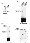
Requirement of ATP hydrolysis for formation of a stable P-TEFb–Tat–TAR ribonucleoprotein complex. (A) The Cdk9–HA-CycT1Δ1 heterodimer was affinity purified from 293T cells transiently transfected with an HA-tagged CycT1Δ1 cDNA by α-HA IP followed by HA peptide elution. The purified fraction containing the heterodimer and free HA-CycT1Δ1 was incubated with 32P-labeled wild-type TAR RNA (lanes labeled w) or the loop mutant TAR+31/+34 (lanes labeled m) in the absence (−) or presence (+) of Tat and/or ATP as indicated. The reactions were analyzed by EMSA. Lanes 9 and 12 also contained anti-Cdk9 antibodies (α-Cdk9), which caused the retardation of the Cdk9–HA-CycT1Δ1–Tat–TAR complex and also partially destabilized the complex, probably because of the polyclonal nature of the antibodies. (B) Silver-stained SDS-polyacrylamide gel showing the presence of Cdk9–HA-CycT1Δ1 and free HA-CycT1Δ1 in the α-HA IP fraction (lane 1). A nonspecific protein (∗) was revealed in a control fraction prepared in parallel from untransfected 293T cells (lane 2). (C) Free HA-CycT1Δ1 present in the α-HA IP fraction formed an ATP-independent complex with Tat and TAR in gel shift reactions. Recombinant CycT1Δ1 (rT1Δ1) was used as a reference (lanes 5 to 7) for determining the identity of the ATP-independent complex formed with the α-HA IP fraction (lanes 8 to 10). (D) Requirement of ATP hydrolysis for P-TEFb–Tat–TAR assembly. All reaction mixtures contained the α-HA IP fraction, Tat, and 32P-labeled wild-type TAR RNA. [γ-S]ATP was used in place of ATP in lane 3.
FIG. 2
FIG. 2
The P-TEFb–Tat–TAR complex is stabilized by C-terminal truncation of CycT1 in addition to P-TEFb phosphorylation. (A) Diagram showing the domain structures of CycT1FL and the C-terminal truncation mutant constructs CycT1Δ1 to -Δ4. The C-terminal boundary of TRM is located between amino acids 250 and 262 at the C-terminal edge of the conserved cyclin box domain (amino acids 1 to 272). The N-terminal boundary of TRM is unclear. (B) The C-terminal truncation of CycT1 did not affect its association with Cdk9. HA-CycT1FL or HA-CycT1Δ1 to -Δ4 and their associated Cdk9 proteins were affinity purified from 293T cells transiently expressing the various HA-tagged CycT1 proteins by anti-HA IP. The levels of Cdk9 and HA-CycT1 in these preparations were analyzed by Western blotting with anti-Cdk9 and anti-HA antibodies. A faint band right below CycT1FL is a nonspecific cross-reactive protein. (C) Equal amounts of Cdk9–HA-CycT1FL and Cdk9–HA-CycT1Δ1 to -Δ4 were tested for their abilities to form ternary complexes with Tat and 32P-labeled wild-type (lanes w) or the loop mutant (lanes m) TAR RNAs in a gel mobility shift assay.
FIG. 3
FIG. 3
Autophosphorylation of Cdk9 is required for efficient P-TEFb–Tat–TAR complex formation. (A) P-TEFb complexes containing either HA-tagged wild-type Cdk9 (WT) or a kinase-defective Cdk9 (D167N) were affinity purified by α-HA IP and normalized for their CycT1 levels by Western blotting with anti-CycT1 antibodies. (B) The kinase activity of Cdk9 is required for P-TEFb–Tat–TAR formation. The two P-TEFb complexes prepared for panel A were compared in a gel mobility shift assay for forming P-TEFb–Tat–TAR complexes in the presence (+) or absence (−) of ATP. Since the HA tag was attached to Cdk9, no free CycT1 was present in these α-HA IP fractions. (C) Autophosphorylation of P-TEFb is required for P-TEFb–Tat–TAR complex formation. Purified Cdk9-HA–CycT1 immobilized on anti-HA antibody beads was incubated with ATP in a kinase reaction mixture. Upon removal of ATP, the phosphorylated complex was eluted from the beads and incubated with Tat and 32P-labeled wild-type TAR RNA in a reaction mixture without ATP. An equal amount of unphosphorylated P-TEFb was used as a control. (D) Phosphorylation of CycT1 by Cdk9 is not required for P-TEFb–Tat–TAR formation. Cdk9–HA-CycT1FL and Cdk9–HA-CycT1Δ1 to -Δ4 complexes were prepared, and their levels were normalized as described for Fig. 2B. Equal amounts of these complexes were analyzed in in vitro kinase reactions. Note that HA-CycTΔ1 was not phosphorylated by Cdk9. However, Cdk9–HA-CycT1Δ1 formed an ATP-dependent complex with Tat-TAR.
FIG. 4
FIG. 4
P-TEFb phosphorylation does not affect P-TEFb–Tat binding but induces conformational changes in P-TEFb for better TAR recognition. (A) Equal amounts of in vitro-phosphorylated P-TEFb (Cdk9-HA–CycT1; lane 4), unphosphorylated P-TEFb (lanes 1 to 3), and alkaline phosphatase (AP; 10 U)-treated P-TEFb (lane 5) complexes were incubated with wild-type (WT) GST–Tat(1-48) proteins bound to glutathione-Sepharose beads. After washes, the amount of P-TEFb bound to Tat was examined by Western blotting with anti-CycT1 antibodies (α-CycT1). The GST–Tat(1-48, C22G) mutant protein, which contains a point mutation in the Tat activation domain, was used as a control (lane 3). (B) Alkaline phosphatase treatment inhibited the formation of the Cdk9–T1Δ1-Tat-TAR complex but not the complex containing free T1Δ1. The binding and gel shift conditions are the same as described for lane 11 of Fig. 1A, except that 10 U of AP was included in the binding reaction mixture in lane 3 prior to electrophoresis. (C) P-TEFb phosphorylation resulted in changes in trypsin sensitivity in the CycT1 C-terminal region. Purified Cdk9-Flag–HA-CycT1 complexes were first subjected to in vitro kinase reactions in the presence or absence of ATP, and then trypsin was added to the reaction mixtures. The cleaved products were detected by Western blotting with an anti-HA monoclonal antibody (α-HA) which recognizes the HA tag at the N terminus of CycT1. The position of a cross-reactive protein doublet is indicated by an ∗.
FIG. 5
FIG. 5
The CycT1 C-terminal region interacts with the N-terminal region to inhibit P-TEFb–Tat–TAR complex formation. (A) HA-tagged recombinant CycT1Δ1 proteins were incubated with equal amounts of GST or GST–CycT1-C (CycT1 C-terminal fragment, amino acids 402 to 701) bound to glutathione-Sepharose beads. After washes, bound CycT1Δ1 was detected by Western blotting with the anti-HA monoclonal antibody 12CA5. Twenty percent of the CycT1Δ1 used in the binding reaction mixture was shown as input. (B) Binding of CycT1-C to recombinant CycT1Δ1 (rT1Δ1) and the Cdk9–CycT1Δ1 complex (Cdk9/T1Δ1) in trans did not impede their interactions with Tat and TAR. Recombinant CycT1Δ1 (∼200 ng) or Cdk9-CycT1Δ1 (∼300 ng) was incubated in the presence or absence of CycT1-C (∼400 ng) for 10 min, followed by the addition of Tat (100 ng) and 32P-labeled TAR RNA. The reaction products were analyzed by gel mobility shift assay. (C) Binding of CycT1-C to Cdk9-CycT1FL disrupted the intramolecular interaction in CycT1 and stabilized the P-TEFb–Tat–TAR complex. Reaction mixtures containing CycT1-C (∼400 ng), Cdk9-CycT1FL (∼300 ng), Tat (100 ng), TAR, and ATP were analyzed as described for panel B. Lane 1′ is an eight-times-longer exposure of lane 1 and shows the position of Cdk9-CycT1FL–Tat–TAR in the gel.
FIG. 6
FIG. 6
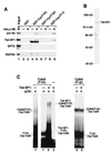
Tat-SF1 interacts with the C-terminal region of CycT1 and represses the autoinhibitory activity of this region. (A) The C-terminal region of CycT1 interacted with Tat-SF1 and RNA Pol IIa in HeLa nuclear extracts (NE). HeLa nuclear extracts were incubated with equal amounts of GST, GST-CycT1FL, GST-CycT1Δ1, and GST-CycT1-C proteins bound to glutathione-Sepharose beads. After washes, the bound proteins were analyzed by Western blotting with antibodies specific for Tat-SF1, Pol IIa, RAP30, and SPT5. Five percent of the nuclear extracts used in the binding reaction mixture were shown as input. The nonspecific bands shown in lanes 4 and 5 in anti-Tat-SF1 and anti-RAP30 antibody panels are cross-reactive bacterial proteins. (B) Silver-stained SDS-polyacrylamide gel showing Flag-tagged Tat-SF1 affinity purified from transfected 293T cells. (C) Binding of Tat-SF1 to the C-terminal region of CycT1 enhanced the P-TEFb–TAR interaction. Equal amounts of Cdk9–HA-CycT1FL (Cdk9/T1FL; lanes 1′, 1, and 2) and Cdk9–HA-CycT1Δ1 (Cdk9/T1Δ1; lanes 4 and 5) were incubated with Tat, 32P-labeled TAR, and ATP in the presence or absence of purified Tat-SF1. Reaction products were analyzed by gel mobility shift assay. Lane 1′ is an eight-times-longer exposure of lane 1 and shows the position of Cdk9–T1FL-Tat-TAR.
FIG. 7
FIG. 7
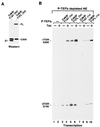
The C-terminal region of CycT1 is required for efficient HIV-1 transcriptional elongation. (A) P-TEFb complexes containing either HA-tagged CycT1Δ1 or CycT1FL were normalized for their Cdk9 and CycT1 levels by Western blotting with anti-Cdk9 and anti-HA antibodies. (B) Equal amounts of the two P-TEFb complexes were added to transcription reaction mixtures containing P-TEFb-depleted HeLa nuclear extracts (NE) as well as the DNA templates pHIV+TAR-G400 and pHIVΔTAR-G100 in the presence (+) or absence (−) of Tat protein. The amount of P-TEFb analyzed in lanes 3 to 6 was three times (3×) higher than that in lanes 7 to 10. +TAR-G400 and ΔTAR-G100 are RNase T1-resistant RNA fragments transcribed from the two G-less DNA cassettes (400 and 100 bp) inserted, respectively, into pHIV+TAR-G400 and pHIVΔTAR-G100 at a position ≈1 kb downstream of the HIV-1 promoter region.
FIG. 8
FIG. 8
Model for the assembly of the P-TEFb–Tat–TAR complex through relief of two built-in autoinhibitory mechanisms in P-TEFb. See the text for details. It is important to point out that in this diagram the order of events depicted between the first and last steps is purely hypothetical.

Similar articles

Cited by

References

    1. Bieniasz P D, Grdina T A, Bogerd H P, Cullen B R. Recruitment of a protein complex containing Tat and cyclin T1 to TAR governs the species specificity of HIV-1 Tat. EMBO J. 1998;17:7056–7065. - PMC - PubMed
    1. Chen D, Fong Y, Zhou Q. Specific interaction of Tat with the human but not rodent P-TEFb complex mediates the species-specific Tat activation of HIV-1 transcription. Proc Natl Acad Sci USA. 1999;96:2728–2733. - PMC - PubMed
    1. Chun R F, Jeang K T. Requirements for RNA polymerase II carboxyl-terminal domain for activated transcription of human retroviruses human T-cell lymphotropic virus I and HIV-1. J Biol Chem. 1996;271:27888–27894. - PubMed
    1. Cujec T P, Cho H, Maldonado E, Meyer J, Reinberg D, Peterlin B M. The human immunodeficiency virus transactivator Tat interacts with the RNA polymerase II holoenzyme. Mol Cell Biol. 1997;17:1817–1823. - PMC - PubMed
    1. Cujec T P, Okamoto H, Fujinaga K, Meyer J, Chamberlin H, Morgan D O, Peterlin B M. The HIV transactivator TAT binds to the CDK-activating kinase and activates the phosphorylation of the carboxy-terminal domain of RNA polymerase II. Genes Dev. 1997;11:2645–2657. - PMC - PubMed

Publication types

MeSH terms

Substances

LinkOut - more resources