. 2000 Aug;74(16):7508-17.
doi: 10.1128/jvi.74.16.7508-7517.2000.

Characterization of the vaccinia virus H3L envelope protein: topology and posttranslational membrane insertion via the C-terminal hydrophobic tail

Affiliations

Characterization of the vaccinia virus H3L envelope protein: topology and posttranslational membrane insertion via the C-terminal hydrophobic tail

F G da Fonseca et al. J Virol. 2000 Aug.

Abstract

The vaccinia virus H3L open reading frame encodes a 324-amino-acid immunodominant membrane component of virus particles. Biochemical and microscopic studies demonstrated that the H3L protein was expressed late in infection, accumulated in the cytoplasmic viral factory regions, and associated primarily with amorphous material near immature virions and with intracellular virion membranes. Localization of the H3L protein on the surfaces of viral particles and anchorage via the hydrophobic tail were consistent with its extraction by NP-40 in the absence of reducing agents, its trypsin sensitivity, its reactivity with a membrane-impermeable biotinylation reagent, and its immunogold labeling with an antibody to a peptide comprising amino acids 247 to 259. The H3L protein, synthesized in a coupled in vitro transcription/translation system, was tightly anchored to membranes as determined by resistance to Na(2)CO(3) (pH 11) extraction and cytoplasmically oriented as shown by sensitivity to proteinase K digestion. Further studies demonstrated that membrane insertion of the H3L protein occurred posttranslationally and that the C-terminal hydrophobic domain was necessary and sufficient for this to occur. These data indicated that the H3L protein is a member of the C-terminal anchor family and supported a model in which it is synthesized on free ribosomes and inserts into the membranes of viral particles during their maturation.


Figures

FIG. 1
FIG. 1
Synthesis of the H3L protein. BS-C-1 cells in six-well plates were mock infected (UN) or infected with 10 PFU of vaccinia virus per cell for 0, 2, 4, 6, 12, 18, or 24 h. The cells were collected by centrifugation, lysed with SDS and mercaptoethanol, passed several times through a 25- by 5-mm needle, and analyzed by SDS-PAGE. The separated proteins were transferred to a membrane and probed with rabbit polyclonal H3L peptide antibody followed by an antirabbit horseradish peroxidase conjugate. The bands detected by chemiluminescence are shown. The positions and masses of markers are indicated on the right.
FIG. 2
FIG. 2
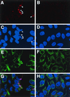
Localization of the H3L protein in infected cells. Confocal microscopy of uninfected HeLa cells (B, D, F, and H) and cells infected with vaccinia WR virus (A, C, E, and G). After 8 h, cells were fixed, permeabilized, and triple labeled with anti-H3L peptide antibody and rhodamine-conjugated anti-rabbit IgG, anti-PDI and fluorescein isothiocyanate-conjugated anti-mouse IgG, and Hoechst dye. Fluorescence due to the H3L antibody (A and B), DNA (C and D), and PDI (E and F) and a merged image of all three (G and H) are shown. Arrowheads, viral factories.
FIG. 3
FIG. 3
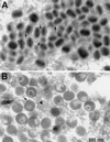
Immunogold electron microscopy. BS-C-1 cells that had been infected for 24 h were fixed in paraformaldehyde, cryosectioned, and incubated with H3L peptide antibody and then with 10-nm gold particles conjugated to protein A. (A) Field containing large numbers of IMV. (B) Field containing large numbers of IV. Arrows, gold particles associated with IMV or IV; arrowhead, gold particles in amorphous material in the factory area.
FIG. 4
FIG. 4
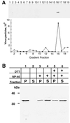
Association of the H3L protein with purified vaccinia virus particles and extraction with nonionic detergent. (A) Western blot of purified virus particles. Sucrose gradient-purified intracellular virions were centrifuged in a performed CsCl gradient. Fractions (0.5 ml) were collected from the top of the tube, diluted, and centrifuged to pellet virus particles. The pellets were suspended, and part of the suspension was used to calculate the number of virus particles from the absorbance at 260 nm; the remainder was analyzed by SDS-PAGE and immunoblotted with H3L peptide antibody followed by anti-rabbit IgG horseradish peroxidase conjugate. CsCl fraction numbers are indicated. (B) Extraction of H3L protein with NP-40 detergent. Purified vaccinia virions were incubated in Tris buffer containing 0.5% NP-40 or 0.5% NP-40 and 50 mM DTT. After centrifugation, the supernatant (S) and pellet (P) fractions were analyzed by SDS-PAGE and immunoblotting with the H3L peptide antibody. The masses and positions of markers are indicated at the left.
FIG. 5
FIG. 5
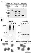
Topology of the H3L protein. (A) Trypsin sensitivity of the virion-associated H3L protein. Sucrose gradient-purified virus particles were treated with trypsin and then collected by centrifugation. Equivalent portions of the supernatant and pellet fractions were analyzed by SDS-PAGE and Western blotting with the H3L peptide antibody. Masses of protein markers are indicated on the left. (B) Biotinylation of membrane-bound H3L protein. Sucrose gradient-purified virus particles were incubated with sulfo-NHS-LC-biotin for 30 min. At the end of the reaction, the mixture was layered over a sucrose cushion and centrifuged to separate virus particles from residual unlinked biotin. A portion of the biotinylated virus particles was analyzed by SDS-PAGE (lane 2); another portion was extracted with NP-40 and separated into soluble (lane 3) and insoluble (lane 4) fractions. Lane 1, nonbiotinylated virions; lane 5, soluble proteins that were biotinylated after NP-40 extraction and centrifugation. The electrophoretically separated proteins were transferred to a membrane and probed with NeutrAvidin (avidin). The membrane was then stripped and immunoblotted with the anti-H3L antibody followed by anti-rabbit IgG horseradish peroxidase conjugate (anti-H3). (C) Decoration of vaccinia virus particles with the antibody to the H3L protein. Sucrose gradient-purified vaccinia virions were adsorbed to grids and incubated with the H3L peptide antibody and protein A-gold (right) or with protein A-gold alone (left). After negative staining, the grids were examined by electron microscopy.
FIG. 6
FIG. 6
In vitro synthesis of the H3L protein and association with microsomal membranes: sensitivity to Na2CO3 and proteinase K. (A) Proteinase sensitivity of the membrane-associated H3L protein. The H3L ORF, regulated by a bacteriophage T7 promoter, was transcribed and translated in a reticulocyte lysate in the presence of [35S]methionine. Reactions were carried out in the absence (lane 2) or presence (lanes 1 and 3 to 7) of a DNA template or in the absence (lanes 1 and 3) or presence (lanes 2 and 4 to 7) of canine microsomal membranes (micro). The mixtures were layered over a sucrose cushion and centrifuged and supernatant (S) and pellet (P) fractions were obtained. Some of the pellet fractions were treated with proteinase K alone for 1 min (lane 5) or 2 min (lane 6) or with Triton X-100 plus proteinase K (lane 7). The samples were analyzed by SDS-PAGE and autoradiography. (B) Resistance of membrane-bound H3L protein to Na2CO3 extraction. Reactions were carried out and analyzed as for panel A except that in lane 5 the microsomes were incubated in Na2CO3 and then centrifuged through a sucrose cushion before SDS-PAGE. The masses and positions of markers are indicated on the left.
FIG. 7
FIG. 7
Hydrophobicity and posttranslational insertion of the H3L protein into membranes. (A) Partition of the H3L protein in the Triton X-114 detergent phase. The H3L protein was synthesized in vitro in the absence of microsomal membranes as described in the legend to Fig. 6. Triton X-114 detergent was added to the reaction mixture, and the mixture was subjected to temperature-induced phase separation. The [35S]methionine-labeled proteins in the total extract (T) and in the aqueous (A) and detergent (D) phases were analyzed by SDS-PAGE and autoradiography. Lane 2, control in which no template was added. (B) Posttranslational insertion of the H3L protein into membranes. Transcription/translation in the absence or presence of DNA or microsomal membranes was carried out as described in the legend to Fig. 6. In lane 3, 200 μg of cycloheximide/ml was added to stop translation before the addition of microsomal membranes to demonstrate posttranslational membrane insertion of H3L. In lane 4, cycloheximide was added at the start of the reaction to demonstrate complete inhibition of translation. The masses and positions of markers are indicated at the left.
FIG. 8
FIG. 8
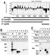
Membrane insertion of truncated H3L proteins. (A) Hydrophilicity plot of H3L protein. Bars beneath the plot, full-length H3L protein, an N-terminal (NT) truncation, a C-terminal (CT) truncation, and a C-terminal peptide. (B) Insertion of truncated H3L proteins into membranes. Transcription/translation in the presence or absence of microsomal membranes and analysis of supernatant and pellet fractions by SDS-PAGE and autoradiography were carried out as described in the legend to Fig. 6. Lane 1, no DNA was added to the reaction mixture; lanes 2 to 5, the full-length (FL) protein was synthesized; lanes 6 to 8, the C-terminal truncated species (CTr) was synthesized; lanes 9 to 11, the N-terminal truncated species (NTr) was synthesized. Soluble (S) and pellet (P) fractions were analyzed by SDS-PAGE and autoradiography. (C) Insertion of a C-terminal peptide (CT pep) into membranes. Transcription/translation reactions in the absence or presence of microsomal membranes were carried out using a template encoding the C-terminal peptide. Where indicated (+), proteinase K treatment was carried out after the separation of supernatant and pellet fractions. Masses and positions of markers are indicated at the left.

Similar articles

Cited by

References

    1. Betakova T, Wolffe E J, Moss B. Membrane topology of the vaccinia virus A17L envelope protein. Virology. 1999;261:347–356. - PubMed
    1. Betakova T, Wolffe E J, Moss B. Vaccinia virus A14.5L gene encodes a hydrophobic 53-amino-acid virion membrane protein that enhances virulence in mice and is conserved among vertebrate poxviruses. J Virol. 2000;74:4085–4092. - PMC - PubMed
    1. Blobel G, Dobberstein B. Transfer of proteins across membranes. II. Reconstruction of functional rough microsomes from heterologous components. J Cell Biol. 1975;67:852–862. - PMC - PubMed
    1. Bordier C. Phase separation of integral membrane proteins in Triton X-114 solution. J Biol Chem. 1981;256:1604–1607. - PubMed
    1. da Fonseca F G, Weisberg A, Wolffe E J, Moss B. Effects of deletion or stringent repression of the H3L envelope gene on vaccinia virus replication. J Virol. 2000;74:7518–7528. - PMC - PubMed

Publication types

MeSH terms

Substances

LinkOut - more resources