. 2000 Jul 17;19(14):3576-85.
doi: 10.1093/emboj/19.14.3576.

Measles virus matrix protein specifies apical virus release and glycoprotein sorting in epithelial cells

Affiliations

Measles virus matrix protein specifies apical virus release and glycoprotein sorting in epithelial cells

H Y Naim et al. EMBO J. .

Abstract

In polarized epithelial cells measles virus (MV) is predominantly released at the apical cell surface, irrespective of the sorting of its two envelope glycoproteins F and H. It has been reported previously that the viral matrix (M) protein modulates the fusogenic capacity of the viral envelope glycoproteins. Here, extant MV mutants and chimeras were used to determine the role of M protein in the transport of viral glycoproteins and release of progeny virions in polarized epithelial CaCo2 cells. In the absence of M, envelope glycoproteins are sorted to the basolateral surface, suggesting that they possess intrinsic basolateral sorting signals. However, interactions of M with the glycoprotein cytoplasmic tails allow M-glycoprotein co-segregation to the apical surface, suggesting a vectorial function of M to retarget the glycoproteins for apical virion release. Whereas this may allow virus airway shedding, the intrinsic sorting of the glycoproteins to the basolateral surface may account for systemic host infection by allowing efficient cell-cell fusion.


Figures

None
Fig. 1. Structures of MV cDNAs encoding standard, recombinant, mutant and chimeric viruses. The portions of the plasmids giving rise to anti-genomic RNAs are shown. The constructions of p(+)MV and its derivatives p(+)MVΔM, p(+)MVΔtails, p(+)MGV and p(+)MG/FV were reported (Radecke et al., 1995; Cathomen et al., 1998a,b; Spielhofer et al., 1998). The white boxes represent the reading frames of the six MV genes N, P, M, F, H and L. The black boxes represent the non-translated gene boundary regions. The gray boxes represent reading frames of VSV-G and the hybrid VSV-G/MV-F, respectively. The extent of deletions (Δ) of M, as well as F and H tails, and the replacement of the G tail by that of F are indicated. The dark gray box represents the position of the GFP reading frame in the MV genome sequence. Strong expression of GFP was ensured by creating an additional independent transcription unit upstream of N; a duplicate of the N/P intergenic region was placed between the GFP and the N reading frames. The vertical gray lines represent the transmembrane regions in F, H, G and G/F reading frames.
None
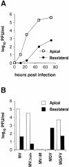
Fig. 2. Polarized release of MV and mutant viruses in CaCo2 cells. (A) Release of MV at increasing times p.i. CaCo2 cells were grown on transwell filters until a tight monolayer was reached. Cells were then infected with standard MV, and medium was collected from the apical and the basolateral domains at 12, 24, 48 and 70 h p.i. Infectious viruses released to either side were plaque assayed. (B) Effects of F- and H-tail truncation, M deletion and envelope glycoprotein exchange on virus budding. At 50 h p.i. (MV, MVΔtails and MVΔM) and 70 h p.i. (MGV and MG/FV) virus shed into the media from the apical and basolateral domains was determined by plaque assays.
None
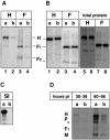
Fig. 3. Sorting of infectious MV and the envelope glycoproteins F and H in CaCo2 cells. Infected cells were radiolabeled at (A) 30 h p.i. and (B) 60 h p.i., for 3 h and chased for another 3 h. Monoclonal anti-F or anti-H antibodies were applied from either the apical or the basolateral domains; captured proteins were immunoprecipitated by protein A–Sepharose. To analyze total proteins, additional antibodies were added to the corresponding cell lysates and immunoprecipitated (B, lanes 5–8). (C) Monoclonal anti-SI antibody was used to capture surface SI at the apical surface. (D) Labeled particles were isolated from infected cultures after 3 h pulse and 3 h chase at 30–36 h p.i. and at 60–66 h p.i. Viruses were partially purified by pelleting through a 30% sucrose cushion; the asterisk indicates the position of N and possibly F proteins. Proteins were analyzed by 10–12% SDS–PAGE and quantitated by phosphoimaging. (E) Localization of viral glycoproteins at the surface by immunofluorescence and confocal vertical sections at 36 h p.i. (a) and 66 h p.i. (b). Monoclonal anti-F antibody coupled to Cy3 was used. Bar, 10 µm.
None
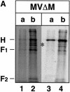
Fig. 4. Sorting of F and H in MVΔM-infected CaCo2 cells. Infection and pulse–chase were exactly as for standard MV. (A) In this experi ment both anti-H and anti-F antibodies were added consecutively from either the apical or the basolateral domains. The asterisk indicates uncleaved F0 protein (shown as double bands). (B) Localization of viral proteins by immunofluorescence and confocal vertical sections at 36 h p.i. (a) and 66 h p.i. (b). Monoclonal anti-F antibody coupled to Cy3 was used. Bar, 10 µm.
None
Fig. 5. Envelope and M protein sorting in MGV, MG/FV and standard MV-infected CaCo2 cells. (A) MGV- and MG/FV-infected cells were pulsed and chased at 70 h p.i., and polyclonal anti-VSV antibody was used. To analyze total G and G/F proteins, additional antibodies were added to the corresponding cell lysates and immunoprecipitated (lanes 5–8); the faster bands in lanes 7 and 8 represent the immature high-mannose population. (B) Localization of M, G and G/F by confocal vertical sections; M was stained with FITC (green), G and G/F were stained with Cy3 (red). (C) Localization of M and H in CaCo2 cells infected with standard MV. Cells were fixed and permeabilized at 66 h p.i. and double stained first with polyclonal anti-H coupled to FITC (green signal in b and c), then with monoclonal anti-M coupled to Cy3 (red signal in a and c). To demonstrate the polarization of the CaCo2 cells, antibody to DPPIV, which sorts to the apical domain (d), and antibody to gp120 (e), which sorts to the basolateral domain, were used in MV-infected cells. Bar, 10 µm.
None
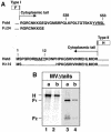
Fig. 6. Sorting of F and H with cytoplasmic tail deletion. (A) Scheme of F- and H-tail deletions. The underlined letters highlight the potential basolateral sorting signals and their positions in the cytoplasmic domains. (B) Polarity of FΔtail and HΔtail at 66 h p.i. Proteins were expressed by MVΔtails, labeled and immunoprecipitated as with standard MV in Figure 3. (C) Immunofluorescence analysis at 66 h p.i. of HΔtail coupled to FITC (green, b and c) and M with Cy3 (red, a and c). Bar, 10 µm.

Similar articles

Cited by

References

    1. Alfalah M., Jacob,R., Preuss,U., Zimmer,K.P., Naim,H. and Naim,H.Y. (1999) O-linked glycans mediate apical sorting of human intestinal sucrase-isomaltase through association with lipid rafts. Curr. Biol., 9, 593–596. - PubMed
    1. Billeter M.A., Cattaneo,R., Spielhofer,P., Kaelin,K., Huber,M., Schmid,A., Baczko,K. and ter Meulen,V. (1994) Generation and properties of measles virus mutations typically associated with subacute sclerosing panencephalitis. Ann. N Y Acad. Sci., 724, 367–377. - PubMed
    1. Blau D.M. and Compans,R.W. (1995) Entry and release of measles virus are polarized in epithelial cells. Virology, 210, 91–99. - PubMed
    1. Bohn W., Rutter,G., Hohenberg,H., Mannweiler,K. and Nobis,P. (1986) Involvement of actin filaments in budding of measles virus: studies on cytoskeletons of infected cells. Virology, 149, 91–106. - PubMed
    1. Brown D.A. and Rose,J.K. (1992) Sorting of GPI-anchored proteins to glycolipid-enriched membrane subdomains during transport to the apical cell surface. Cell, 68, 533–544. - PubMed

Publication types

MeSH terms