. 2000 Jul 10;150(1):193-203.
doi: 10.1083/jcb.150.1.193.

Morphogenic effects of ezrin require a phosphorylation-induced transition from oligomers to monomers at the plasma membrane

Affiliations

Morphogenic effects of ezrin require a phosphorylation-induced transition from oligomers to monomers at the plasma membrane

A Gautreau et al. J Cell Biol. .

Abstract

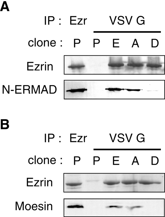
ERM (ezrin, radixin, moesin) proteins act as linkers between the plasma membrane and the actin cytoskeleton. An interaction between their NH(2)- and COOH-terminal domains occurs intramolecularly in closed monomers and intermolecularly in head-to-tail oligomers. In vitro, phosphorylation of a conserved threonine residue (T567 in ezrin) in the COOH-terminal domain of ERM proteins disrupts this interaction. Here, we have analyzed the role of this phosphorylation event in vivo, by deriving stable clones producing wild-type, T567A, and T567D ezrin from LLC-PK1 epithelial cells. We found that T567A ezrin was poorly associated with the cytoskeleton, but was able to form oligomers. In contrast, T567D ezrin was associated with the cytoskeleton, but its distribution was shifted from oligomers to monomers at the membrane. Moreover, production of T567D ezrin induced the formation of lamellipodia, membrane ruffles, and tufts of microvilli. Both T567A and T567D ezrin affected the development of multicellular epithelial structures. Collectively, these results suggest that phosphorylation of ERM proteins on this conserved threonine regulates the transition from membrane-bound oligomers to active monomers, which induce and are part of actin-rich membrane projections.


Figures

Figure 1
Figure 1
T567D ezrin has a reduced N-ERMAD binding activity in vitro, and does not oligomerize with moesin at the membrane in vivo. Throughout this study, clones producing VSV G-tagged wt ezrin, T567A ezrin, T567D ezrin (E, A, and D cells, respectively) or clones obtained after transfection of the empty plasmid (P cells) were compared. (A) Denatured extracts from P, E, A, and D clones were used to immunoprecipitate ezrin or VSV G-tagged wt, T567A, and T567D ezrin. The immunoprecipitates were probed with ezrin antibodies or with biotinylated ezrin N-ERMAD (1–309). (B) Ezrin from P, E, A, or D cell membrane extracts was immunoprecipitated with either anti-ezrin antibodies or anti-VSV G antibodies as indicated. After SDS-PAGE, the immunoprecipitates were stained by Coomassie blue to reveal ezrin (top), or immunoblotted with moesin-specific antibodies (bottom). Moesin associated with ezrin can be seen in the Coomassie blue–stained gel as a faint band just below the strong ezrin band.
Figure 3
Figure 3
In the cytosol, T567D ezrin has a functional C-ERMAD. Various VSV G-tagged cDNAs, wt, T567A, and T567D ezrin, or an ezrin construct lacking a C-ERMAD due to the deletion of the 29 COOH-terminal amino acids (Δ29), were cotransfected with myc-tagged wt ezrin cDNA into LLC-PK1 cells to detect oligomerization. Total, cytosol, or membrane lysates (top) or VSV G immunoprecipitates of these lysates (bottom) were analyzed by immunoblotting with anti-VSV G or anti-Myc antibodies as indicated on the right of each panel. Δ29 ezrin was strongly enriched in the membrane fraction, and completely defective in oligomer formation. In contrast, T567D ezrin was correctly distributed between the cytosolic and the membrane fraction, and exhibited a strongly reduced amount of oligomers specifically at the membrane.
Figure 2
Figure 2
T567D ezrin and endogenous phosphorylated ERM proteins are preferentially monomeric at the plasma membrane. A, Membrane or cytosolic extracts from P, E, A, and D cells were resolved by gel filtration chromatography on a superose-6 column. Fractions 15–41 were analyzed by SDS-PAGE and immunoblotted with antiezrin antibodies (P) or anti-VSV G antibodies (E, A, and D). T567D ezrin exhibited a strongly reduced amount of oligomers at the membrane, but not in the cytosol. B, Total, cytosolic, and membrane extracts were immunoblotted with 297S mAb, recognizing all three ERM proteins when phosphorylated on this conserved threonine (pERM), or with a mixture of antibodies specific for ERM. Ezrin and radixin comigrated at ∼80 kD, and moesin migrated at 75 kD. Phosphorylated ERM proteins were strongly enriched in the membrane fraction. C, LLC-PK1 cells were pretreated with calyculinA, a protein phosphatase inhibitor, and the membrane extract was resolved by gel filtration chromatography. Oligomeric and monomeric fractions were pooled, and immunoblotted as in B. Monomers were preferentially phosphorylated over oligomers.
Figure 4
Figure 4
Morphology of LLC-PK1 cells producing T567D ezrin. (A) Clones were examined by phase-contrast optics. P, E, and A cells grew in typical LLC-PK1 islets (only the E control is presented). D colonies exhibited a number of morphological changes. In D colonies, cells were not always adherent to each other. The periphery of D colonies was irregular with wide lamellipodia (arrowheads). Those lamellipodia were occasionally formed at the extremity of long extensions (arrow). In most D cells, the membrane area around the nuclei was highly refractile. Bar, 25 μm. (B) Scanning EM examination of E and D cell morphology. Extensive membrane ruffling was observed in the cell central area, probably corresponding to the refractility observed by phase-contrast optics. Bars: (A) 25 μm; (B) 1 μm.
Figure 5
Figure 5
Scanning EM analysis of microvilli in clones producing T567A and T567D ezrin. Monolayers of P, E, and A cells exhibited comparable microvilli in density and length. In D cultures, above a layer of flat cells, which contain comparable microvilli to P, E, and A cells, some round cells containing numerous and long microvilli were frequently observed. In addition, tufts of microvilli occasionally emerged from flat D cells (right). Bars, 1 μm.
Figure 6
Figure 6
Ezrin association with the actin cytoskeleton requires phosphorylation of T567. (A) Localization of ezrin variants with anti-VSV G antibodies by immunofluorescence and confocal microscopy in E, A, or D cells. Cells were also stained after extraction with a Triton X-100 buffer that preserves cytoskeleton-associated material (csk). A single apical section is shown. Wt, T567A, and T567D ezrin were observed in microvilli. T567D ezrin was also present in the membrane ruffles it induced. After extraction of ezrin-soluble pool, wt and T567D ezrin staining were preserved, whereas T567A ezrin staining was strongly reduced. Bar, 5 μm. (B) Western blot analysis of ezrin cytoskeletal association. Similar fractions of soluble material (Sol), extracted with the Triton X-100 buffer, and insoluble material (Ins), were immunoblotted with anti-ezrin antibodies for P cell extract or with anti-VSV G antibodies for E, A, and D cell extracts. A densitometric analysis was performed and the Ins/Sol ratio was calculated from data obtained from two to four independent experiments with three different A and D clones (mean ± SEM). (C) Soluble and insoluble fractions from LLC-PK1 cells were equalized for their ERM content and immunoblotted with either 297S mAb (pERM) or ERM antibodies. Phosphorylated ERM proteins are enriched in the insoluble fraction.
Figure 7
Figure 7
Production of T567A and T567D ezrin affects the development of multicellular epithelial structures. (A) Morphogenesis of suspension cysts examined by phase-contrast optics. Aggregates of LLC-PK1 cells in suspension are able to form hollow epithelial cysts. P, E, and A cells were not affected in this process, whereas D cells formed loose aggregates in which individual cells could still be distinguished at the periphery. (B) Tubulogenesis assay examined by Nomarski optics. In three-dimensional collagen type I, in the presence of HGF, P cells are able to differentiate into multicellular tubules. Production of wt ezrin potentiated growth and branching morphogenesis of tubules. Production of both T567A and T567D ezrin impaired tubulogenesis. A cells exhibited a growth defect, whereas D cells grew in disorganized colonies. Bars, 50 μm.

Similar articles

Cited by

References

    1. Algrain M., Turunen O., Vaheri A., Louvard D., Arpin M. Ezrin contains cytoskeleton and membrane binding domains accounting for its proposed role as a membrane–cytoskeletal linker. J. Cell Biol. 1993;120:129–139. - PMC - PubMed
    1. Andréoli C., Martin M., Le Borgne R., Reggio H., Mangeat P. Ezrin has properties to self-associate at the plasma membrane. J. Cell Sci. 1994;107:2509–2521. - PubMed
    1. Berryman M., Franck Z., Bretscher A. Ezrin is concentrated in the apical microvilli of a wide variety of epithelial cells whereas moesin is found primarily in endothelial cells. J. Cell Sci. 1993;105:1025–1043. - PubMed
    1. Berryman M., Gary R., Bretscher A. Ezrin oligomers are major cytoskeletal components of placental microvillia proposal for their involvement in cortical morphogenesis. J. Cell Biol. 1995;131:1231–1242. - PMC - PubMed
    1. Bretscher A. Regulation of cortical structure by the ezrin-radixin-moesin protein family. Curr. Opin. Cell Biol. 1999;11:109–116. - PubMed

Publication types

MeSH terms