. 2000 Aug;74(15):6911-21.
doi: 10.1128/jvi.74.15.6911-6921.2000.

Characterization of an essential RNA secondary structure in the 3' untranslated region of the murine coronavirus genome

Affiliations

Characterization of an essential RNA secondary structure in the 3' untranslated region of the murine coronavirus genome

B Hsue et al. J Virol. 2000 Aug.

Abstract

We have previously identified a functionally essential bulged stem-loop in the 3' untranslated region of the positive-stranded RNA genome of mouse hepatitis virus. This 68-nucleotide structure is composed of six stem segments interrupted by five bulges, and its structure, but not its primary sequence, is entirely conserved in the related bovine coronavirus. The functional importance of individual stem segments of this stem-loop was characterized by genetic analysis using targeted RNA recombination. We also examined the effects of stem segment mutations on the replication of mouse hepatitis virus defective interfering RNAs. These studies were complemented by enzymatic and chemical probing of the stem-loop. Taken together, our results confirmed most of the previously proposed structure, but they revealed that the terminal loop and an internal loop are larger than originally thought. Three of the stem segments were found to be essential for viral replication. Further, our results suggest that the stem segment at the base of the stem-loop is an alternative base-pairing structure for part of a downstream, and partially overlapping, RNA pseudoknot that has recently been shown to be necessary for bovine coronavirus replication.


Figures
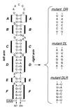
FIG. 1
FIG. 1
Mutational analysis of the proposed bulged stem-loop structure in the MHV 3′ UTR (12). Nucleotide numbering begins at the 3′ end of the genome, excluding the poly(A) tail; the N gene stop codon is boxed. The six stem segments of the structure are designated A through F. Shown at the right are strand replacement mutants for stem segment D.
FIG. 2
FIG. 2
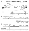
Plasmid construction and schemes for mutant selection. (A) Parent plasmid pBL85, used for construction of vectors for stem segment mutants, encodes a DI RNA composed entirely of MHV components, except for a 48-nt linker region (hatched rectangle). pBL85 was derived from pB36 (32) by creation of an MluI site near the end of the N gene and an EcoRV site in the 3′ UTR. Portions of sequence containing these two sites, as well as the encoded carboxy terminus of the N protein, are shown; the original wild-type nucleotide and amino acid residues are indicated in parentheses. Nucleotide numbering begins at the 3′ end of the genome, excluding the poly(A) tail; the N gene stop codon is boxed. Plasmid pBL122, constructed for generation of the positive-strand transcript used for enzymatic and chemical probing, contains the MluI-SacI fragment derived from pBL85. All restriction sites shown, except for BstEII, are unique in the plasmids in which they appear. The positions of primers BL66, BL67, PM28, and PM112, used for RT-PCR analysis of negative-strand DI RNA, are indicated. (B) Scheme for construction of MHV mutants by targeted recombination using the temperature-sensitive and thermolabile mutant Alb4 as the recipient virus. In this case, recombinants generated by the indicated crossover event can be selected on the basis of having regained wild-type thermal stability. (C) Scheme for targeted recombination using the interspecies chimeric virus fMHV, which grows only in feline cells. In this case, recombinants generated by the indicated crossover event can be selected on the basis of having regained the ability to grow in murine cells. In both panels B and C, the star represents mutations in the 3′ UTR transduced from the synthetic donor RNA into the recipient genome.
FIG. 3
FIG. 3
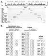
Composition and viability of stem segment mutants. (A) Nucleotide changes in stem segment mutants. The primary sequence of the wild-type (WT) bulged stem-loop structure is shown at the top, with stem segments labeled as in Fig. 1. Nucleotide numbering begins at the 3′ end of the genome, excluding the poly(A) tail. The loci of restriction sites MluI, EcoRV, and BstEII in the plasmid vector are indicated. For each mutant, only those bases that differ from the wild type are shown. The first letter of the name of each mutant (M or B) indicates the origin (MHV or BCoV, respectively) of the remainder of the 3′ UTR downstream of the stem-loop. The second letter (A, B, C, D, E, or F) indicates the stem segment. The final letters (L, R, or both) indicate which arm of the stem segment has been mutagenized. (B) Summary of the replicative ability of stem segment mutants in recombinant viruses or in DI RNAs. On the left are listed those donor RNA mutations for which viable recombinant viruses containing the mutations could (+) or could not (−) be obtained. For each mutation that scored negatively by the Alb4 targeted recombination strategy, an attempt was made to incorporate it into the viral genome by selection with fMHV. In the center are listed DI RNA replication results for stem mutations constructed in the background of the MHV 3′ UTR. For all replication results that were discrepant with the recombinant virus results, wild-type recombinant DI RNAs were detected by negative-strand-specific RT-PCR. On the right are listed DI RNA replication results for stem mutations constructed in the background of the BCoV 3′ UTR.
FIG. 4
FIG. 4
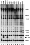
Replicative ability of DI RNA stem-loop mutants constructed with the background of the BCoV 3′ UTR. Mouse 17Cl1 cells infected with wild-type MHV were mock transfected (lane 1) or transfected with each indicated DI RNA and then labeled with 134 μCi of [33P]orthophosphate per ml in the presence of 20 μg of actinomycin D per ml from 10 to 12 h postinfection (12). Purified cytoplasmic RNA was denatured with formaldehyde and formamide, separated by electrophoresis through 1% agarose containing formaldehyde, and visualized by autoradiography. Control DI RNAs were pB36 (lane 2) (32), the original wild-type MHV DI progenitor of all DI RNAs in this study; pBL34 (lane 3), the original nonreplicating DI containing a chimeric MHV-BCoV stem-loop in the 3′ UTR (12); and pBL72 (lane 4), a DI RNA with the wild-type MHV stem-loop in the background of the BCoV 3′ UTR (12).
FIG. 5
FIG. 5
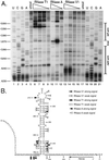
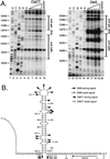
Enzymatic structural probing of the bulged stem-loop structure. A 264-nt RNA encompassing the stem-loop region of the MHV 3′ UTR was transcribed in vitro from plasmid pBL122 (Fig. 2A), purified, renatured, and digested with various concentrations of RNases. Positions of cleavage sites were determined by primer extension with a 5′-end-labeled primer, PM165, complementary to nt 199 to 216. (A) Enzymatic cleavage sites generated by single-stranded nucleases RNase T1 (G specific) and RNase A (U and C specific) and double-stranded nuclease RNase V1. Lanes 1 to 4 and 18 to 21, sequencing ladders generated with end-labeled PM165 and terminated with ddATP, ddGTP, ddCTP, or ddTTP, respectively; lanes 5 and 17, undigested RNA; lanes 6 to 9, RNA digested with 15, 10, 5.0, and 1.0 U of RNase T1, respectively; lanes 10 to 12, RNA digested with 0.01, 0.001, and 0.0001 U of RNase A, respectively; lanes 13 to 16, RNA digested with 0.5, 0.3, 0.1, and 0.05 U of RNase V1, respectively. Ten-nucleotide intervals and the position of the stem-loop are indicated to the left and right of the autoradiogram, respectively. Each primer extension product from nuclease-digested RNA terminates one base downstream of the corresponding nucleotide in the sequencing ladder because all three RNases cut 3′ to their target bases. (B) Summary of observed enzymatic cleavage sites superimposed on the originally proposed stem-loop structure. Nucleotide numbering begins at the 3′ end of the genome, excluding the poly(A) tail; the N gene stop codon is boxed, and the position of the primer PM165 is shown. Bases indicated in italics at the 5′ end of the synthetic RNA are those derived from the polylinker of the transcription vector. Artifactual RNase T1 signals at A293 and U295 are not included in this diagram.
FIG. 6
FIG. 6
Chemical structural probing of the bulged stem-loop structure. A 264-nt RNA encompassing the stem-loop region of the MHV 3′ UTR was transcribed in vitro from plasmid pBL122 (Fig. 2A), purified, renatured, and reacted with various concentrations of chemical reagents. Positions of base modification were determined by primer extension as for Fig. 5. (A) Modification sites generated by single-stranded RNA-specific reagents CMCT (G and U specific) and DMS (A and C specific). Lanes 1 to 4 and 11 to 14, sequencing ladders generated with end-labeled PM165 and terminated with ddATP, ddGTP, ddCTP, or ddTTP, respectively; lanes 5 and 15, unmodified RNA; lanes 6 to 9, RNA modified with 4.2, 8.4, 12.6, and 16.8 mg of CMCT per ml, respectively; lanes 16 to 19, RNA modified with 0.5, 1.0, 1.5, and 2.0% DMS, respectively; lanes 10 and 20, control reactions in which the highest concentration of each reagent was added to RNA after addition of quenching reagents. Ten-nucleotide intervals and the position of the stem-loop are indicated to the left and right of each autoradiogram, respectively. Each primer extension product from chemically modified RNA is positioned one base downstream of the corresponding nucleotide in the sequencing ladder because primer extension terminates at the nucleotide immediately 3′ to the modified base. (B) Summary of observed chemical modification sites superimposed on the originally proposed stem-loop structure. Nucleotide numbering begins at the 3′ end of the genome, excluding the poly(A) tail; the N gene stop codon is boxed, and the position of the primer PM165 is shown. Bases indicated in italics at the 5′ end of the synthetic RNA are those derived from the polylinker of the transcription vector. Artifactual DMS signals at U234, G257, U270, G271, and G294 are not included in this diagram.
FIG. 7
FIG. 7
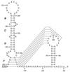
Refined model of the stem-loop structure based on genetic and structural experiments. Nucleotide numbering begins at the 3′ end of the genome, excluding the poly(A) tail, and the N gene stop codon is boxed. Dashed lines indicate alternative participation of nt 231 to 238 either in stem F or in one stem of a highly conserved pseudoknot that has been demonstrated to be essential in the 3′ UTR of BCoV (56).

Similar articles

Cited by

References

    1. An S, Maeda A, Makino S. Coronavirus transcription early in infection. J Virol. 1998;72:8517–8524. - PMC - PubMed
    1. Blackwell J L, Brinton M A. BHK cell proteins that bind to the 3′ stem-loop structure of the West Nile virus genome RNA. J Virol. 1995;69:5650–5658. - PMC - PubMed
    1. Blackwell J L, Brinton M A. Translation elongation factor-1 alpha interacts with the 3′ stem-loop region of West Nile virus genomic RNA. J Virol. 1997;71:6433–6444. - PMC - PubMed
    1. Blight K J, Rice C M. Secondary structure determination of the conserved 98-base sequence at the 3′ terminus of hepatitis C virus genome RNA. J Virol. 1997;71:7345–7352. - PMC - PubMed
    1. Chang R-Y, Hofmann M A, Sethna P B, Brian D A. A cis-acting function for the coronavirus leader in defective interfering RNA replication. J Virol. 1994;68:8223–8231. - PMC - PubMed

Publication types

LinkOut - more resources