. 2000 Jul;20(14):5077-86.
doi: 10.1128/MCB.20.14.5077-5086.2000.

Tat modifies the activity of CDK9 to phosphorylate serine 5 of the RNA polymerase II carboxyl-terminal domain during human immunodeficiency virus type 1 transcription

Affiliations

Tat modifies the activity of CDK9 to phosphorylate serine 5 of the RNA polymerase II carboxyl-terminal domain during human immunodeficiency virus type 1 transcription

M Zhou et al. Mol Cell Biol. 2000 Jul.

Abstract

Tat stimulates human immunodeficiency virus type 1 (HIV-1) transcriptional elongation by recruitment of carboxyl-terminal domain (CTD) kinases to the HIV-1 promoter. Using an immobilized DNA template assay, we have analyzed the effect of Tat on kinase activity during the initiation and elongation phases of HIV-1 transcription. Our results demonstrate that cyclin-dependent kinase 7 (CDK7) (TFIIH) and CDK9 (P-TEFb) both associate with the HIV-1 preinitiation complex. Hyperphosphorylation of the RNA polymerase II (RNAP II) CTD in the HIV-1 preinitiation complex, in the absence of Tat, takes place at CTD serine 2 and serine 5. Analysis of preinitiation complexes formed in immunodepleted extracts suggests that CDK9 phosphorylates serine 2, while CDK7 phosphorylates serine 5. Remarkably, in the presence of Tat, the substrate specificity of CDK9 is altered, such that the kinase phosphorylates both serine 2 and serine 5. Tat-induced CTD phosphorylation by CDK9 is strongly inhibited by low concentrations of 5, 6-dichloro-1-beta-D-ribofuranosylbenzimidazole, an inhibitor of transcription elongation by RNAP II. Analysis of stalled transcription elongation complexes demonstrates that CDK7 is released from the transcription complex between positions +14 and +36, prior to the synthesis of transactivation response (TAR) RNA. In contrast, CDK9 stays associated with the complex through +79. Analysis of CTD phosphorylation indicates a biphasic modification pattern, one in the preinitiation complex and the other between +36 and +79. The second phase of CTD phosphorylation is Tat-dependent and TAR-dependent. These studies suggest that the ability of Tat to increase transcriptional elongation may be due to its ability to modify the substrate specificity of the CDK9 complex.


Figures

FIG. 1
FIG. 1
Tat-stimulated transcription from HIV-1 LTR. In vitro transcription reactions were performed with the purified PICs, and the transcripts were labeled with [α-32P]UTP. The runoff transcripts are 168 nt, as indicated. (A) Tat stimulated transcription from the wild-type HIV-1 LTR. (B) Tat was not able to activate transcription from the TAR mutant (TM26) HIV-1 LTR. A comparison of the transcription activities of wild-type HIV-1 LTR (lanes 1 to 3) and TM26 (lanes 4 to 6) is shown.
FIG. 2
FIG. 2
CDK7 and CDK9, but not CDK8, are components of the HIV-1 PIC. Association reactions (30 μl) were performed with 15 μl of HeLa nuclear extract, 1.0 μg of biotinylated HIV-1 LTR templates, and 1.0 μg of poly(dI-dC) in the absence (lanes 1 and 2) or presence (lanes 3 and 4) of Tat. PICs were purified with streptavidin-coated magnetic beads. Western blot analysis of the purified HIV-1 PICs was then done with anti-CDK9, anti-CDK7, anti-cyclin H, anti-Mat1, anti-p62, anti-CDK8, and anti-Tat antibodies. An HIV-1 LTR TATA box mutant (Mut) was used as a parallel control (lanes 2 and 4). WT, wild type.
FIG. 3
FIG. 3
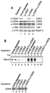
The effects of the CTD kinase activities of CDK7 and CDK9 on HIV-1 transcription. (A) Western blot analysis of mock-depleted, CDK7-depleted, CDK9-depleted, or CDK7- and CDK9-depleted extracts with anti-CDK7, anti-CDK9, anti-CDK8, anti-TBP, or anti-CTD of RNAP II antibody. Panels 3, 4, and 5 demonstrate that depletions did not change the level of CDK8, other general transcription factors, or RNAP II. (B) The effects of the CTD kinase activities of CDK7 and CDK9 on HIV-1 transcription. Biotinylated HIV-1 LTR templates were incubated with mock-depleted, CDK8-depleted, CDK7-depleted, CDK9-depleted, and CDK7- and CDK9-depleted extracts in the absence (lanes 1 to 5) or presence (lanes 6 to 10) of Tat, and PICs were then purified with streptavidin-coated magnetic beads. In vitro transcription was done with the purified PICs, and transcripts were labeled with [α-32P]UTP and fractionated on 6% denaturing polyacrylamide gel containing 7 M urea in 1× TBE buffer (top). The Western blot analysis of kinase-depleted extracts was done with anti-CDK8 antibody (bottom).
FIG. 4
FIG. 4
Phosphorylation of RNAP II CTD in HIV-1 PICs. Biotinylated HIV-1 LTR templates were incubated with mock-depleted, CDK7-depleted, CDK9-depleted, or CDK7- and CDK9-depleted extracts in the absence (lanes 1 to 4) or presence (lanes 5 to 8) of Tat, and PICs were then purified with streptavidin-coated magnetic beads. Kinase reactions were performed with the purified PICs, and phosphorylated RNAP II was labeled with [γ-32P]ATP and immunoprecipitated (IP) with anti-CTD monoclonal antibody 8WG16.
FIG. 5
FIG. 5
CDK9 and CDK7 phosphorylated serine 2 and serine 5, respectively, of the RNAP II CTD in HIV-1 PICs. Biotinylated HIV-1 LTR templates were incubated with mock-depleted, CDK7-depleted, CDK9-depleted, or CDK7- and CDK9-depleted extracts in the absence (lanes 1 to 4) or presence (lanes 5 to 8) of Tat. PICs were then purified with streptavidin-coated magnetic beads. The PICs were then incubated with 50 μM ATP for 10 min in order to have RNAP II CTD phosphorylated and washed extensively. Western blot analysis of the PICs was done with anti-CTD monoclonal antibodies 8WG16, H5, or H14.
FIG. 6
FIG. 6
CTD phosphorylation by Tat-modified CDK9 is sensitive to DRB. Biotinylated HIV-1 LTR templates were incubated with CDK7-depleted extract in the presence of Tat, and PICs were then purified with streptavidin-coated magnetic beads. The inhibition assays were performed by incubating the purified PICs with ATP in the presence of different concentrations of DRB. Western blot analyses of the complexes were done with anti-CTD monoclonal antibody H5 or H14, and the activities were determined by direct quantitation using the Molecular Dynamics ImageQuant. The top curve (♦) indicates the inhibition of serine 2 phosphorylation, while the bottom curve (▪) indicates the inhibition of serine 5 phosphorylation.
FIG. 7
FIG. 7
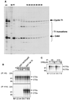
Stepwise walking of RNAP II elongation complexes and TAR-dependent rephosphorylation of RNAP II CTD during elongation. The purified PICs were incubated with 50 μM ATP for 10 min and then washed extensively. The PICs were walked to position U14 by incubation with 50 μM CTP, GTP, and UTP for 5 min at 30°C and then washed extensively. The TECs stalled at U14 were walked stepwise along the DNA by repeated incubation with different sets of three NTPs and then washed extensively to remove the unincorporated NTPs. (A) Western blot analysis of PICs and stalled TECs. (B and C) TAR-dependent rephosphorylation of RNAP II CTD during elongation. (D) Transcription facilitated by TAR-dependent rephosphorylation of RNAP II CTD during elongation.
FIG. 8
FIG. 8
The recruitment of the Tat–P-TEFb complex by TAR binding during elongation. Biotinylated wild-type (WT) or TAR mutant (TM26) HIV-1 LTR templates were incubated with CDK9-depleted extract, and PICs were purified with streptavidin-coated magnetic beads. The transcription complexes were walked stepwise along the templates to +79 (as described in Materials and Methods). The runoff transcription was then performed by incubating the TECs stalled at +79 with ATP, CTP, GTP, and [α-32P]UTP. P-TEFb and Tat were added at different sites, as indicated.
FIG. 9
FIG. 9
Tat directly modified the substrate specificity of P-TEFb in a CTD kinase assay. (A) A silver-stained SDS-polyacrylamide gel electrophoresis of recombinant P-TEFb fractions. (B) CTD kinase assay. The assays were performed by mixing 100 ng of GST-CTD, 100 ng of P-TEFb, 10 μM ATP, and 10 μCi of [γ-32P]ATP in the absence or presence of Tat and incubating for 60 min at 23°C. The total reaction mixture volume was 30 μl, and the final conditions were 50 mM Tris-HCl (pH 7.5), 5 mM DTT, 5 mM MnCl2, 4 mM MgCl2, and 10 μM ZnSO4. The phosphorylated GST-CTD was then immunoprecipitated (IP) with anti-CTD monoclonal antibody H5 (top) or H14 (bottom) and fractionated by electrophoresis on SDS–8% polyacrylamide gels. Numbers at left represent molecular masses in kilodaltons. The labeled products were detected by PhosphorImager. Numbers at the top show the amounts of Tat (GST-Tat 72) or a Tat mutant (GST-Tat72Cys22) that were added, expressed in nanograms. Lane M, molecular mass marker. (C) DRB sensitivity assay of serine 2 phosphorylation (lanes 1 to 4) and serine 5 phosphorylation (lanes 5 to 8). Micromolar concentrations of DRB are expressed. Numbers at left, molecular masses in kilodaltons.

Similar articles

Cited by

References

    1. Alonso A, Derse D, Peterlin B M. Human chromosome 12 is required for optimal interactions between Tat and TAR of human immunodeficiency virus type 1 in rodent cells. J Virol. 1992;66:4617–4621. - PMC - PubMed
    1. Bieniasz P D, Grdina T A, Bogerd H P, Cullen B R. Recruitment of a protein complex containing Tat and cyclin T1 to TAR governs the species specificity of HIV-1 Tat. EMBO J. 1998;17:7056–7065. - PMC - PubMed
    1. Bieniasz P D, Grdina T A, Bogerd H P, Cullen B R. Recruitment of cyclin T1/P-TEFb to an HIV type 1 long terminal repeat promoter proximal RNA target is both necessary and sufficient for full activation of transcription. Proc Natl Acad Sci USA. 1999;96:7791–7796. - PMC - PubMed
    1. Boris-Lawrie K A, Brady J N, Kumar A. Sequences within the R region of the long terminal repeat activate basal transcription from the HIV-1 promoter. Gene Expr. 1992;2:215–230. - PMC - PubMed
    1. Chen D, Zhou Q. Tat activates human immunodeficiency virus type 1 transcriptional elongation independent of TFIIH kinase. Mol Cell Biol. 1999;19:2863–2871. - PMC - PubMed

MeSH terms

LinkOut - more resources