

Squamous epithelial proliferation induced by walleye dermal sarcoma retrovirus cyclin in transgenic mice
- PMID: 10811912
- PMCID: PMC18567
- DOI: 10.1073/pnas.110024497
Squamous epithelial proliferation induced by walleye dermal sarcoma retrovirus cyclin in transgenic mice
Abstract
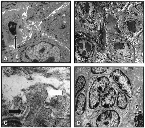
Walleye dermal sarcoma (WDS) is a common disease of walleye fish in the United States and Canada. These proliferative lesions are present autumn through winter and regress in the spring. Walleye dermal sarcoma virus (WDSV), a retrovirus distantly related to other members of the family Retroviridae, has been etiologically linked to the development of WDS. We have reported that the D-cyclin homologue [retroviral (rv) cyclin] encoded by WDSV rescues yeast conditionally deficient for cyclin synthesis from growth arrest and that WDSV-cyclin mRNA is present in developing tumors. These data strongly suggest that the rv-cyclin plays a central role in the development of WDS. To test the ability of the WDSV rv-cyclin to induce cell proliferation, we have generated transgenic mice expressing the rv-cyclin in squamous epithelia from the bovine keratin-5 promoter. The transgenic animals were smaller than littermates, had reduced numbers of hair follicles, and transgenic females did not lactate properly. Following injury the transgenic animals developed severe squamous epithelial hyperplasia and dysplasia with ultrastructural characteristics of neoplastic squamous epithelium. Immunocytochemistry studies demonstrated that the hyperplastic epithelium stained positive for cytokeratin and were abnormally differentiated. Furthermore, the rv-cyclin protein was detected in the thickened basal cell layers of the proliferating lesions. These data are the first to indicate that the highly divergent WDSV rv-cyclin is a very potent stimulator of eukaryotic cell proliferation and to demonstrate the potential of a cyclin homologue encoded by a retrovirus to induce hyperplastic skin lesions.
Figures


Similar articles
-
Walleye retroviruses associated with skin tumors and hyperplasias encode cyclin D homologs.J Virol. 1998 Nov;72(11):8765-71. doi: 10.1128/JVI.72.11.8765-8771.1998. J Virol. 1998. PMID: 9765420 Free PMC article.
-
Retroviral cyclin controls cyclin-dependent kinase 8-mediated transcription elongation and reinitiation.J Virol. 2015 May;89(10):5450-61. doi: 10.1128/JVI.00464-15. Epub 2015 Mar 4. J Virol. 2015. PMID: 25741012 Free PMC article.
-
Transgenic expression of walleye dermal sarcoma virus rv-cyclin (orfA) in zebrafish does not result in tissue proliferation.Mar Biotechnol (NY). 2011 Apr;13(2):142-50. doi: 10.1007/s10126-010-9274-2. Epub 2010 Mar 28. Mar Biotechnol (NY). 2011. PMID: 20349325 Free PMC article.
-
Pathology of tumors in fish associated with retroviruses: a review.Vet Pathol. 2013 May;50(3):390-403. doi: 10.1177/0300985813480529. Epub 2013 Mar 1. Vet Pathol. 2013. PMID: 23456970 Review.
-
Comparative pathogenesis of epsilonretroviruses.J Virol. 2003 Dec;77(23):12385-91. doi: 10.1128/jvi.77.23.12385-12391.2003. J Virol. 2003. PMID: 14610162 Free PMC article. Review. No abstract available.
Cited by
-
An activation domain within the walleye dermal sarcoma virus retroviral cyclin protein is essential for inhibition of the viral promoter.Virology. 2005 Nov 25;342(2):240-51. doi: 10.1016/j.virol.2005.08.011. Epub 2005 Sep 15. Virology. 2005. PMID: 16150476 Free PMC article.
-
Viruses of lower vertebrates.J Vet Med B Infect Dis Vet Public Health. 2001 Aug;48(6):403-75. doi: 10.1046/j.1439-0450.2001.00473.x. J Vet Med B Infect Dis Vet Public Health. 2001. PMID: 11550762 Free PMC article. Review.
-
Walleye dermal sarcoma virus cyclin interacts with components of the mediator complex and the RNA polymerase II holoenzyme.J Virol. 2002 Aug;76(16):8031-9. doi: 10.1128/jvi.76.16.8031-8039.2002. J Virol. 2002. PMID: 12134008 Free PMC article.
-
Exploitation of the Mediator complex by viruses.PLoS Pathog. 2022 Apr 21;18(4):e1010422. doi: 10.1371/journal.ppat.1010422. eCollection 2022 Apr. PLoS Pathog. 2022. PMID: 35446926 Free PMC article. No abstract available.
-
Cyclin-cyclin-dependent kinase regulatory response is linked to substrate recognition.J Biol Chem. 2011 Mar 18;286(11):9713-25. doi: 10.1074/jbc.M110.173872. Epub 2011 Jan 13. J Biol Chem. 2011. PMID: 21233209 Free PMC article.
References
-
- Walker R. Natl Cancer Inst Monogr. 1969;31:195–207. - PubMed
-
- Walker R. Anat Rec. 1947;99:559–560. - PubMed
-
- Yamamoto T, Kelley R K, Nielsen O. Fish Pathol. 1985;20:361–372.
-
- Bowser P R, Wolfe M J, Forney J L, Wooster G A. J Wildlife Dis. 1988;24:292–298. - PubMed
-
- Anders K, Yoshimizu M. Dis Aquat Org. 1994;19:215–232.
Publication types
MeSH terms
Substances
Grants and funding
LinkOut - more resources
Full Text Sources
Molecular Biology Databases
Research Materials
