. 2000 May 15;20(10):3915-25.
doi: 10.1523/JNEUROSCI.20-10-03915.2000.

Do glia have heart? Expression and functional role for ether-a-go-go currents in hippocampal astrocytes

Affiliations

Do glia have heart? Expression and functional role for ether-a-go-go currents in hippocampal astrocytes

A Emmi et al. J Neurosci. .

Abstract

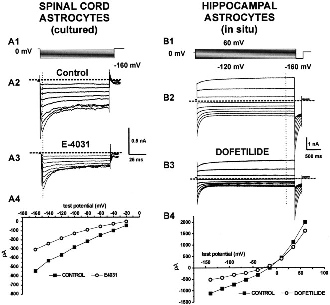
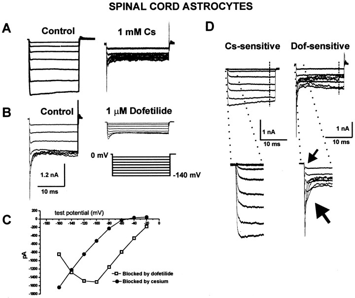
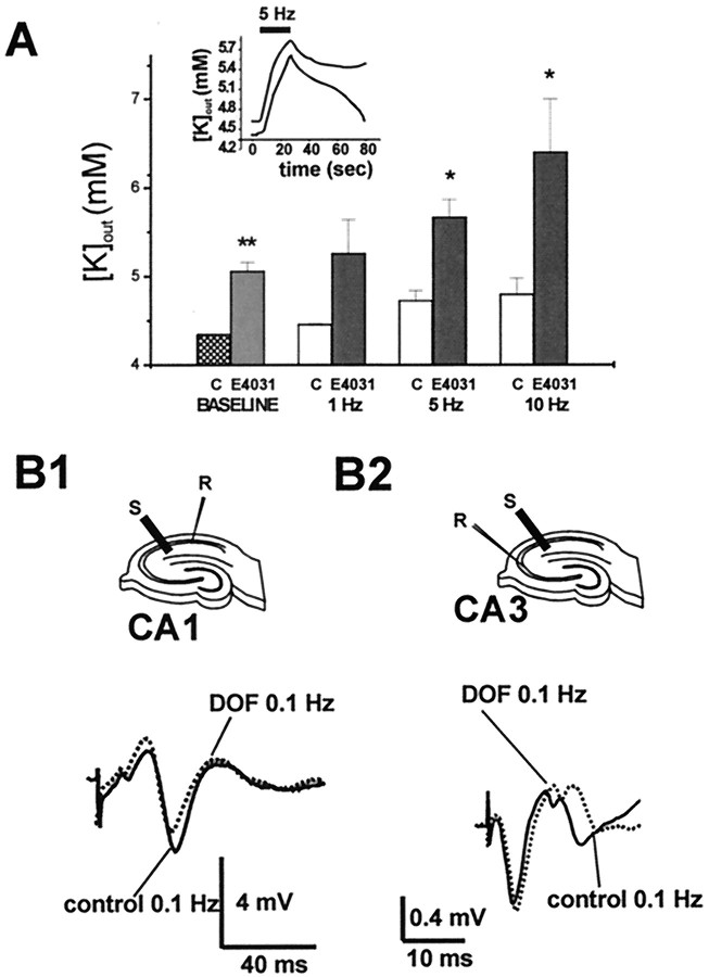
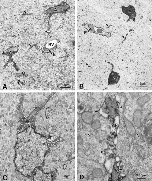
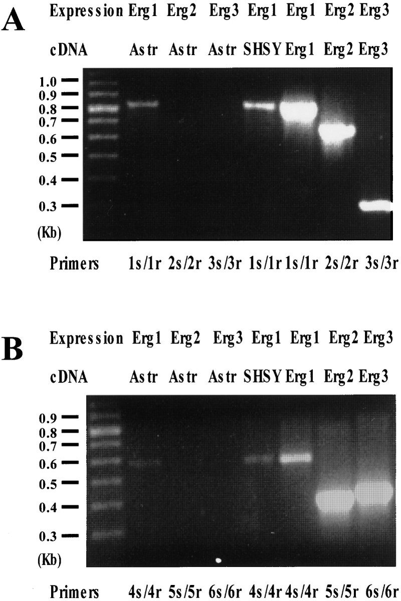
Potassium homeostasis plays an important role in the control of neuronal excitability, and diminished buffering of extracellular K results in neuronal Hyperexcitability and abnormal synchronization. Astrocytes are the cellular elements primarily involved in this process. Potassium uptake into astrocytes occurs, at least in part, through voltage-dependent channels, but the exact mechanisms involved are not fully understood. Although most glial recordings reveal expression of inward rectifier currents (K(IR)), it is not clear how spatial buffering consisting of accumulation and release of potassium may be mediated by exclusively inward potassium fluxes. We hypothesized that a combination of inward and outward rectifiers cooperate in the process of spatial buffering. Given the pharmacological properties of potassium homeostasis (sensitivity to Cs(+)), members of the ether-a-go-go (ERG) channel family widely expressed in the nervous system could underlie part of the process. We used electrophysiological recordings and pharmacological manipulations to demonstrate the expression of ERG-type currents in cultured and in situ hippocampal astrocytes. Specific ERG blockers (dofetilide and E 4031) inhibited hyperpolarization- and depolarization-activated glial currents, and ERG blockade impaired clearance of extracellular potassium with little direct effect on hippocampal neuron excitability. Immunocytochemical analysis revealed ERG protein mostly confined to astrocytes; ERG immunoreactivity was absent in presynaptic and postsynaptic elements, but pronounced in glia surrounding the synaptic cleft. Oligodendroglia did not reveal ERG immunoreactivity. Intense immunoreactivity was also found in perivascular astrocytic end feet at the blood-brain barrier. cDNA amplification showed that cortical astrocytes selectively express HERG1, but not HERG2-3 genes. This study provides insight into a possible physiological role of hippocampal ERG channels and links activation of ERG to control of potassium homeostasis.


Figures

Fig. 1.
Fig. 1.
Pharmacological manipulation reveals ERG-type currents in astrocytes: effects of E4031 or dofetilide. A1, B1, Voltage-clamp protocols used to evoke whole-cell currents.A2, B2, Currents evoked in spinal cord ([K+]out = 40 mm) and in hippocampal slice ([K+]out = 4.35 mm) astrocytes. Note the characteristic time-dependent deactivation of inward currents at negative potentials (A2) and the lack of inactivation after membrane depolarization (B2). A3, B3, Partial blockade of astrocytic currents by the specific ERG channel blocker E4031 (100 nm) or dofetilide (1000 nm). Residual E4031-insensitive currents could be recorded even after prolonged exposures to the drug (>15 min). A4, B4,I–V plots determined by measuring steady-state currents before and after exposure to E4031 or dofetilide; the vertical dotted lines in A2, A3, B2, andB3 represent the time point at which current measurements were taken.
Fig. 2.
Fig. 2.
Coexistence of ERG-like and KIRcurrents: sensitivity to both dofetilide and Cs+.A, Cesium (1 mm) blockade of inward currents in cultured spinal cord astrocytes bathed in elevated [K+]out. Note that that theI–V profile of the subtraction current shown inD (IControlIcs) is characterized by inward going rectification (C, filled symbols). In contrast, dofetilide block revealed a current (B) with complex voltage dependency and s-shaped behavior of theI–V relationship (C). The data points used to construct the I–V curve inC were obtained from the subtraction currents inD and were measured at the end of the voltage steps, as indicated by the dashed lines. D,Cs+- and dofetilide-sensitive currents have different kinetic properties. Inward rectifier currents unmasked by Cs+ blockade were characterized by little time-dependent inactivation, whereas dofetilide-sensitive currents inactivated rapidly at negative potentials (large arrow). Also note the apparent activation (or removal of inactivation) of the dofetilide-sensitive current at the beginning of the hyperpolarizing step (small arrow, bottom right panel).
Fig. 3.
Fig. 3.
Functional role for ERG currents in the hippocampus. A, Effect of ERG blockade by E4031 on CA1 resting potassium level and potassium accumulation induced by neuronal activity (inset). The resting (baseline) [K+]out increased slightly after the drug treatment, whereas larger increases were measured after afferent stimulation at 5 and 10 Hz (*p < 0.01; **p < 0.0001); the [K+]out changes induced by stimulation at low frequency (1 Hz) were not significantly affected by treatment with E-4031. B, Field recordings in CA1 and CA3 (cell body layer), in the presence of 100 nm E4031, fail to reveal any significant increase in neuronal excitability as reflected in population spike amplitude; the result suggests that the K changes produced under these conditions were too small to induce gross changes in neuronal excitability. The traces shown were taken after stimulation at 0.1 Hz, but no significant differences (control vs treatment) were observed after stimulation at higher (1, 5, and 10 Hz) frequencies.
Fig. 4.
Fig. 4.
Immunocytochemical localization of ERG (C1 antibody) channel protein in hippocampal astrocytes. Electron microscopy of biocytin-filled astrocytes (A, C) and immunocytochemistry of ERG (C1) channel protein in astrocytes (B, D) within the hippocampal CA1 subregion. Note the similar morphological features of hippocampal astrocytes of biocytin-filled and ERG-immunopositive cell bodies (A, B,) and their processes (arrows, C, D). ERG immunoreactivity was confined within the cell body cytoplasm of astrocytes and within large primary astrocytic processes and on small branched processes within the neuropil. Note that an oligodendrocyte (O) was immunonegative (A). Pyramidal cells did not show ERG immunoreactivity.
Fig. 5.
Fig. 5.
Immunocytochemical localization of ERG channel protein in astrocytes surrounding blood vessels in hippocampal CA1 (A,D); comparison with biocytin-filled cells (B,C). A, Low-power magnification of an ERG-immunoreactive astrocyte process (AP) forming an astrocytic endfoot (AE) around a capillary (BV). The endothelial cell (E) of the capillary wall does not show ERG immunoreactivity. As comparison, the capillary wall from a biocytin-filled astrocytic end foot is shown in B. D, Immunocytochemical localization of ERG channel protein in astrocytes surrounding blood vessels in hippocampal CA3 subregion; note the immunonegative basal lamina (BL). C, Comparison with the capillary wall of biocytin-filled astrocytes. E, Specificity of the ERG antibody is demonstrated in a control section after preabsorption of the primary antibody.
Fig. 6.
Fig. 6.
Perisynaptic glia, but not neuronal synaptic elements, are immunopositive for ERG protein. A, B, Electron micrographs of synapses in stratum radiatum of the CA1 subregion which are in close apposition to astrocytic processes that are immunopositive for ERG (C1) channel protein (AP). Note the absence of immunoreactivity in presynaptic (T) and postsynaptic (S) components. C, Immunonegative axon terminal (T) forming an asymmetric synapse with a neruonal cell body (P) and a small dendrite, both also immunonegative; D, Electron micrographs of synapses in stratum lucidum of the CA3 subregion showing ERG-immunopositive astrocytic processes (AP) in close apposition to immunonegative presynaptic and postsynaptic elements (MFB, mossy fiber bouton). E, Mossy fiber boutons (MFB) in synaptic contact with dendritic spines (D). Note that large portions of the mossy fiber bouton surface is apposed to astrocytic processes that are immunopositive for ERG channel protein (AP).
Fig. 7.
Fig. 7.
Expression of Erg1, Erg2, and Erg3 in rat astrocytes. A, RNA extracted from cultured rat cortical astrocytes (lanes 2, 3, and 4) or from SH-SY5Y cells (lane 5) were retrotranscribed as described in Materials and Methods. cDNAs from these RT reactions, as well as cloned cDNAs encoding for hErg1 (lane 6), rErg2 (lane 7), and rErg3 (lane 8), were amplified using the following primers: 1s/1r (specific for Erg1) for lanes 2, 5, and 6; 2s/2r (specific for Erg2) for lanes 3 and 7; and 3s/3r (specific for Erg3) for lanes 4 and 8. Molecular weights of the expected bands were: 805, 673, and 316 bp for 1s/1r, 2s/2r, and 3s/3r, respectively. Lane 1 shows the position of the molecular weight DNA markers (100 bp ladder from Pharmacia, Piscataway, NJ). Half (25 μl) of each amplification reaction per lane was loaded on the gel. B, RNA extracted from cultured rat cortical astrocytes (lanes 2, 3, and 4) or from SH-SY5Y cells (lane 5) were retrotranscribed as described in Materials and Methods. cDNAs from these RT reaction, as well as cloned cDNAs encoding for hErg1 (lane 6), rErg2 (lane 7), and rErg3 (lane 8), were amplified using the following primers: 4s/4r (specific for Erg1) for lanes 2, 5, and 6; 5s/5r (specific for Erg2) for lanes 3 and 7; and 6s/6r (specific for Erg3) for lanes 4 and 8. Molecular weights of the expected bands were: 597, 400, and 428 bp for 4s/4r, 5s/5r, and 6s/6r, respectively.Lane 1 shows the position of the molecular weight DNA markers (100 bp ladder from Pharmacia). Half (25 μl) of each amplification reaction per lane was loaded on the gel.

Similar articles

Cited by

References

    1. Ammann D. Ion sensitive electrodes. Springer; Berlin: 1986.
    1. Araque A, Parpura V, Sanzgiri RP, Haydon PG. Tripartite synapses: glia, the unacknowledged partner. Trends Neurosci. 1999;22:208–215. - PubMed
    1. Arcangeli A, Bianchi L, Becchetti A, Faravelli L, Coronnello M, Mini E, Olivotto M, Wanke E. A novel inward-rectifying K+ current with a cell-cycle dependence governs the resting potential of mammalian neuroblastoma cells. J Physiol (Lond) 1995;489:455–471. - PMC - PubMed
    1. Ballanyi K, Branchereau P, Champagnat J, Fortin G, Velluti J. Extracellular potassium, glial and neuronal potentials in the solitary complex of rat brainstem slices. Brain Res. 1993;607:99–107. - PubMed
    1. Bennett SA, Stevenson B, Staines WA, Roberts DC. Periodic acid-Schiff (PAS)-positive deposits in brain following kainic acid-induced seizures: relationships to fos induction, neuronal necrosis, reactive gliosis, and blood–brain barrier breakdown. Acta Neuropathol Berl. 1995;89:126–138. - PubMed

Publication types

MeSH terms