. 2000 Jun;74(11):5040-52.
doi: 10.1128/jvi.74.11.5040-5052.2000.

Loss of G(1)/S checkpoint in human immunodeficiency virus type 1-infected cells is associated with a lack of cyclin-dependent kinase inhibitor p21/Waf1

Affiliations

Loss of G(1)/S checkpoint in human immunodeficiency virus type 1-infected cells is associated with a lack of cyclin-dependent kinase inhibitor p21/Waf1

E Clark et al. J Virol. 2000 Jun.

Abstract

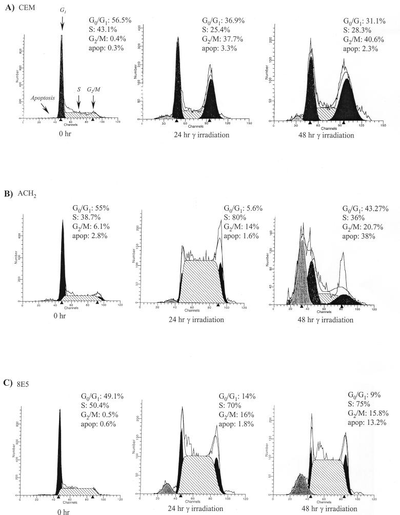
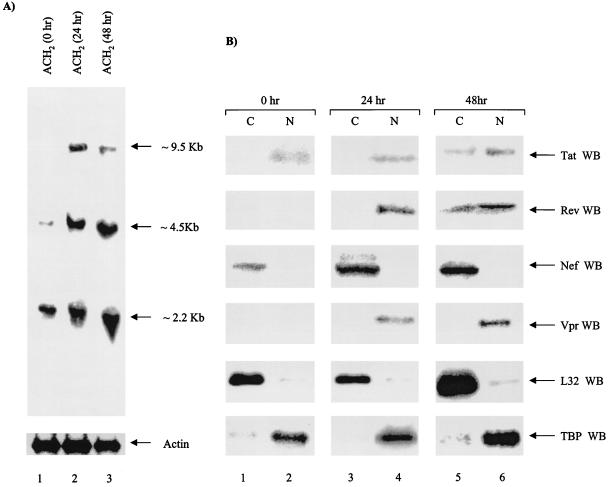
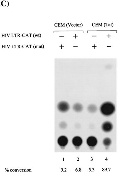
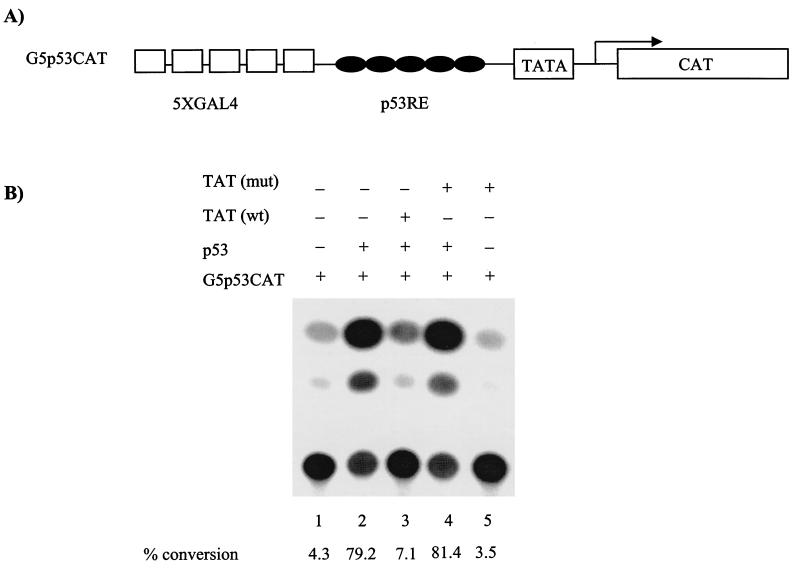
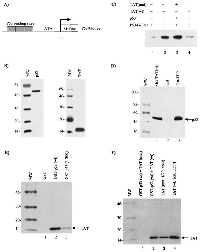
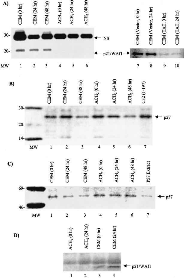
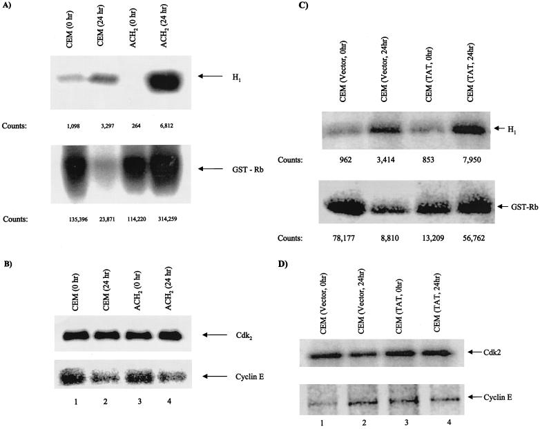
Productive high-titer infection by human immunodeficiency virus type 1 (HIV-1) requires the activation of target cells. Infection of quiescent peripheral CD4 lymphocytes by HIV-1 results in incomplete, labile reverse transcripts and lack of viral progeny formation. An interplay between Tat and p53 has previously been reported, where Tat inhibited the transcription of the p53 gene, which may aid in the development of AIDS-related malignancies, and p53 expression inhibited HIV-1 long terminal repeat transcription. Here, by using a well-defined and -characterized stress signal, gamma irradiation, we find that upon gamma irradiation, HIV-1-infected cells lose their G(1)/S checkpoints, enter the S phase inappropriately, and eventually apoptose. The loss of the G(1)/S checkpoint is associated with a loss of p21/Waf1 protein and increased activity of a major G(1)/S kinase, namely, cyclin E/cdk2. The p21/Waf1 protein, a known cyclin-dependent kinase inhibitor, interacts with the cdk2/cyclin E complex and inhibits progression of cells into S phase. We find that loss of the G(1)/S checkpoint in HIV-1-infected cells may in part be due to Tat's ability to bind p53 (a known activator of the p21/Waf1 promoter) and sequester its transactivation activity, as seen in both in vivo and in vitro transcription assays. The loss of p21/Waf1 in HIV-1-infected cells was specific to p21/Waf1 and did not occur with other KIP family members, such as p27 (KIP1) and p57 (KIP2). Finally, the advantage of a loss of the G(1)/S checkpoint for HIV-1 per se may be that it pushes the host cell into the S phase, which may then allow subsequent virus-associated processes, such as RNA splicing, transport, translation, and packaging of virion-specific genes, to occur.


Figures

FIG. 1
FIG. 1
Effect of gamma irradiation on HIV-infected and uninfected cells. The cells were grown to the mid-log phase of growth, serum starved for 3 days in 1% FBS, gamma irradiated, and processed at various time points for FACS analysis. (A) Parental CEM (12D7) cells; (B and C) HIV-1-integrated latently infected cells with wild-type (ACH2) and reverse transcription mutant (8E5) proviral genomes. Each panel shows cell cycle histogram profiles and percentages of cell numbers at various stages of the cell cycle. apop, percentage of cells that were apoptosing at each 0-, 24-, and 48-h time point. Apoptosis, G1, S, and G2/M peaks are indicated by arrows. Apoptotic cells are cells that have exited the cell cycle and are about to apoptose. We normally gate on live cells for flow cytometry, but the cell cycle parameters were calculated after the apoptotic cells were additionally gated out. More information on gating will be provided upon request.
FIG. 2
FIG. 2
Gene expression and translation of HIV-1-infected cells following gamma irradiation. (A) Twenty micrograms of total RNA was separated on an RNA-formaldehyde-agarose gel, transferred, and probed with full-length labeled HIV-1 genomic RNA messages corresponding to a 2.2-kb collection of doubly spliced regulatory proteins, such as Tat, Nef, Rev, and Vpr. The ∼4.5-kb messages correspond to the Gag, Pol, and Env singly spliced messages, and the ∼9.5-kb messages represent full genomic RNA that is packaged into the virion. The blot was later stripped and used to probe with a 40-mer anti-sense actin probe (bottom). (B) Both cytoplasmic (50-μg) and nuclear (20-μg) extracts were processed from ACH2 cells and Western blotted for viral Tat, Nef, Rev, Vpr, cytoplasmic (L32) (C), and nuclear (TBP) (N) proteins. Forty-eight-hour samples were trichloroacetic acid precipitated prior to separation on a 4 to 20% Tris-glycine gel. WB, Western blotting.
FIG. 3
FIG. 3
Effect of Tat on G1/S checkpoint in CEM cells. Both CEM (Vector) and CEM (Tat) cells were grown in the presence of 100 μg of hygromycin/ml to the mid-log phase of growth. The cells were serum starved for 3 days prior to gamma irradiation. Following gamma irradiation, the cells were put in complete medium with 10% FBS and processed for FACS analysis at various time points. (A and B) Histograms and numbers of cells at various stages of the cell cycle. (C) Transfection of both cell types with either wild-type (wt) LTR-CAT or a TAR mutant (mut) (TM26) LTR-CAT in CEM (Vector) and CEM (Tat) cells. Five micrograms of each DNA was transfected (by electroporation) into the cells and processed 18 h later for CAT assay. Fifty micrograms of total protein was used for the CAT assay, and acetylated products were run on thin-layer chromatography, exposed on a PhosphorImager cassette and counted with Molecular Dynamics software. +, present; −, absent.
FIG. 3
FIG. 3
Effect of Tat on G1/S checkpoint in CEM cells. Both CEM (Vector) and CEM (Tat) cells were grown in the presence of 100 μg of hygromycin/ml to the mid-log phase of growth. The cells were serum starved for 3 days prior to gamma irradiation. Following gamma irradiation, the cells were put in complete medium with 10% FBS and processed for FACS analysis at various time points. (A and B) Histograms and numbers of cells at various stages of the cell cycle. (C) Transfection of both cell types with either wild-type (wt) LTR-CAT or a TAR mutant (mut) (TM26) LTR-CAT in CEM (Vector) and CEM (Tat) cells. Five micrograms of each DNA was transfected (by electroporation) into the cells and processed 18 h later for CAT assay. Fifty micrograms of total protein was used for the CAT assay, and acetylated products were run on thin-layer chromatography, exposed on a PhosphorImager cassette and counted with Molecular Dynamics software. +, present; −, absent.
FIG. 4
FIG. 4
In vivo effect of Tat on p53-activated transcription. CEM (12D7) cells were grown to mid-log phase and transfected with a reporter (G5P53CAT) and activator (CMV-p53) in the presence (+) or absence (−) of Tat expression vector. (A) Diagram of the reporter gene used for transfection studies, which contains five GAL4 binding sites and five DNA-responsive elements. (B) Transfection experiment in CEM cells using electroporation method. The samples were processed similarly to those in Fig. 3C.
FIG. 5
FIG. 5
In vitro analysis of Tat on P53-activated transcription. In vitro transcription assays were performed with a supercoiled G-free cassette DNA (P53/G-Free), CEM (12D7) whole-cell extract, and purified p53 and Tat proteins. (A) Diagram of the reporter G-Free plasmid. (B) Coomassie blue stain of p53 and Tat proteins used for in vitro transcription assays. (C) In vitro transcription with p53 (lane 2 was treated with monoclonal antibody PAb 421 to increase DNA binding) in the presence (+) or absence (−) of either mutant (lane 3, Tat 41 mutant [25]) (mut) or wild-type (lane 4) (wt) Tat. Neither wild-type nor mutant Tat protein alone activated P53/G-Free transcription (data not shown). (D) In vitro binding of GST-TBP (positive control), GST, or GST-Tat to 35S-labeled p53 (TNT; Promega). Samples were bound overnight and washed the next day, and bound p53 proteins were run on a 4 to 20% Tris-glycine gel, dried, and exposed to a PhosphorImager cassette. (E) Binding of 35S-labeled Tat to GST-p53 wild type or a mutant containing p53 residues 1 to 300. As previously reported (27), Tat binds to the C-terminal domain of p53. (F) Binding of either the wild-type or a 41 mutant of Tat to GST-p53. The experiments shown in panels E and F were performed in the presence of 50 μg of ethidium bromide/ml (to eliminate nonspecific binding to nucleic acids) and washed with TNE150 plus 0.1% NP-40 buffer prior to being loaded on SDS-PAGE.
FIG. 6
FIG. 6
Late-G1-phase cdkI KIP family members in infected and uninfected cells. Both CEM and ACH2 cells were gamma irradiated, and nuclear extracts were processed at various time points for cdkI levels. (A, B, and C) Western blots with anti-p21 (Waf1) monoclonal antibody (MAb) (A), anti-p27 MAb (B), and anti-p57 MAb (C). All samples were processed on a 10 to 20% Tricine gel (Novex), transferred to a polyvinylidene difluoride membrane, and Western blotted with appropriate antibodies. Antigen-antibody-associated complexes were detected with 125I-protein G (Amersham). The samples in panel A, lanes 7 to 10, were processed from CEM vector control and CEM Tat-expressing cells. (D) Nuclear extracts from CEM and ACH2 after gamma irradiation and Western blotting for p21/Waf1 protein.
FIG. 7
FIG. 7
Immunoprecipitation of cyclin E-associated complex in both infected and uninfected cells. Infected and uninfected cell extracts from gamma-irradiated cells were treated for immunoprecipitation with polyclonal rabbit anti-cyclin E antibody at 4°C. The immune complexes were pelleted the next day with protein A plus protein G beads, washed, and used for kinase assays with histone H1 or GST-Rb as substrates. (A) Labeled products resolved by SDS–4 to 20% PAGE and their corresponding counts with the Molecular Dynamics PhosphorImager software. (B) Straight Western blots for cdk2 and cyclin E in infected and uninfected cells. (C and D) Similar to panels A and B except that CEM (Vector) and CEM (Tat) were used as the starting materials for immunoprecipitations.
FIG. 8
FIG. 8
Proposed model of Tat-p53 interaction and its functional consequences for p21/Waf1 levels. Upon DNA damage, the primary effect of p53 response at G1/S is upregulation of the p21/Waf1 gene product and its subsequent binding to the cyclin-cdk complex. The functional consequence of the p21-cyclin-cdk complex is to stop cells at the G1/S border prior to entry of the cells into the S phase. In HIV-infected cells, Tat inactivates p53 function, thus downregulating p21 expression and allowing loss of the G1/S checkpoint.

Similar articles

Cited by

References

    1. Biggs J R, Kraft A S. Inhibitors of cyclin-dependent kinase and cancer. J Mol Med. 1995;73:509–514. - PubMed
    1. Bohan C A, Kashanchi F, Ensoli B, Buonaguro L, Boris-Lawrie K A, Brady J N. Analysis of Tat transactivation of human immunodeficiency virus transcription in vitro. Gene Expr. 1992;2:391–407. - PMC - PubMed
    1. Brugarolas J, Chandrasekaran C, Gordon J I, Beach D, Jacks T, Hannon G J. Radiation-induced cell cycle arrest compromised by p21 deficiency. Nature. 1995;377:552–557. - PubMed
    1. Bunz F, Dutriaux A, Lengauer C, Waldman T, Zhou S, Brown J P, Sedivy J M, Kinzler K W, Vogelstein B. Requirement for p53 and p21 to sustain G2 arrest after DNA damage. Science. 1998;282:1497–1501. - PubMed
    1. Chang I J, McNulty E, Martin M. Human immunodeficiency viruses containing heterologous enhancer/promoters are replication competent and exhibit different lymphocyte tropisms. J Virol. 1993;67:743–752. - PMC - PubMed

Publication types

MeSH terms