

doi: 10.1073/pnas.080502497.
A self-recombining bacterial artificial chromosome and its application for analysis of herpesvirus pathogenesis
Affiliations
- PMID: 10781094
- PMCID: PMC18325
- DOI: 10.1073/pnas.080502497
Item in Clipboard
A self-recombining bacterial artificial chromosome and its application for analysis of herpesvirus pathogenesis
Proc Natl Acad Sci U S A.
.
Abstract
A self-recombining bacterial artificial chromosome (BAC) containing the 142-kb pseudorabies virus genome was constructed such that the viral genome is rapidly excised from the BAC vector backbone on delivery into mammalian cells. The recombination is mediated by loxP sites in the plasmid and Cre recombinase encoded within the BAC vector. A synthetic intron inserted in the middle of the cre ORF completely inhibits recombination in Escherichia coli, but is spliced out after delivery of the plasmid into mammalian cells. Recombination is efficient, and pure virus lacking the BAC vector backbone is immediately isolated from transfected mammalian cells without the need of serial passage or plaque purification.
Figures
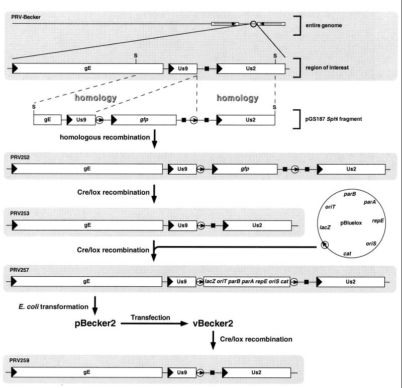
Construction of the loxP recombinant virus and insertion of the pBluelox vector into the viral genome are illustrated. The PRV-Becker 142-kb genome is diagrammed at top, with a portion of the right end of the genome expanded to show the gE, Us9, and Us2 genes. For clarity, only this region of all subsequent viral genomes is illustrated. All recombinations were carried out in transfected PK15 cells. Transformation of PRV257 DNA into E. coli yielded the full-length clone, pBecker2. Transfection of pBecker2 back into PK15 cells yielded vBecker2, the latter of which is indistinguishable from PRV257. Viral genes are represented as open rectangles with an arrowhead denoting direction of transcription. Open circles represent loxP sites with the enclosed arrow indicating relative orientation. Polyadenylation sequences are denoted by solid squares, and viral inverted repeats are shown as open rectangles with enclosed arrows (additional notations: lacZ, β-galactosidase gene; cat, chloramphenicol-resistance gene; repE, parA, and parB, replication and partitioning genes; oriS, BAC origin of replication; oriT, origin of transfer; S, SphI site).
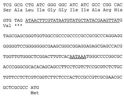
Sequence of the loxP insertion from PRV253. A portion of the Us9 ORF is shown with translation immediately upstream the loxP site, and the codon encoding the start methionine of Us2 is included at the end of the sequence. The loxP site is underlined, and the polyadenylation consensus for the Us9 gene is double underlined.
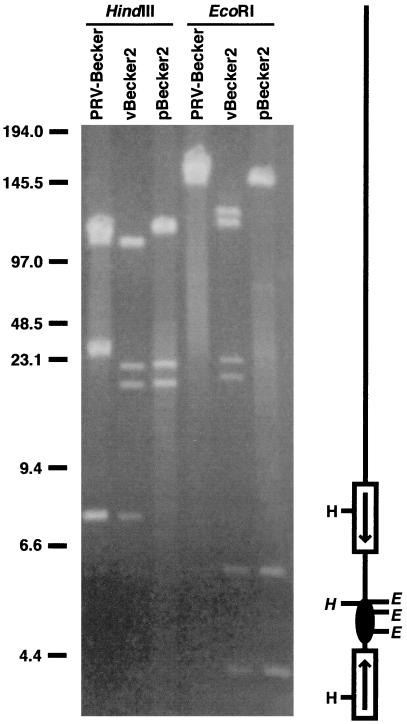
Pulse-field gel electrophoresis of HindIII- and EcoRI-digested viral nucleocapsid (PRV-Becker and vBecker2) and E. coli plasmid (pBecker2) DNA. To the right of the gel is a diagram of the viral DNA, indicating positions of HindIII sites at left and EcoRI sites at right. The BAC vector is shown as a solid ellipse. Restriction sites within the BAC vector insertion are shown in italics, which includes one HindIII site and all three EcoRI sites, and are absent from PRV-Becker nucleocapsid DNA. The viral inverted repeats (shown as open rectangles with enclosed arrows) isomerize during viral replication in mammalian cells, as evident in the EcoRI digestion of vBecker2 (reviewed in ref. 23). The ends of the viral DNA are ligated together in the pBecker2 plasmid from E. coli. Size standards are indicated in kb.
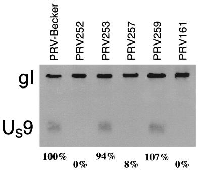
Detection of Us9 expression in infected PK15 cells by Western blot analysis. Lysates were probed with an anti-Us9 monoclonal antibody, and reprobed with an anti-gI polyclonal antiserum. The latter provided a gel loading control. Percentages listed at bottom are quantitation of Us9 signal relative to gI signal, as described in Materials and Methods. The quantitation was an average of two gels, only one of which is shown. PRV161 harbors a deletion in the Us9 gene.
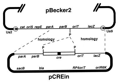
Construction of pBecker3 by insertion of the intron-containing Cre-expression cassette into pBecker2 is illustrated. A portion of the 156-kb pBecker2 plasmid is shown above, with the BAC vector region and loxP sites (circled arrows) oriented down. The pCREin vector is shown below with the Cre cassette and homologous flanking regions, the latter derived from pBluelox, oriented up. Homologous recombination in E. coli between the two plasmids yielded pBecker3. The pGS284 allelic exchange vector, which is the source of the pCREin backbone, and the E. coli allelic exchange method are described in ref. (notations are as in Fig. 1, with the addition of: sacB, levansucrase gene; bla, ampicillin-resistance gene; RP4oriT, origin of transfer; oriR6K, conditional origin of replication; P, PacI site).
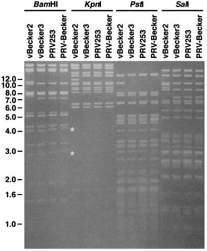
Gel electrophoresis of BamHI-, KpnI-, PstI-, and SalI-digested vBecker2, vBecker3, PRV253, and PRV-Becker viral nucleocapsid DNA. Restriction fragments unique to vBecker2 are all accounted for by the pBluelox insertion (data not shown). The restriction fragment length polymorphisms observed at ≈3.0 kb and ≈4.0 kb in the BamHI samples (marked by the white asterisks) map to the viral inverted repeats immediately flanking either side of the unique-short region, which contain small direct repeats that fluctuate in copy number during viral replication in mammalian cells (24, 25). Size standards are indicated in kb.
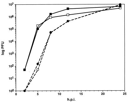
Single-step growth curves of wild-type PRV-Becker and vBecker3 in PK15 cells. Virus was harvested from both the media and cells at 2, 5, 8, 12, and 24 h postwash (squares, cells; circles, supernatants; open symbols, PRV-Becker; solid symbols, vBecker3).
Similar articles
-
Construction of a self-excisable bacterial artificial chromosome containing the human cytomegalovirus genome and mutagenesis of the diploid TRL/IRL13 gene.J Virol. 2002 Mar;76(5):2316-28. doi: 10.1128/jvi.76.5.2316-2328.2002. J Virol. 2002. PMID: 11836410 Free PMC article.
-
Construction of an infectious bacterial artificial chromosome clone of a pseudorabies virus variant: Reconstituted virus exhibited wild-type properties in vitro and in vivo.J Virol Methods. 2018 Sep;259:106-115. doi: 10.1016/j.jviromet.2018.06.004. Epub 2018 Jun 9. J Virol Methods. 2018. PMID: 29894711
-
[Construction of an infectious clone of pseudorabies virus strain ZJ genome maintained as a bacterial artificial chromosome].Bing Du Xue Bao. 2010 Jul;26(4):330-5. Bing Du Xue Bao. 2010. PMID: 20836388 Chinese.
-
New tools to convert bacterial artificial chromosomes to a self-excising design and their application to a herpes simplex virus type 1 infectious clone.BMC Biotechnol. 2016 Aug 31;16(1):64. doi: 10.1186/s12896-016-0295-4. BMC Biotechnol. 2016. PMID: 27580861 Free PMC article.
-
Genetic engineering of herpes simplex virus and vector genomes carrying loxP sites in cells expressing Cre recombinase.Virology. 2000 Feb 1;267(1):102-10. doi: 10.1006/viro.1999.0108. Virology. 2000. PMID: 10648187
Cited by
-
Infection of human cytomegalovirus in cultured human gingival tissue.Virol J. 2006 Oct 5;3:84. doi: 10.1186/1743-422X-3-84. Virol J. 2006. PMID: 17022821 Free PMC article.
-
Murine cytomegalovirus open reading frame M27 plays an important role in growth and virulence in mice.J Virol. 2001 Feb;75(4):1697-707. doi: 10.1128/JVI.75.4.1697-1707.2001. J Virol. 2001. PMID: 11160668 Free PMC article.
-
Murine gammaherpesvirus 68 open reading frame 31 is required for viral replication.J Virol. 2004 Jun;78(12):6610-20. doi: 10.1128/JVI.78.12.6610-6620.2004. J Virol. 2004. PMID: 15163752 Free PMC article.
-
Frequent coinfection of cells explains functional in vivo complementation between cytomegalovirus variants in the multiply infected host.J Virol. 2005 Aug;79(15):9492-502. doi: 10.1128/JVI.79.15.9492-9502.2005. J Virol. 2005. PMID: 16014912 Free PMC article.
-
Construction of a self-excisable bacterial artificial chromosome containing the human cytomegalovirus genome and mutagenesis of the diploid TRL/IRL13 gene.J Virol. 2002 Mar;76(5):2316-28. doi: 10.1128/jvi.76.5.2316-2328.2002. J Virol. 2002. PMID: 11836410 Free PMC article.
References
-
- O'Connor M, Peifer M, Bender W. Science. 1989;244:1307–1312. - PubMed
-
- Horsburgh B C, Hubinette M M, Qiang D, MacDonald M L, Tufaro F. Gene Ther. 1999;6:922–930. - PubMed
-
- Saeki Y, Ichikawa T, Saeki A, Chiocca E A, Tobler K, Ackermann M, Breakefield X O, Fraefel C. Hum Gene Ther. 1998;9:2787–2794. - PubMed
Publication types
MeSH terms
Substances
Grants and funding
LinkOut - more resources
Full Text Sources
Other Literature Sources
Miscellaneous
