Enterotoxigenic Escherichia coli secretes active heat-labile enterotoxin via outer membrane vesicles
- PMID: 10777535
- PMCID: PMC4347834
- DOI: 10.1074/jbc.275.17.12489
Enterotoxigenic Escherichia coli secretes active heat-labile enterotoxin via outer membrane vesicles
Abstract
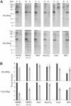
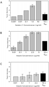
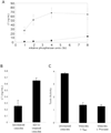
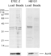
Escherichia coli and other Gram-negative bacteria produce outer membrane vesicles during normal growth. Vesicles may contribute to bacterial pathogenicity by serving as vehicles for toxins to encounter host cells. Enterotoxigenic E. coli (ETEC) vesicles were isolated from culture supernatants and purified on velocity gradients, thereby removing any soluble proteins and contaminants from the crude preparation. Vesicle protein profiles were similar but not identical to outer membranes and differed between strains. Most vesicle proteins were resistant to dissociation, suggesting they were integral or internal. Thin layer chromatography revealed that major outer membrane lipid components are present in vesicles. Cytoplasmic membranes and cytosol were absent in vesicles; however, alkaline phosphatase and AcrA, periplasmic residents, were localized to vesicles. In addition, physiologically active heat-labile enterotoxin (LT) was associated with ETEC vesicles. LT activity correlated directly with the gradient peak of vesicles, suggesting specific association, but could be removed from vesicles under dissociating conditions. Further analysis revealed that LT is enriched in vesicles and is located both inside and on the exterior of vesicles. The distinct protein composition of ETEC vesicles and their ability to carry toxin may contribute to the pathogenicity of ETEC strains.
Figures


Similar articles
-
The release of outer membrane vesicles from the strains of enterotoxigenic Escherichia coli.Microbiol Immunol. 1995;39(7):451-6. doi: 10.1111/j.1348-0421.1995.tb02228.x. Microbiol Immunol. 1995. PMID: 8569529
-
Porcine Enterotoxigenic Escherichia coli Strains Differ in Their Capacity To Secrete Enterotoxins through Varying YghG Levels.Appl Environ Microbiol. 2020 Nov 24;86(24):e00523-20. doi: 10.1128/AEM.00523-20. Print 2020 Nov 24. Appl Environ Microbiol. 2020. PMID: 32561576 Free PMC article.
-
Enterotoxigenic Escherichia coli vesicles target toxin delivery into mammalian cells.EMBO J. 2004 Nov 24;23(23):4538-49. doi: 10.1038/sj.emboj.7600471. Epub 2004 Nov 18. EMBO J. 2004. PMID: 15549136 Free PMC article.
-
Context-dependent activation kinetics elicited by soluble versus outer membrane vesicle-associated heat-labile enterotoxin.Infect Immun. 2011 Sep;79(9):3760-9. doi: 10.1128/IAI.05336-11. Epub 2011 Jun 27. Infect Immun. 2011. PMID: 21708992 Free PMC article.
-
Adhesion of enterotoxigenic Escherichia coli in humans and animals.Ciba Found Symp. 1981;80:142-60. doi: 10.1002/9780470720639.ch10. Ciba Found Symp. 1981. PMID: 6114818 Review.
Cited by
-
Interspecies communication in the gut, from bacterial delivery to host-cell response.J Physiol. 2012 Feb 1;590(3):433-40. doi: 10.1113/jphysiol.2011.220822. Epub 2011 Nov 21. J Physiol. 2012. PMID: 22106176 Free PMC article. Review.
-
Biogenesis of outer membrane vesicles in Serratia marcescens is thermoregulated and can be induced by activation of the Rcs phosphorelay system.J Bacteriol. 2012 Jun;194(12):3241-9. doi: 10.1128/JB.00016-12. Epub 2012 Apr 6. J Bacteriol. 2012. PMID: 22493021 Free PMC article.
-
CexE Is a Coat Protein and Virulence Factor of Diarrheagenic Pathogens.Front Microbiol. 2020 Jun 30;11:1374. doi: 10.3389/fmicb.2020.01374. eCollection 2020. Front Microbiol. 2020. PMID: 32714302 Free PMC article.
-
Outer Membrane Vesicles of Gram-Negative Bacteria: An Outlook on Biogenesis.Front Microbiol. 2021 Mar 4;12:557902. doi: 10.3389/fmicb.2021.557902. eCollection 2021. Front Microbiol. 2021. PMID: 33746909 Free PMC article. Review.
-
Mucosal immunization with Vibrio cholerae outer membrane vesicles provides maternal protection mediated by antilipopolysaccharide antibodies that inhibit bacterial motility.Infect Immun. 2010 Oct;78(10):4402-20. doi: 10.1128/IAI.00398-10. Epub 2010 Aug 2. Infect Immun. 2010. PMID: 20679439 Free PMC article.
References
Publication types
MeSH terms
Substances
Grants and funding
LinkOut - more resources
Full Text Sources
Other Literature Sources

