Disruption of the fibroblast growth factor-2 gene results in decreased bone mass and bone formation
- PMID: 10772653
- PMCID: PMC300831
- DOI: 10.1172/JCI8641
Disruption of the fibroblast growth factor-2 gene results in decreased bone mass and bone formation
Abstract
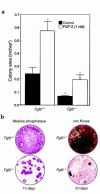
Basic fibroblast growth factor (FGF-2), an important modulator of cartilage and bone growth and differentiation, is expressed and regulated in osteoblastic cells. To investigate the role of FGF-2 in bone, we examined mice with a disruption of the Fgf2 gene. Measurement of trabecular bone architecture of the femoral metaphysis of Fgf2(+/+) and Fgf2(-/-) adult mice by micro-CT revealed that the platelike trabecular structures were markedly reduced and many of the connecting rods of trabecular bone were lost in the Fgf2(-/-) mice. Dynamic histomorphometry confirmed a significant decrease in trabecular bone volume, mineral apposition, and bone formation rates. In addition, there was a profound decreased mineralization of bone marrow stromal cultures from Fgf2(-/-) mice. This study provides strong evidence that FGF-2 helps determine bone mass as well as bone formation.
Figures


Similar articles
-
Reduced expression and function of bone morphogenetic protein-2 in bones of Fgf2 null mice.J Cell Biochem. 2008 Apr 15;103(6):1975-88. doi: 10.1002/jcb.21589. J Cell Biochem. 2008. PMID: 17955502
-
Impaired osteoclast formation in bone marrow cultures of Fgf2 null mice in response to parathyroid hormone.J Biol Chem. 2003 Jun 6;278(23):21258-66. doi: 10.1074/jbc.M302113200. Epub 2003 Mar 28. J Biol Chem. 2003. PMID: 12665515
-
Knockout of nuclear high molecular weight FGF2 isoforms in mice modulates bone and phosphate homeostasis.J Biol Chem. 2014 Dec 26;289(52):36303-14. doi: 10.1074/jbc.M114.619569. Epub 2014 Nov 11. J Biol Chem. 2014. PMID: 25389287 Free PMC article.
-
Disruption of the Fgf2 gene activates the adipogenic and suppresses the osteogenic program in mesenchymal marrow stromal stem cells.Bone. 2010 Aug;47(2):360-70. doi: 10.1016/j.bone.2010.05.021. Epub 2010 May 25. Bone. 2010. PMID: 20510392 Free PMC article.
-
Regulation of osteoblast differentiation: a novel function for fibroblast growth factor 8.Endocrinology. 2006 May;147(5):2171-82. doi: 10.1210/en.2005-1502. Epub 2006 Jan 26. Endocrinology. 2006. PMID: 16439448
Cited by
-
FGF signaling in the developing endochondral skeleton.Cytokine Growth Factor Rev. 2005 Apr;16(2):205-13. doi: 10.1016/j.cytogfr.2005.02.003. Epub 2005 Apr 1. Cytokine Growth Factor Rev. 2005. PMID: 15863035 Free PMC article. Review.
-
Nuclear isoforms of fibroblast growth factor 2 are novel inducers of hypophosphatemia via modulation of FGF23 and KLOTHO.J Biol Chem. 2010 Jan 22;285(4):2834-46. doi: 10.1074/jbc.M109.030577. Epub 2009 Nov 20. J Biol Chem. 2010. PMID: 19933269 Free PMC article.
-
Prolyl isomerase Pin1-mediated conformational change and subnuclear focal accumulation of Runx2 are crucial for fibroblast growth factor 2 (FGF2)-induced osteoblast differentiation.J Biol Chem. 2014 Mar 28;289(13):8828-38. doi: 10.1074/jbc.M113.516237. Epub 2014 Feb 7. J Biol Chem. 2014. PMID: 24509851 Free PMC article.
-
Identification of genes associated with the differentiation potential of adipose-derived stem cells to osteocytes or myocytes.Mol Cell Biochem. 2015 Feb;400(1-2):135-44. doi: 10.1007/s11010-014-2269-y. Epub 2014 Nov 11. Mol Cell Biochem. 2015. Retraction in: Mol Cell Biochem. 2015 Oct;408(1-2):297. doi: 10.1007/s11010-015-2500-5 PMID: 25385480 Retracted.
-
The Regulatory Role of Signaling Crosstalk in Hypertrophy of MSCs and Human Articular Chondrocytes.Int J Mol Sci. 2015 Aug 14;16(8):19225-47. doi: 10.3390/ijms160819225. Int J Mol Sci. 2015. PMID: 26287176 Free PMC article. Review.
References
-
- Gospodarowicz D. Basic science and pathology fibroblast growth factor chemical structure and biologic function. Clin Orthop Rel Res. 1990;257:231–248. - PubMed
-
- Hurley, M.M., and Florkiewicz, R. 1996. Fibroblast growth factor and vascular endothelial fibroblast growth factor families. In Principles of bone biology. Bilezikian, J.P., Raisz, L.G., and Rodan, G.A., editors. Academic Press. San Diego, CA. 627–645.
-
- Hauschka PV, Mavrakos AE, Iafrati MD, Doleman SE, Klagsbrun M. Growth factors in bone matrix. J Biol Chem. 1986;261:12665–12674. - PubMed
-
- Shing Y, et al. Heparin affinity: duplication of a tumour derived capillary endothelial cell growth factor. Science. 1984;223:1926–1928. - PubMed
-
- Hurley MM, Kessler M, Gronowicz G, Raisz LG. The interaction of heparin and basic fibroblast growth factor on collagen synthesis in 21-day fetal rat calvariae. Endocrinology. 1992;130:2675–2681. - PubMed
Publication types
MeSH terms
Substances
Grants and funding
LinkOut - more resources
Full Text Sources
Other Literature Sources
Molecular Biology Databases

