

Infectious agents are not necessary for murine atherogenesis
- PMID: 10770809
- PMCID: PMC2193142
- DOI: 10.1084/jem.191.8.1437
Infectious agents are not necessary for murine atherogenesis
Abstract
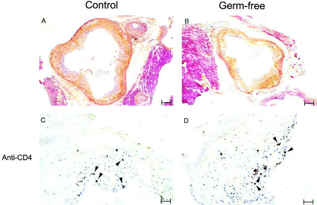
Recent work has revealed correlations between bacterial or viral infections and atherosclerotic disease. One particular bacterium, Chlamydia pneumoniae, has been observed at high frequency in human atherosclerotic lesions, prompting the hypothesis that infectious agents may be necessary for the initiation or progression of atherosclerosis. To determine if responses to gram-negative bacteria are necessary for atherogenesis, we first bred atherosclerosis-prone apolipoprotein (apo) E(-/)- (deficient) mice with animals incapable of responding to bacterial lipopolysaccharide. Atherogenesis was unaffected in doubly deficient animals. We further tested the role of infectious agents by creating a colony of germ-free apo E(-/)- mice. These animals are free of all microbial agents (bacterial, viral, and fungal). Atherosclerosis in germ-free animals was not measurably different from that in animals raised with ambient levels of microbial challenge. These studies show that infection is not necessary for murine atherosclerosis and that, unlike peptic ulcer, Koch's postulates cannot be fulfilled for any infectious agent in atherosclerosis.
Figures


Similar articles
-
Chlamydia pneumoniae infection does not induce or modify atherosclerosis in mice.Circulation. 2001 Jun 12;103(23):2834-8. doi: 10.1161/01.cir.103.23.2834. Circulation. 2001. PMID: 11401941
-
Murine models of Chlamydia pneumoniae infection and atherosclerosis.J Infect Dis. 1997 Apr;175(4):883-90. doi: 10.1086/513986. J Infect Dis. 1997. PMID: 9086145
-
Chlamydia pneumoniae infection accelerates the progression of atherosclerosis in apolipoprotein E-deficient mice.J Infect Dis. 1999 Jul;180(1):238-41. doi: 10.1086/314855. J Infect Dis. 1999. PMID: 10353889
-
Mouse models of Chlamydia pneumoniae infection and atherosclerosis.Am Heart J. 1999 Nov;138(5 Pt 2):S516-8. doi: 10.1016/s0002-8703(99)70290-5. Am Heart J. 1999. PMID: 10539863 Review. No abstract available.
-
Mouse models of C. pneumoniae infection and atherosclerosis.J Infect Dis. 2000 Jun;181 Suppl 3:S508-13. doi: 10.1086/315629. J Infect Dis. 2000. PMID: 10839749 Review.
Cited by
-
Mechanisms of NLRP3 priming in inflammaging and age related diseases.Cytokine Growth Factor Rev. 2020 Oct;55:15-25. doi: 10.1016/j.cytogfr.2020.08.003. Epub 2020 Aug 24. Cytokine Growth Factor Rev. 2020. PMID: 32883606 Free PMC article. Review.
-
Linking immune-mediated arterial inflammation and cholesterol-induced atherosclerosis in a transgenic mouse model.Proc Natl Acad Sci U S A. 2000 Nov 7;97(23):12752-7. doi: 10.1073/pnas.220427097. Proc Natl Acad Sci U S A. 2000. PMID: 11050173 Free PMC article.
-
Microbiome and Cardiovascular Disease.Handb Exp Pharmacol. 2022;270:311-334. doi: 10.1007/164_2020_356. Handb Exp Pharmacol. 2022. PMID: 32185503
-
The role of innate immunity in atherogenesis.J Lipid Res. 2009 Apr;50 Suppl(Suppl):S388-93. doi: 10.1194/jlr.R800100-JLR200. Epub 2008 Dec 22. J Lipid Res. 2009. PMID: 19106070 Free PMC article. Review.
-
Far from the eyes, close to the heart: dysbiosis of gut microbiota and cardiovascular consequences.Curr Cardiol Rep. 2014 Nov;16(11):540. doi: 10.1007/s11886-014-0540-1. Curr Cardiol Rep. 2014. PMID: 25303894 Free PMC article. Review.
References
-
- Fabricant C.G., Fabricant J., Minick C.R., Litrenta M.M. Herpesvirus-induced atherosclerosis in chickens. Fed. Proc. 1983;42:2476–2479. - PubMed
-
- Epstein S.E., Speir E., Zhou Y.F., Guetta E., Leon M., Finkel T. The role of infection in restenosis and atherosclerosisfocus on cytomegalovirus Lancet 348Suppl.1996. 13 17 - PubMed
-
- Danesh J., Collins R., Peto R. Chronic infections and coronary heart diseaseis there a link? Lancet. 1997;350:430–436. - PubMed
MeSH terms
Substances
LinkOut - more resources
Full Text Sources
Other Literature Sources
Molecular Biology Databases
